
Remember me


A molecular docking study determined the binding affinity and interaction of bioactive compounds obtained from the GC-MS and LC-MS analysis of M. pudica L. with the target protein DPP-4 (PDB ID: 3VJM). According to the docking results, all the selected compounds showed favourable binding affinities toward DPP-4, with scores ranging between − 9.34 and − 4.38 kcal/mol. The data of the co-crystal ligand and the top 10 compounds are shown in Table 1. The compound with the highest binding affinity was Cassiaoccidentalin B among the docked compounds, followed by Isoorientin, Isoquercetin and Apigetrin. These findings suggest that these compounds may have potent inhibitory potential against DPP-4. The docking score for the co-crystallized ligand (control) was − 8.74 kcal/mol, signifying that several test compounds showed better or equivalent binding affinity than the reference ligand. For all the docked compounds, the interactions with DPP-4 included H-bonding, Pi-Pi stacking and Pi-cation interactions, which are crucial for ligand binding. Cassiaoccidentalin B formed H-bonds and Pi-Pi interactions with critical residues GLU 205, GLU 206, ARG 125, HIS 740, TYR 547 and PHE 357, implying strong binding stability. Isoorientin was primarily H-bonded with GLU 205, TYR 547, TYR 456, ASP 556 and GLN 553, denoting significant interaction with critical binding site residues. For Isoquercetin, hydrogen bonding and Pi-Pi stacking were observed with residues ARG 669, GLU 206, SER 209, TYR547, HIS 740, TYR 666 and ARG 125, which are known to be involved in DPP-4 inhibition. The interaction of Vitexin and Orientin with DPP-4 was established through hydrogen bonding and Pi-Pi interactions involving residues ARG 125, GLU 205 and GLU 206. The co-crystal ligand showed a docking score of -8.74 kcal/mol and interacted with GLU 205 and PHE 357 through hydrogen bonding and Pi-Pi interactions, as shown in Fig. 2. Of the three, Cassiaoccidentalin B, Isoorientin, and Isoquercetin had better-docked scores, implying their strong potential as DPP-4 inhibitors. These compounds exhibited more interactions than were recorded for the co-crystal ligand, thus enhancing their binding stability.
Table 1 Top-scored 10 compounds against DPP-4 and their amino acid interactions between ligands and DPP-4Fig. 2
Represents the interaction between the ligand and the receptor. The left side represents the two-dimensional (2D) aspect, while the right represents the three-dimensional (3D) complex protein-ligand interaction. Interaction of compounds cassiaoccidentalin B and DPP-4 (a, b); the interaction of compounds isoorientin and DPP-4 (c, d); the interaction of compounds Isoquercetin and DPP-4 (e, f); the interaction of compounds co-crystal ligand (Prolyl thiazolidine) and DPP-4 (g, h)
Molecular Dynamics SimulationBased on the molecular docking analysis, the top-three binding scored ligands with DPP-4 protein complexes, Cassiaoccidentalin B (-9.34 kcal/mol), Isoorientin (-9.19 kcal/mol), and isoquercetin (-8.81 kcal/mol), were selected for further molecular dynamics simulation studies using the DESMOND tool for 100 ns. The selection of the above three ligands is not only for binding score but also for amino acid interactions in the target protein. The Root Mean Square Deviation (RMSD) analysis gives an idea of the stability of the protein-ligand complex over the 100 ns simulation. The protein backbone RMSD was stable within an acceptable range for all three ligands, Cassiaoccidentalin B, Isoorientin and Isoquercetin, indicating that the protein remained intact. For Cassiaoccidentalin B, the RMSD initially showed slight fluctuations and then stabilized around 2.5 Å after 20 ns, which means sustained binding interaction. The ligand induces similar stability as Isoorientin and Isoquercetin, with RMSD values stabilizing at approximately 2.3 Å and 2.6 Å, respectively. From these observations regarding ligand RMSD values, it can be concluded that all three ligands were secured in the binding pocket with very minute fluctuation values, signifying strong interactions with the target protein (Figs. 3(a)-3(c)). Root Mean Square Fluctuation (RMSF) analysis was carried out to assess the flexibility of individual residues within the protein structure. The fluctuations were observed primarily at the N- and C-terminal regions, which is expected due to their inherent flexibility. Moderates fluctuate in loop regions while secondary structural elements, α-helices and β-sheets remain stable. Of all the ligands, Cassiaoccidentalin B showed slightly higher fluctuation in residues surrounding the active site. However, such variations did not affect the stability of the binding interaction. Isoorientin and Isoquercetin exhibited similar RMSF patterns with pronounced stability in the central areas of the protein (Figs. 3(d)-3(f)). The analysis of the protein-ligand interaction showed that all three ligands maintained consistent interactions throughout the simulation. Cassiaoccidentalin B had the highest number of hydrogen bonds and hydrophobic interactions, indicating a strong binding affinity. Isoorientin and Isoquercetin also exhibited stable hydrogen bonds and hydrophobic contacts, but to a slightly lesser extent. The water-mediated interactions were critical in helping the ligands bind stably in the binding pocket, which was responsible for their sustained binding during the simulation (Figs. 3(g)-3(i)). The trajectory analysis of protein-ligand interactions confirmed that all three compounds have significant potential for DPP-4 inhibition. Detailed mapping of ligand-protein interactions revealed constant engagement of critical amino acid residues involved in the binding process with the ligands. For instance, Cassiaoccidentalin B displayed interactions with critical residues for DPP-4 inhibition, namely SER 630, GLU 205 and PHE 357 (Figs. 3(j)-3(l)). Stable interactions with these residues were also observed for Isoorientin and Isoquercetin, thus confirming their binding potential. The timeline representation of interactions showed that these contacts persisted throughout the 100 ns simulation run, reinforcing these bioactive compounds’ stability and possible efficacy in targeting DPP-4. These results suggest that Cassiaoccidentalin B, Isoorientin and Isoquercetin are potential candidates for DPP-4 inhibition, with Cassiaoccidentalin B showing the most significant binding interactions (Figs. 3(m)-3(o)). Molecular modeling studies serve as an essential tool for discovering potential DPP-4 inhibitors. By modeling the interactions between potential inhibitors and the DPP-4 enzyme, researchers can predict which compounds are likely to bind efficiently and inhibit the enzyme’s function. This information is subsequently utilized to direct additional in vitro and in vivo investigations, ultimately facilitating the expedited identification and development of novel antidiabetic drugs.
Fig. 3
Plotting the root mean square deviation (RMSD) research for a 100-nanoseconds molecular dynamics (MD) simulation of the docked complex between cassiaoccidentalin B and DPP-4 (a), Isoorientin and DPP-4 (b), Isoquercetin and DPP-4 docked complex (c). Root mean square fluctuation of the cassiaoccidentalin B for characterizing changes in the ligand atom positions (d), root mean square fluctuation of the isoorientin for characterizing changes in the ligand atom positions (e), root mean square fluctuation of the isoquercetin for characterizing changes in the ligand atom positions (f). Percentage of amino acid and water-mediated interactions in MD simulations with cassiaoccidentalin B (g), Isoorientin (h), Isoquercetin (i). The timeline representation of the cassiaoccidentalin B (j) DPP-4-cassiaoccidentalin B contacts concerning the amino acids in the target (m). The timeline representation of the isoorientin (k) DPP-4-isoorientin contacts concerning the amino acids in the target (n). The timeline representation of the isoquercetin (l) DPP-4-isoquercetin contacts concerning the amino acids in the target (o)
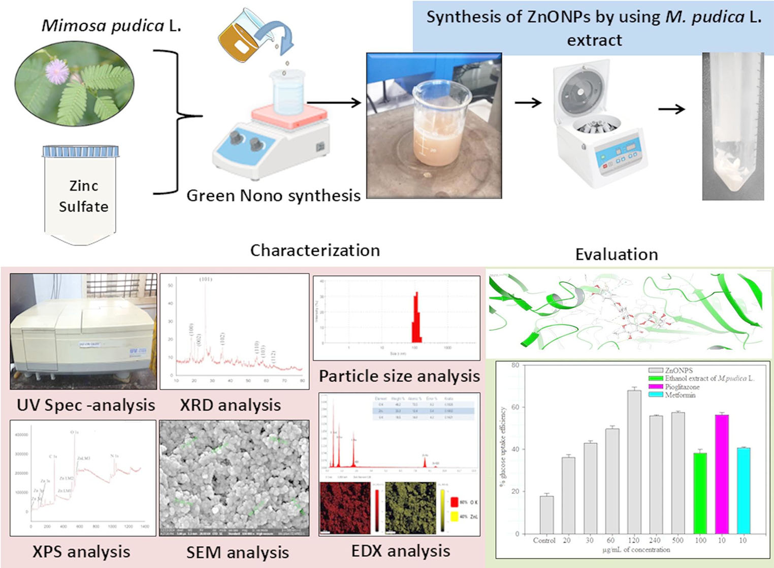
UV-visible Spectra AnalysisWhen M. pudica ethanol extract was combined with zinc sulfate precursor, a white precipitate developed, indicating the synthesis of ZnONPs with an 86.3% yield. The synthesized ZnONPs were sonicated for 10 min after becoming soluble in deionized water to examine the UV-visible spectra. The UV-visible wavelength range was 200–500 nm. The UV-visible spectra indicated the effective synthesis of ZnONPs, which showed a prominent absorption peak around 360 nm (Fig. 4). The absorption peak of ZnONPs coincides with the synthesized ZnONPs from Papaver somniferum L [41]., Alnus nepalensis D. Don [42] and Talaromyces islandicus-mediated ZnONPs [36]. Our current research validated these outcomes. The absorption band that extends to longer wavelengths may result from the movement of the electronic cloud across the ZnONPs framework. UV-visible spectroscopy analysis was conducted on the ethanol extract of M. pudica, revealing a peak at a wavelength of 610 nm (Fig. 4). The M. pudica extract contains alkaloids, flavonoids, proteins, and antioxidant components, and these compounds act as reducing agents, effectively converting zinc ions (Zn2+) into ZnO. The presence of these compounds in the plant extract could donate the electrons that could reduce zinc metal salt, resulting in the formation of ZnONPs. This study proved that ZnONPs may be generated in massive quantities using chemically comparable plant-mediated production methodologies.
Fig. 4
UV–visible absorption spectra of ethanol extract of M. pudica-mediated synthesized ZnONPs and ethanol extract of Mimosa pudica
FTIR AnalysisThe functional groups in the M. pudica extract that could act as reducing agents as well as stabilizing agents during the synthesis of ZnONPs were identified using FTIR spectroscopy. The present study identified that zinc sulfate was reduced into ZnONPs by the bioactive compounds of M. pudica. Fig. 5; Table 2 illustrate the FTIR spectral peak and their assignments, signifying the green-synthesized ZnONPs and the ethanol extract from the roots of M. pudica. The FTIR spectra of the M. pudica extract-mediated synthesized ZnONPs exhibited a broad peak around 3315 cm–1, indicating the stretching and bending vibrations of the − OH group attributable to water adsorption on the metal’s surface. The absorption peaks observed at about 2976–2887 cm–1 indicated a C–H stretching vibration that might be obtained from plant bioactive compounds. The bioactive components in the plant extract facilitate the reduction of metal ions into nanoscale structures. On the other hand, the peak displayed at 1651 cm–1 corresponds to the stretching vibration properties of aromatic rings. The absorption peaks obtained at 1046 cm− 1 and 880 cm–1 correspond to the C–H in-plane and out-of-plane bending stretching. The peak detected at approximately 600 cm–1 signified the stretching vibrations of ZnO. Literature study suggested that C–H, C–O–C, and C = C groups of heterocyclic compounds could have stabilizing properties [43]. The metal (Zinc)-oxygen (ZnO) stretching absorption was often detected in the 400–800 cm− 1 [44]. These results agree with the FTIR spectra of ZnONPs previously published in various plant extracts [42]. The conversion of zinc ions into zinc oxide nanoparticles and their stabilization in an aqueous medium is ascribed to the bioactive constituents of plant extract, such as phenolic compounds, proteins, and steroids, as indicated by FTIR analysis results [45].
Fig. 5
FTIR spectra of ethanol extract of M. pudica (a) and biosynthesized ZnONPs (b)
Table 2 Positions of FTIR absorption peaks and their functional assignment groups for the ethanol extract of M. pudica root and their derived ZnONPsXRD AnalysisThe XRD analysis confirmed the crystallinity of the synthesized ZnONPs using an X-ray diffractometer. Fig. 6 displays the XRD pattern of the ethanol extract of M. pudica -extract mediated synthesized ZnONPs. The crystallinity of the powder resulted from the synthesis using ethanol extract from M. pudica. The diffraction peaks of the synthesized ZnONPs at 2θ values are 18.54°, 21.86°, 26.17°, 35.49°, 54.61°, 58.12°, and 64.13°, likened to planes such as (100), (002), (101), (102), (110), (103), and (112) are lattice planes. The diffraction peaks revealed that the synthesized ZnONPs are essentially crystalline. All prominent diffraction peaks in the XRD spectra corresponded with those documented in the Joint Committee on Powder Diffraction Standards (JCPDS, card No. 89-7102) [46], which confirmed the crystallographic Wurtzite structure [47]. Impurities of diffraction peaks were not detected, which proved the high purity of the products. The following Debye–Scherrer equation was used to measure the size of synthesized ZnONPs.
Fig. 6
XRD pattern of lyophilized powder of ZnONPs
D = kλ/βscosθ.
D represents the crystallite size, while k is contingent upon size. Debye–Scherrer constant (0.94 for spherical particles), λ represents the wavelength of incident X-radiation (1.548 Å), and βs denotes the full width at half maximum. The mean diameter of the synthesized ZnONPs was ascertained to be 45 nm. The observed size of the ZnONPs is consistent with the report of Shah Faisal et al. 2022 [48]. This discovery is analogous to previously documented dimensions, such as 11.9 nm [49], demonstrating consistency and reliability in the synthesis and characterization methodologies.
Particle Size AnalysisThe primary purpose of dynamic light scattering (DLS) analysis is to ascertain the particle sizes in various suspensions. The degree of particle aggregation in aqueous media was evaluated by the mean hydrodynamic particle diameter (d. nm) [50]. The intensity-weighted mean diameter (z-average) and zeta potential of ZnONPs mediated by M. pudica extract are determined using DLS, as illustrated in Figs. 7(a) and 7(b). The synthesized ZnONPs exhibit a particle size of around 50 to 100 nm, a polydispersity index of approximately 0.4, with a zeta potential of -0.153 mV. The results indicate that the nanoparticles are highly stable and monodisperse. Our DLS findings for ZnONPs are consistent with previous studies utilizing leaf extracts from Elaeagnus angustifolia L. for the synthesis of ZnONPs [51]. The XRD and this result show that the produced ZnONPs have high crystallinity. Since the generated ZnONPs particle size is about 100 nm, it is considered an excellent outcome.
Fig. 7
DLS analysis of the ethanol extract of M. pudica-mediated synthesized ZnONPs. Dynamic particle size distribution (a), and zeta potential (b) of ZnONPs
XPS AnalysisFig. 8 illustrates the high-resolution XPS survey spectra of the ethanol root extract of M. pudica-mediated synthesized ZnONPs. The substance’s composition was confirmed by the presence of carbon (C 1s), oxygen (O 1s), and nitrogen (N 1s). The survey study confirmed the presence of Zn through Zn (3p, 3d, 3s, LM1, LM2, and LM). The C 1s signal indicates that the C (H, C), C–N, C–O, C = O, and C–O–C groups from M. pudica extract phytocompounds exhibit a peak at 288 eV. The O1s peak at 587 eV arises from the–C–O–H group of M. pudica extract phytocompounds, while the N1s signal at 534 eV is attributed to the presence of the C–N bond in M. pudica extract phytocompounds. Zn exhibits many peaks at 15 eV (Zn 3d), 91 eV (Zn 3p), 114 eV (Zn 3s), 479 eV (Zn LM1), 501 eV (Zn LM2), and 558 eV (Zn LM3). These peaks signify the existence of Zn in the form of Zn (II) oxide [52].
Fig. 8 FESEM and EDAX
FESEM analysis was conducted to examine the dimensions and morphology of ZnONPs. Figs. 9(a)-9(d) illustrate the morphological characteristics at various magnifications and scales of synthesized ZnONPs, predominantly exhibiting a spherical morphology. The average size of ZnONPs was roughly 40 to 100 nm. The elemental composition and chemical analysis of synthesized ZnONPs were assessed using EDAX (Fig. 10(a)). The research examined Zn, O, and C within the ZnONPs. EDAX mapping revealed around 40% zinc and 60% oxygen (Figs. 10(b) and 10(c)). This study indicates that the presence of carbon signifies the integration of the capping agent in the ZnONPs. The reference indicated a comparable outcome for SEM analysis [53].
Fig. 9
Field emission scanning electron microscope images of biologically synthesized ZnONPs (a)–(d)
Fig. 10
EDAX pattern of synthesized ZnONPs (a), and EDAX elemental mapping of synthesized ZnONPs (b) and (c)
Cellular Viability Studies of ZnONPsThe MTT assay determined the in vitro cellular viability against the tested samples. It describes cellular proliferation, viability, and cytotoxicity and tracks how well cells respond to different stimuli following treatment in culture media. The cytotoxic effect of ZnONPs at various concentrations (20, 30, 60, 120, 240, and 500 µg) in 3T3-L1 adipocytes for 24 h is depicted in Fig. 11. The findings showed that the cell viability of 3T3-L1 adipocytes declines as the concentration of ZnONPs increases. 3T3-L1 adipocytes may maintain cellular viability at higher ZnONPs concentrations. Therefore, a 500 µg concentration of ZnONPs is primarily responsible for the observed cell viability of 73.47 ± 1.59% on 3T3-L1 adipocytes. Cell viability was observed at 84.56 ± 1.42% at 120 µg ZnONPs. This study proved no cytotoxic effects of M. pudica ethanol extract-mediated synthesized ZnONPs. Similarly, no cytotoxic effects were reported by Cyperus Rotundus grass extracts-mediated synthesis of zinc oxide nanostructures [54].
Fig. 11
The ability of cellular viability of synthesized ZnONPs at different concentrations. Data are presented as mean ± standard deviation (n = 3 in each group)
In Vitro Glucose Utilization Efficiency of ZnONPsUsing 6-NBDG, the impact of M. pudica-extract-mediated ZnONPs on glucose utilization efficiency was measured. Differentiated 3T3-L1 adipocytes were preincubated with the M. pudica L. extract (100 µg), varying concentrations of ZnONPs (20, 30, 60, 120, 240 and 500 µg), pioglitazone (10 µg), and metformin (10 µg) for 24 h. In comparison to 100 µg M. pudica L. extract (38.28 ± 1.64%), 10 µg pioglitazone (56.32 ± 1.08%), and 10 µg metformin (40.65 ± 0.39%), the adipocytes treated with 120 µg of ZnONPs demonstrated the highest uptake of glucose (6-NBDG) (68 ± 1.55%). 3T3-L1 adipocytes treated with ZnONPs exhibit a dose-dependent rise in glucose absorption, a highly promising finding that can be attributed to the impact of ZnONPs (Fig. 12). Additionally, in 3T3-L1 adipocytes, the effect of glucose uptake on 60 µg of ZnONPs was higher than that of metformin (49.68 ± 1.49%) and comparable to 10 µg pioglitazone (56.32 ± 1.08%). A class of glucose transporter proteins (GLUT4) facilitates glucose uptake in skeletal muscle and adipose tissue. When insulin levels are low, GLUT4 stays lodged in the vesicles [55]. Following its binding to the receptor, insulin phosphorylates the insulin receptor substrate (IRS), which triggers the activation of protein kinase B via a sequence of signaling cascade molecules [56]. As a result, GLUT4 can move from the vesicle to the plasma membrane. As a result, the free glucose diffuses into the cell more easily along its concentration gradient [55]. Numerous plant-derived compounds have been shown to improve glucose translocation and increase the insulin-dependent pathway [57,58,59]. Enhanced blood glucose levels and enhanced serum DPP-4 activity levels are hallmarks of type 1 and type 2 diabetes. Based on the results of our study, DPP-4 inhibition enhanced GLUT4 expression and membrane insertion, increasing adipocytes’ glucose absorption. These DDP-4 inhibitors of M. pudica bioactive substances may reduce insulin resistance in the liver or muscle cells. Although plant extract-mediated manufactured ZnONPs show potential in diabetes management, their limitations encompass conflicting findings across research, possible interactions between metal ions and drugs, and the necessity for more rigorous clinical trials to elucidate processes, delivery, and dosing comprehensively. Additionally, the complexity of plant extracts and the possibility for diversity in preparation and extraction procedures might lead to conflicting outcomes.
Fig. 12
In vitro glucose utilization efficiency of ethanol extract of M. pudica-mediated synthesized ZnONPs in 3T3-L1 adipocytes. Data are presented as mean ± standard deviation (n = 3 in each group). *p < 0.01 as compared to control, ±p < 0.01 as compared to metformin and control, #p < 0.05 as compared to 60 µg & 60 µg of ZnOPs and control
Comments (0)