BV2 Microglia Cells Culture
BV2 microglia cell line was used in this study. The cells were cultured in DMEM/F12 (Life Technologies, San Jose, CA, USA) supplemented with 10% FBS and 1% Penicillin and Streptomycin antibiotics in a 37 °C CO2 incubator.
Errα siRNA Transfection
For transfection, cells were seeded into 24-well plates and transfected with si-Errα (sequence 5′-GAGCAUCCCAGGCUUCUCAd′T d′T-3′)(Addgene, Cambridge, MA) by use of Lipofectamine 3000 reagent (ThermoFisher).
Cell Viability Analysis by Cell Counting kit-8 (CCK8) Assay
BV2 microglia cells (5 × 10³ cells/well) with different treatments were cultured in 96-well plates for 24 h. Absorbance at 450 nm was measured via the Microplate Reader (Thermo Fisher Scientific, San Jose, CA, USA) after incubating the cells with 10 μL sterile CCK8 dye (0.5 mg/mL, Sigma, St. Louis, USA) for 1, 2, and 3 h at 37 °C. Cell viability was calculated using the formula: (Absorbance of Test- Blank Absorbance)/(Absorbance of Control- Blank Absorbance) × 100%.
Enzyme-Linked Immunosorbent Assay (ELISA)
The mouse serum level of IL-10, IL-1β, TNF-α, NSE, S100β, iron, and Ferritin were measured by ELISA (IL-10, Solarbio, SEKM-0010; IL-1β, KeyGEN, KGC1201; TNF-α, Proteintech, KE10002;NSE, JINGMEI Bio, JM-02346M1; S100β, JONLN Bio, JL20189; Iron, Solarbio, BC1735; Ferritin, Elabscience, E-EL-M0491) according to the manufacturer’s protocols. In brief, serum samples were added to the capture antibody-coated wells of a 96-well microplate and were incubated overnight at 4 °C. After washing with the provided washing buffers, matched biotin-labeled detection antibody was then added to the wells for incubation. Horseradish peroxidase and 3,3′,5,5′-tetramethylbenzidine were used for detection. The reaction was stopped by the addition of 2 M sulfuric acid, and the absorbance at 450 nm was measured using a microtiter plate reader. A standard curve was obtained by serial dilutions that covered the entire detection range of the assay.
Western Blotting
Cells or tissues were lysed with the CytoblusterTM protein extraction reagent (Novagen) or the Membrane Protein Extraction Reagent (Thermo Fisher Scientific) with a protease inhibitor cocktail (Thermo Fisher Scientific). The concentration of protein was measured using the Pierce™ BCA Protein Assay Kit (Thermo Fisher Scientific).
Specifically, totally, it was determined by pipetting 10 μL each of standard and sample into a 96-well microplate, followed by the addition of Coomassie Plus Reagent. After mixing on a plate shaker for 30 s, the plate was incubated at room temperature for 10 min, and the absorbance was measured at 562 nm using a Synergy H1 microplate reader (BioTek Instruments, Winooski, VT, USA) (Supplymentary Fig. 3).
Protein samples were separated on 10% SDS-PAGE and transferred to PVDF membrane with the pre-chilled transfer buffer (25 mM Tris–Hcl, 193 mM glycine, 0.1% SDS, and 20% methanol). The filter papers (Bio-Rad) and sponge sheets used for the transfer were soaked in transfer buffer before use. The PVDF membrane was soaked in methanol and then washed in transfer buffer before use. The membrane was then blocked with 5% non-fat milk and incubated with antibody and the reference protein anti-β-actin antibody (1:5000 dilution; ZhongShan, Beijing, China), followed by horseradish peroxidase-conjugated against rabbit antibody (1:2000 dilution; ZhongShan, Beijing, China). Antibodies carried out in Western blot analyses were listed in Supplemental Table S1, followed by the corresponding biotinylated secondary antibodies. Blotting signals were revealed with ECL Plus (Beytime, Shanghai, China).
RNA Isolation and Quantitative Real-Time PCR (qPCR)
Total RNA was isolated from mouse hippocampus and BV2 cells using the Cell Total RNA Isolation Kit (RE-03111, Foregene, Chengdu, China). After digestion and lysis with Buffer cRL1, the cells were filtered by DNA-cleaning column to collect cell lysis supernatant and remove genomic DNA in the system. After adding 1.6 times Buffer cRL2, the mixture was transferred to the RNA-only column for purification. After centrifugation, 500 ul Buffer RW1 was used to remove protein, 700 ul Buffer RW2 was used to desalt twice, and then an empty tube centrifugation was applied to remove the residual ethanol. NOX1, NRF2, GPX4, and FTH1 were evaluated using qRT-PCR analysis (PrimeScript™ RT reagent Kit with gDNA Eraser, RR047 A, Takara, Kyoto, Japan; TB Green® Premix Ex Taq™ II, RR820 A, Takara, Kyoto, Japan). 18 s was used as internal controls. Primers used in the study are shown in Supplemental Table S2. PCR conditions for mRNA amplification were set as 95 °C for 30 s, followed by 40 cycles of 95 °C for 10 s and 60 °C for 30 s. mRNA transcription levels were normalized to endogenous 18 s and were quantified using the delta-delta CT method.
Flow Cytometry
Tissues were chopped and subject to enzymatic digestion in Hanks’ Balanced Salt Solution (HBSS) containing 2.5 mg/mL collagenase D (Roche) and 0.1 mg/mL DNase I (Roche) for 20 min at 37 °C with gentle shaking every 5 min. The supernatants were then passed through 100-mm filters. After washing once with MACS buffer (PBS pH 7.4 plus 2% FCS and 2 mM EDTA), erythrocytes were depleted using ACK lysis buffer, and cells were resuspended in HBSS. To perform BODIPY-C11 staining, cells were resuspended in 100 µl Hanks Balanced Salt Solution (HBSS, Gibco 14–025–092), containing 5 mM BODIPYI 581/591 C11(Thermo Fisher, D3861) and incubated for 15 min at 37 ℃ in a tissue culture incubator. Cells were washed and resuspended in 200 µl fresh HBSS and analyzed immediately with a flow cytometer (Deflex, Beckman). To detect Fe2+, cells were stained with FerroOrange (1 µM, MKBio, MX4559) at 37 ℃ for 30 min. GMFI was calculated. The data was analyzed using FlowJo version 10.1 software (Tree Star, Ashland, USA).
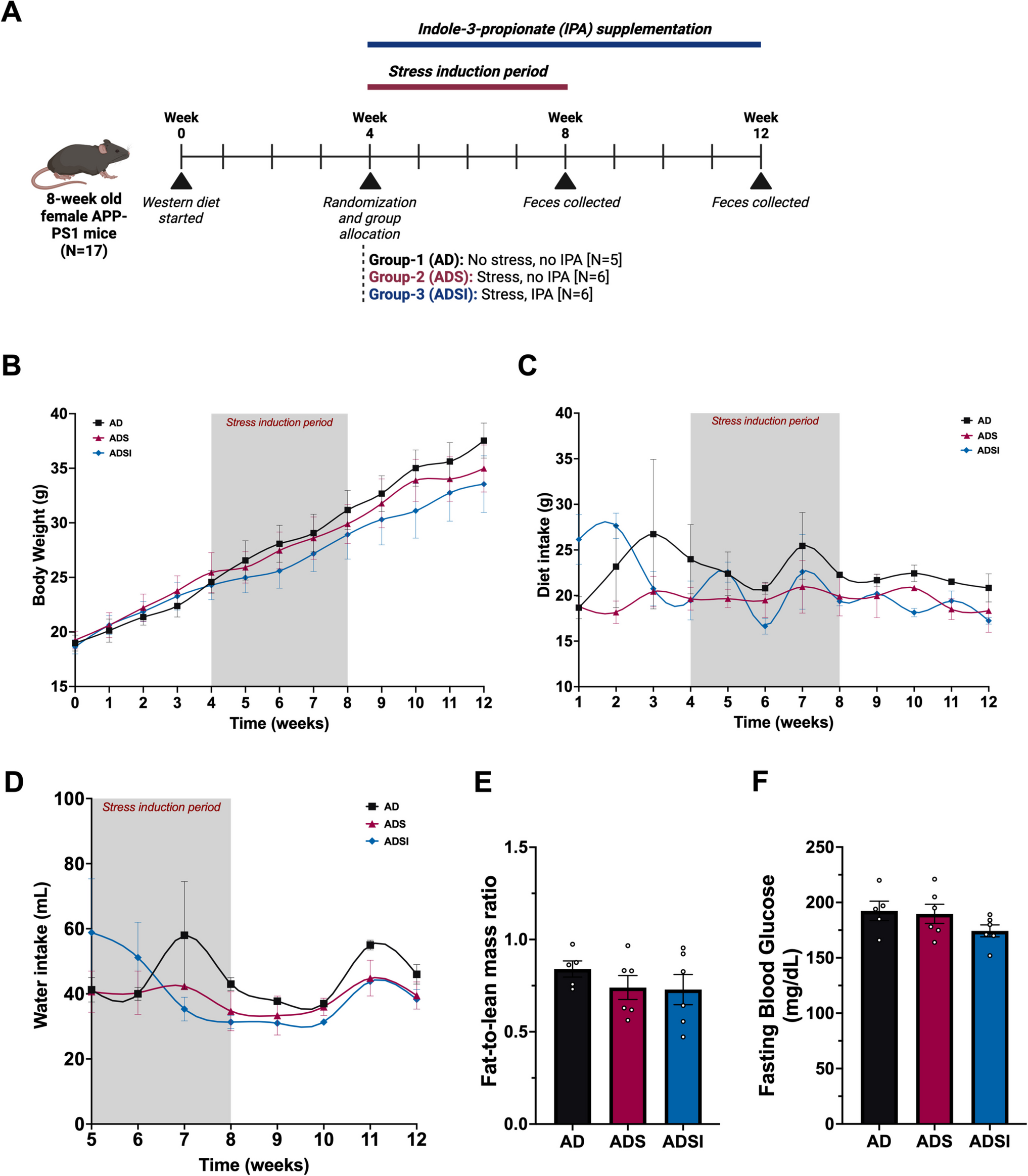
Animal study
The animal experimental protocols were approved by the Animal Ethics Committee of Shenzhen Huarui Model Organisms Biotechnology Co.. Male mice (6–8 weeks of age, 23–28 g) were housed in a specific pathogen-free SPF animal room with a 14 h light/10 h dark cycle.
Errα KO mouse model
Errαtransgenic mice were generated from C57BL/6 (Suzhou Cyagen Organisms Co.). gRNAs for CRISPER CAS-9 are listed in Supplemental Table S3.
SABD Mouse Model Establishment
A mouse model of SABD was constructed by Cecal Ligation and Puncture (CLP) and combined with neurological score (≤ 6 points). Neurological score evaluated the changes in neural reflex in mice, including corneal reflex, auricle reflex, tail flick reflex, righting reflex, and escape reflex. The occurrence of the above reflexes within 1 s was recorded as 2 points (normal reflexes), the occurrence of the above reflexes within 1–10 s was recorded as 1 point (weakened reflexes), the non-reflexes were recorded as 0 points, and the sum of the scores (0–10 points) was recorded. If the score was less than 6 points, the modeling of the mice with SABD was considered successful. We carried out experiments with male mice. From the moment of surgical completion, we established a comprehensive observation protocol with high-frequency monitoring of mouse survival status, recording survival data hourly throughout the 48-h observation period. During each observation interval, we systematically documented the survival status of each mouse, immediately recording the precise time of death when mortality occurred and maintaining continuous monitoring for surviving mice. This granular approach enabled us to capture subtle survival changes and provide comprehensive insights into the progression of sepsis-associated physiological dynamics.
Immunocytochemistry/Immunofluorescence Staining
BV2 microglia cells/mouse hippocampus tissues were fixed in 4% formalin overnight, rinsed in PBS, and transferred to 70% ethanol before standard processing to obtain paraffin-embedded Sects. (5 µm). Immunofluorescence staining was performed with the antibodies listed in Supplemental Table S4. Nuclei were stained with DAPI. The slides were observed under Zeiss Laser scanning confocal microscopes (LSM 700, 880 & 900) and were quantified by the Image-Pro Plus software (Media Cybernetics, Inc., MD, USA). In this study, we used inducible nitric oxide synthase (iNOS) as a specific marker for M1-type microglia and Arginase-1 (Arg-1) as a specific marker for M2-type microglia. iNOS is typically associated with the pro-inflammatory phenotype, while Arg-1 is closely related to the anti-inflammatory phenotype and tissue repair. These markers enabled us to accurately distinguish and quantify the changes in microglial polarization states [24].
Hematoxylin and Eosin (H&E) Stain
Sections of mouse hippocampus (5 µm) were subsequently dewaxed, rehydrated, and incubated in hematoxylin (Biyun Tian, #C0105S) for 8 min. After that, the sections were incubated by the differentiation fluid (Biyun Tian, #C0105S) for 1–5 s, Returned Blue Liquid for 1 min (Biyun Tian, #C0105S), eosin (Biyun Tian, #C0105S) for 1 min and dehydrated. The placenta sections through the sagittal midline were chosen for imaging by Zeiss scanning microscope.
Effect of Errα on NF-κB Pathway in Mouse Hippocampus and BV2 Cells
The expression of P-IKKα, P-IκBα, IκBα, and IKKβ in Errα KO mouse hippocampus and ErrαsiRNA BV2 cells were detected by Western bloting. Antibodies carried out were listed in Supplemental Table S5, followed by the corresponding biotinylated secondary antibodies.
Transmission Electron Microscope (TEM)
The tissue blocks were soaked in 2.5% glutaraldehyde, fixed with phosphate buffer for 2 h or more, rinsed with 0.1 M phosphate solution for 15 min (three times), and fixed with 1% Osmium tetroxide for 1–2 h. After that, they were rinsed with 0.1 M phosphoric buffer for 15 min (three times). Six paired experimental conditions (6 experimental groups and 6 corresponding control groups) were established, totaling 12 groups. Sequentially, 50% ethanol (15–20 min), 70% ethanol (15–20 min), 90% ethanol (15–20 min), 90% ethanol and 90% acetone (1:1) (15–20 min), 90% acetone (15–20 min), and 100% acetone at room temperature (15–20 min) for dehydration. Pure acetone embedding solution (2:1) at room temperature for 3–4 h, pure acetone embedding solution (1:2) at room temperature overnight, and pure embedding solution (EPON812) at 37 °C for 2–3 h. Finally, overnight in a 37 °C oven, then 12 h in a 45 °C oven, and 48 h in a 60 °C oven. Ultramicrotome (Leica EM UC7) was used to cure slices. The slices were double stained with 2% uranium acetate and lead citrate and then observed and photographed by transmission electron microscope HT7800 (80 kV). Fifteen electron micrographs were captured per group for systematic comparison.
Statistical Analysis
All the experimental data were expressed as the mean ± SD (standard deviation) and was analyzed by statistical software (SigmaPlot 11.0; Systat Software, Inc., USA and SigmaStat 2.03; Jandel Scientific, San Jose, CA, USA). Non-parametric analysis of variance on the rank test was used to identify group differences, followed by Mann–Whitney U-test as the post-test. A probability value of < 0.05 was considered statistically significant.


Comments (0)