
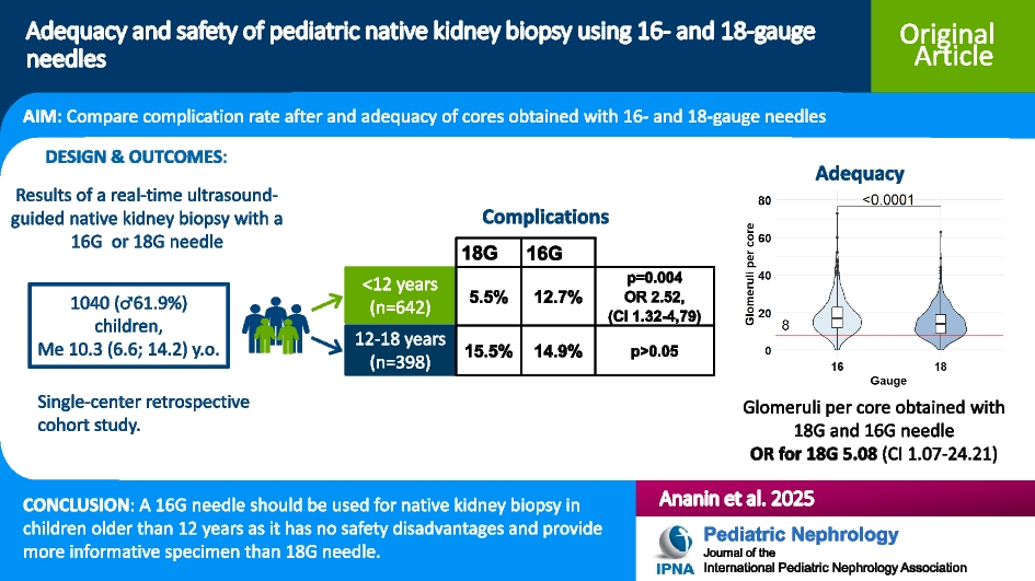
Remember me


A 16-year-old Caucasian female presented with vomiting and abdominal pain two days after initiating cefuroxime treatment for Escherichia coli pyelonephritis. Her medical history was unremarkable, aside from a previous episode of Escherichia coli (E. coli) pyelonephritis two months earlier, which was her first ever episode.
On examination she was afebrile (36.6 °C), tachycardic (150 bpm) and her blood pressure was 97/65 mmHg (< 5th percentile for age). Her height was 151.7 cm, weight 55 kg, BMI 23.9 (81.3%, normal BMI percentile is 5th–85th). Physical examination revealed pallor, abdominal and costovertebral angle tenderness.
Laboratory investigation demonstrated leukopenia (2.3, normal range (3.6–10) × 109/L), normal platelets count (172 × 106/L), plasma electrolytes were within normal range except for mild hyponatremia (134, normal range 135–145 mEq/L), minimal hyperglycemia (107, normal range 65–105 mg/dL), significantly elevated inflammatory markers (C-reactive protein 45.5 mg/dl (normal range 0–0.5 mg/dl), along with prolonged prothrombin time (18.9, normal range 10–14 s) and markedly increased D-dimer levels (4067, normal range 69–580 ng/ml). Additionally acute kidney injury was noted, with elevated plasma creatinine and decreased eGFR (1.11 mg/dl and 56.4 ml/min/1.73 m2 calculated using the Schwartz equation (normal range 0.5–0.9 mg/dl, 112 ± 13 ml/min/1.73 m2). Urinalysis was positive for leukocytes and nitrites. Taken together, the clinical and laboratory abnormalities were consistent with urosepsis. Therapeutic management included the initiation of ampicillin and gentamicin, in conjunction with repeated administration of intravenous fluids. Abdominal ultrasound demonstrated a calculus with minimal hydronephrosis. This finding was confirmed on non-contrast computed tomography (NCCT). Notably, an unexpected presence of air within the collecting system of the right kidney was observed, consistent with class 1 emphysematous pyelonephritis (EPN), defined as gas confined to the collecting system only [1] (Fig. 1).
Fig. 1
Left: Abdominal ultrasound demonstrating right kidney calculus, with minimal hydronephrosis. Right: Non-contrast computed tomography showing air in the collecting system of the right kidney, consistent with class 1 emphysematous pyelonephritis, defined as gas confined to the collecting system only [1]
Due to lack of clinical improvement and septic shock a 6/24 Fr double J stent was placed, resulting in the passage of turbid urine. The following day, she improved clinically, and her creatinine level normalized. Blood and urine cultures were positive for ampicillin-sensitive E. coli.
A week later, the patient underwent right ultrasound-guided-mini percutaneous-nephrolithotomy (PCNL). Kidney stone chemical analysis revealed mixed composition of calcium-oxalate-monohydrate (50%), protein (40%) and calcium-phosphate-carbonate (10%). A comprehensive metabolic evaluation for nephrolithiasis risk factors were negative, with no hyperoxaluria, hyperuricosuria or hypercalciuria. Citrate levels were within the normal range as well.
During a three-year follow-up period, the patient remained free of recurrent nephrolithiasis or pyelonephritis episodes.
Comments (0)