
Remember me


This study was approved by the Ethics in Research Committee at the Faculty of Dentistry, Ain Shams University, Egypt (approval number FDASU-ReclE121904). A total of 21 human mandibular premolar teeth extracted for orthodontic or periodontal reasons were collected for this study. All teeth showed fully developed apices and had no caries, cracks, restorations, erosion, abrasion, or fractures. They had similar root form and root canal shape, averaging cervico-occlusal length of the crown about 8.0 ± 0.5 mm and root length of about 14.5 ± 0.5 mm. Teeth were carefully cleaned and stored in distilled water at room temperature until used.
Using parallelometer dental surveyor, each root was embedded in Acrostone acrylic resin (Acrostone dental and medical supplies, Heliopolis, Egypt) perpendicular to the floor with zero angle tilt up to 2.0 mm short of the cemento-enamel junction (CEJ), using a circular polyvinyl chloride (PVC) cylinder (25.0 mm in diameter/ 20.0 mm high). The set (tooth, cylinder, and resin) remained stable for 72 h to ensure full setting of the resin. Decapitation of the crowns was done 2.0 mm above the level of CEJ with a low-speed diamond disc operating at 25,000 rpm (Dental Fix, Mississauga, Canada) [37].
Root canals were prepared using ProTaper gold rotary files (Dentsply international Inc., Johnson, Tennessee, USA) 1.0 mm short of the apex. Afterward, the canals were irrigated intensely using 10.0% sodium hypochlorite solution Clorel (Alexandria detergents and chemicals Co., Alexandria, Egypt) and dried with absorbent paper points ProTaper gold paper points (Dentsply international Inc., Johnson, Tennessee, USA). Using the cold lateral compaction technique, filling root canals were carried out with ProTaper gold conform Fit gutta-percha (Dentsply international Inc., Johnson, Tennessee, USA) and resin-based sealer (ADSEAL; META BIOMED, Chungcheongbuk-do, Korea) [14].
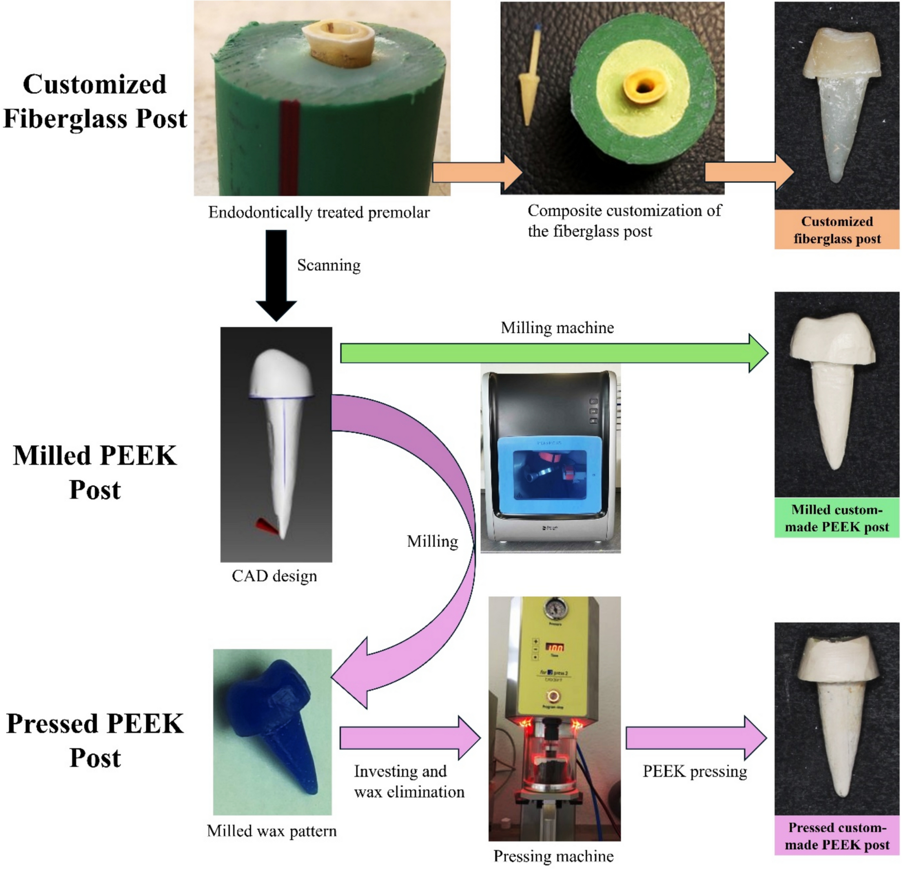
Post space drilling was done using size #2 and #3 LARGO pesso reamer (Dentsply Maillefer, Ballaigues, Switzerland) to remove 8.0 mm of the gutta-percha. Without using coolant, this process was implemented because the heat generated by the drill enhanced gutta-percha removal. To give the drilling space a uniform conformity, the Rely-X fiber post blue drill (3M ESPE, Seefeld, Germany) was used to finish the preparation of the root canals (8.0 mm in length and 0.9 mm in width apically, 5.72° (10.0%) taper). After the preparation of each root, the corresponding teeth were allocated randomly to three groups (n = 7 per group) according to the type of material and fabrication technique used to generate the post–core: group M; milled PEEK, group P; pressed PEEK, group C; fiberglass posts customized with resin composite (control). For the purpose of standardization, all the steps were carried out by the same well-trained operator.
The teeth of groups M and P were scanned using CEREC Omnicam intraoral scanner (CEREC premium SW4.4; Dentsply Sirona, Bensheim, Germany), with a total scanning depth of 10.0 mm [38]. Using Sirona connect application, the produced scans were exported to inLab software 15.1 (Dentsply Sirona, Bensheim, Germany), where the post–core systems were designed. A cement space of 85.0 μm was selected. The margins of the core were designed 1.0 mm away from the external enamel margin of the tooth with a height of 4.0 mm.
For the M group, the designs were milled from BioHPP PEEK blanks (Bredent GmbH & Co., Senden, Germany) into final restorations using the inLab MCX5 milling machine (Dentsply Sirona, Bensheim, Germany). In the P group, the computer-aided design (CAD) files were milled into wax patterns (CopraWax, Whitepeaks Dental Solutions GmbH, Hamminkeln, Germany) using the inLab MCX5 milling machine. The wax patterns were then invested using a special phosphate-bonded investment material (Gilvest powder and liquid; SRL Dental GmbH, Ludwigshafen, Germany). Preheating for melting the wax and managing the expansion of the investment material was performed in the laboratory’s pre-heating oven at 800.0 °C. After complete melting, the temperature was maintained at 400.0 °C for 20 min. The pressing procedure for BioHPP PEEK granules (Bredent GmbH & Co., Senden, Germany) was carried out fully automatically using the For2press system (For2press system; Bredent GmbH & Co., Senden, Germany) and completed within 35 min, after which the restorations were devested (Fig. 1).
Fig. 1
Schematic workflow diagram illustrating steps followed for post–core production of different systems14
For the C group, seven blue-coded Rely-X fiber posts (3M ESPE, Seefeld, Germany) measuring 1.9 mm at the coronal end and 0.9 mm at the apical end were used. The surfaces of the posts were thoroughly cleaned by immersion in 70.0% alcohol for 1 min, dried using sterile gauze, and then coated with a layer of silane coupling agent (Calibra; Dentsply Maillefer, Petrópolis, RJ, Brazil). Customization was carried out using Amaris resin composite (VOCO, Cuxhaven, Germany). The resin composite was applied in layers to the post surface, and the post/composite assembly was subsequently inserted into the root canal, which had been previously coated with a water-soluble gel (KY Gel; Johnson & Johnson, Sao Paulo, Brazil) to serve as a separating medium. After customization, the fiber post head was trimmed 4.0 mm above the coronal end of the specimen to facilitate core fabrication. For standard composite core production, a mold was created with transparent CharmFlex silicone bite registration material (Dentkist Inc., Gyeonggi-do, Korea). The mold was formed over one of the milled PEEK cores [39] from a previous M group. A Universal Tofflemire Matrix Retainer and its band (House Brand Dentistry, Concord, Canada) were placed around the coronal part of the prepared tooth and core. The bite registration material was injected with a small intraoral tip around the core up to the border of the band and left to set completely. For core production, the tofflemire band and holder were used to encircle the teeth to ensure standardized positioning of the mold, and the mold was then positioned over the trimmed fiber post within the confines of the band and filled with a core build-up material (Dentocore; Itena, Villepinte, France), followed by light curing of the core, using LED.H device (Woodpecker; Muenster, Germany) for 40 s according to the manufacturer’s instructions (Fig. 1).
It is known that PEEK has an inert surface, making the bonding procedure a challenging step [25, 40]. Hence, for M and P groups, the cementation process for BioHPP posts followed the manufacturer’s guidelines. Air abrasion was performed first, with the posts being blasted with 110.0 μm aluminum oxide at 2.5 bar pressure from a distance of 3.0 cm. The posts were then coated with a Visiolink primer layer (Bredent GmbH & Co., Senden, Germany) and left for a few minutes to allow solvent evaporation, and light-cured for 90 s. For group C, before cementation, the root canals were rinsed with 2.0 mL of distilled water to remove any residues of the water-soluble gel. They were then dried with a triple syringe using oil-free air, and absorbent paper points were used to ensure complete dryness. The posts were sandblasted with aluminum oxide for 5 s at a distance of 30.0 mm and 2.0 bar pressure and then cleaned in an ultrasonic machine.
All post–core systems were cemented using self-etching resin cement G-CEM dual cure capsules (GC Asia, Changi, Singapore). Initial Tac curing for 5 s was carried out using LED.H light curing device, excess cement was then removed, final curing was carried out for additional 20 s on each surface. Afterward, specimens underwent thermocycling, which involved immersion in two water tanks with temperatures of 5.0 °C (cold) and 55.0 °C (warm), alternating every 15 s, with a 5-s water drip in between, for a total of 5,000 cycles. This procedure simulates approximately 6 months of clinical use [40,41,43].
A universal testing machine (WTS; Jinan, Shandong, China) with a 1.0 mm diameter blunt-end tip was used to apply a progressively increasing load along the long axis of the specimens at a crosshead speed of 1.0 mm/min until fracture occurred. Through magnifying loops assessment (Carl Zeiss, Oberkochen, Germany) and periapical radiograph evaluation, the mode of failure of each specimen was assessed and categorized according to Falcao et al. [44] classification (Table 1), as either favorable (restorable) or non-favorable (catastrophic), based on the restorability of the tooth [45, 46]. Horizontal or oblique fractures at the crestal bone level (Types 0, 1, 2, 3, and 4) were considered favorable, while more apical root fractures (Type 5) were deemed catastrophic and non-restorable [45, 46].
Table 1 Classification of tooth failure mode after load application until fracture [44]Numerical part (finite element analysis)A sample from the M group, along with its designed restoration, was used to create a 3D model of the mandibular premolar based on data obtained from a computed tomography (CT) scan performed with the I-CAT Next Generation scanner (Imaging Sciences International, Hatfield, PA, USA). The scan was conducted at a tube voltage of 120.0 kVp, a tube current of 5.0 mA, with a voxel size of 0.2 mm and a field of view measuring 16.0 × 6.0 cm, taking 26.9 s to complete. The resulting digital Imaging and Communications in Medicine (DICOM) file was imported into SOLIDWORKS 18 (Dassault Systèmes, Waltham, Massachusetts, USA), a 3D mechanical CAD and simulation software, to generate the finite element model of the premolar. Using the software’s design tools, a 0.2 mm periodontal ligament (PDL) was created around the root, surrounded by a block of supporting alveolar bone measuring 12.0 × 9.0 × 18.0 mm [3], positioned at an intact bone level 2.0 mm below the cemento-enamel junction (CEJ).
The corresponding post–core designed restoration was exported as a Standard Tessellation Language (STL) file to SOLIDWORKS and adjusted over the tooth to form the finite element model for both milled and pressed PEEK custom-made post–core systems. For the customized fiberglass post, a tapered cone with the same dimensions as the blue Rely-X fiberglass post (8.0 mm in length, 0.9 mm in width apically, with a taper of 5.72° (10.0%)) was drawn centrally within the design of the PEEK post–core, representing the fiberglass post. The space between the post and root dentin was assumed to be filled with customization composite, the coronal part of the restoration was assumed to be core composite, and the cement gap between the post and the root was assumed to be the cement layer in both models (Figs. 2 and 3).
Fig. 2
Components of finite element models; custom-made milled and pressed Polyetheretherketone (PEEK) posts (left), customized fiberglass post (right)17
Fig. 3
The generated 3D mesh of finite element model using SOLIDWORKS, showing dimensions of investing block of bone in mm (left) and a cross-sectional view showing the post dimensions in mm (right)18
Accordingly, in this study, three different post–core systems were compared using two types of geometry models, as shown in Fig. 2. One of the geometry models was a one-piece post–core model to exhibit a PEEK post–core system fabricated by two techniques: pressing and milling. The other geometry model was a fiberglass post, a customization composite, and a separate core model to exhibit a customized fiberglass post system. The shape of all post–core systems in this study was considered the same, to assess only the effect of the post–core materials, excluding the effect of dimensions.
The length of the designed post was approximately half the root length, the other half of the root was filled with gutta-percha (GP) cone. Finally, the elements of the constructed geometry model consist of an alveolar bone, periodontal ligament (PDL), tooth root, GP cone, resin cement, and one-piece post–core (or composite-customized fiberglass post and resin core). The 3D meshes were generated using a blended curvature-based mesher with four Jacobian points and an element edge length of 0.1032 to 0.516 mm on average. Thus, the meshing procedure generated around 230,000 nodes and 150,000 hexahedral elements, as shown in Fig. 3.
In this model, the mechanical properties of all materials were assumed to be homogeneous, isotropic, and linear elastic for simplicity, except for the fiberglass post. The fiberglass post was considered orthotropic due to its varying mechanical properties along the fiber direction (x-direction) compared to the two perpendicular directions (y- and z-directions). The applied values were derived from previous studies, as detailed in Table 2 [7, 47]. All assemblies were assumed to be fully bonded, and the bottom of the alveolar bone was fixed to prevent any rigid dynamic motions. A vertical force of 300.0 N, representing the average occlusal load [49], was applied to a group of ten central nodes on the core’s outer surface, perpendicular to the occlusal plane and parallel to the tooth’s longitudinal axis. To identify regions of highest stress intensity, equivalent von Mises (VME) stresses were calculated for each component of the numerical models, and the resulting stress distributions were analyzed and plotted.
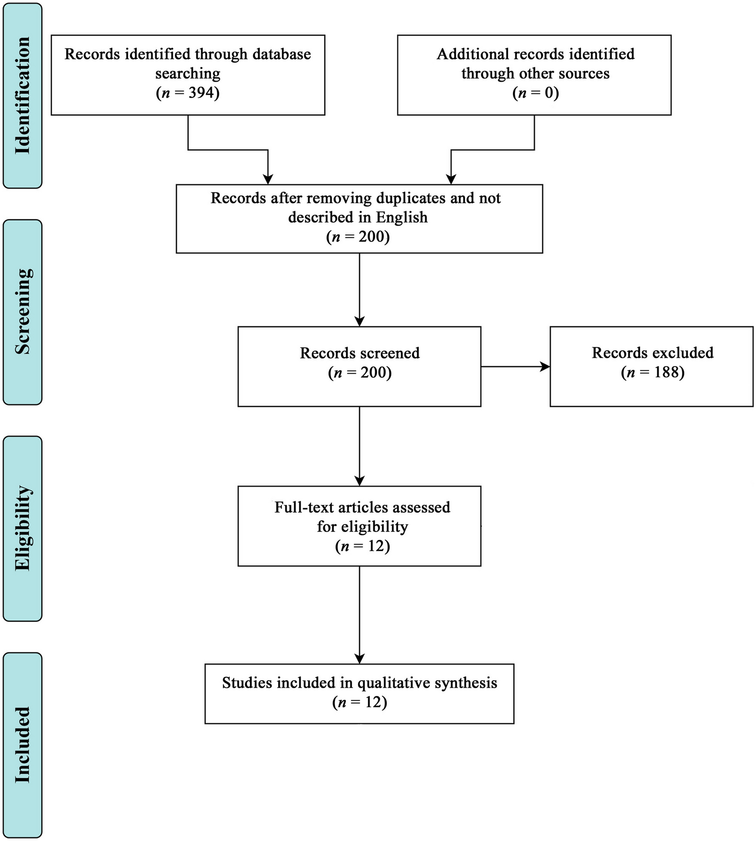
Table 2 The mechanical material properties of the finite element modelsStatistical analysisA power analysis was performed based on the results of Maroulakos et al. 41. The calculated sample size was 21 specimens in total, corresponding to 7 specimens per group. The effect size (f) was determined to be 0.81. Using an alpha (α) level of 0.05 and a beta (β) level of 0.20, corresponding to a statistical power of 80.0%, the total sample size was confirmed to be 21. The sample size calculation was conducted using G*Power software version 3.1.9.4 (IBM Corporation, New York, NY, USA) 50. Categorical data were presented as frequency and percentage values and were analyzed using Fisher’s exact test. The significance level was set at p < 0.05. Statistical analysis was performed using SPSS Statistics Version 26.0 for Windows (IBM Corporation, New York, NY, USA).
Comments (0)