
Remember me


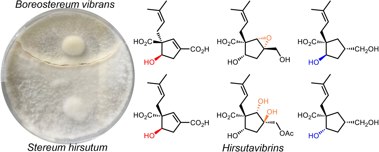

Hirsutavibrin A (1, Fig. 1) was isolated as a colorless oil. High-resolution positive electrospray ionization mass spectrometric [( +)-HRESIMS] analysis of 1 returned a protonated ion peak at m/z 241.10703 [M + H]+, corresponding to the molecular formula of C12H16O5 (mass error 0.08201 ppm) with five degrees of unsaturation. The 1H NMR spectrum of 1 (Table 1) revealed two olefinic protons at δH 6.72 (1H, s) and 5.12 (1H, t, J = 7.6 Hz), in addition to two methyl groups at δH 1.70 (3H, s) and 1.61 (3H, s). The 13C NMR (Table 2) and Distortionless Enhancement by Polarization Transfer (DEPT) spectroscopic data of 1 presented 12 carbon signals, including two CH3 at δC 18.2 (C-11) and 26.3 (C-12), two CH2 at δC 36.0 (C-8) and 40.8 (C-4), three methine carbons at δC 78.4 (C-5), 145.7 (C-2), and 120.6 (C-9), three proton-deficiency carbons at δC 67.4 (C-1), 135.9 (C-10), and 136.6 (C-3), and two carbonyl groups at δC 177.3 (C-7) and 169.1 (C-13). The 1H-1H COSY correlations of H-4/H-5, along with the HMBC correlations from H-2 to C-1, C-3, C-4, C-5, C-13 (Fig. 2) revealed the existence of a five-membered ring in 1, which, together with the two carbonyl groups and two double bonds mentioned above, satisfied the unsaturation degree of compound 1. The 1H–1H COSY correlation of H2-8/H-9, and the HMBC correlations from H3-12 to C-9, C-10, C-11, and from H-8 to C-1 indicated a prenyl group attach to C-1. In addition, the HMBC correlations from H-2 to C-13, from H-8 and H-5 to C-7 enabled the assignment of two carboxylic groups at C-1 and C-3, respectively. Therefore, the planar structure of 1 was established (Fig. 1), which highly resembled vibralactone E [21], except that the CH2OH group (C-13) was replaced by a COOH group in 1.
Fig. 1
Chemical structures of compounds 1–11
Table 1 1H NMR spectroscopic data of compounds 1–6Fig. 2
Key 1H-1H COSY and HMBC correlations of compounds 1–11
The conclusion of the relative configuration of 1 arrived by analyzing the ROESY spectrum (Fig. 3). The key correlations of H-5 (δH 4.22)/H-8a (δH 2.30), H-5/H-4α (δH 2.87), and H-8a/H-4α helped to locate H-5 and the prenyl group orienting the same side of the five-membered ring. Besides, almost all the prenyl group at C-1 from the previously reported vibralactone and its derivatives are β configuration, thus the relative configuration of 1 was determined to be 1R*,5S*. The absolute configuration of 1 was determined by ECD calculations (Supplementary Material, Fig. 4). As a result, the calculated ECD of the stereoisomer 1S,5R was most consistent with the experimental CD data of 1, while the calculated ECD data of 1R,5S stereoisomer showed mirror Cotton effects at specific wavelengths (Fig. 4B). Therefore, the absolute configuration of 1 was assigned as depicted in Fig. 1, which was different from the previously reported vibralactone derivatives.
Fig. 3
Key ROESY correlations of compounds 1–11
Fig. 4
A The four possible stereoisomers of 1 and 2. B Comparisons of the experimental CD and calculated ECD of 1. C Comparisons of the experimental CD and calculated ECD of 2
Hirsutavibrin B (2, Fig. 1) was obtained as a colorless oil. Its molecular formula was determined to be C12H16O5 based on (+)-HRESIMS, which presented an [M + H]+ ion peak at m/z 241.10707 (calcd for C12H17O5, 241.10705). The 13C NMR and DEPT spectroscopic data of 2 (Table 2) showed two CH3 at δC 18.0 and 26.1, two CH2 at δC 30.9 and 39.9, three CH at δC 76.9, 145.1, and 120.7, three non-protonated carbons at δC 64.6, 135.5, and 135.4, and two carbonyl carbons at δC 178.0 and 168.5. The 1D NMR data showed high similarity with those of 1. By interpretating of the 2D NMR spectra, including HSQC, 1H–1H COSY, and HMBC spectra (Fig. 2), 2 was determined to have a planar structure identical to 1. The weak correlation signal of H-5 (δH 4.59)/H-8a (δH 2.60) in the ROESY spectrum indicating that H-5 and the prenyl group resided the opposite sides of the five-membered ring, which helped to establish the relative configuration of 2 as 1R*,5R*. An ECD calculation workflow was applied to determine the absolute configuration of 2. As shown in Fig. 4C, the calculated ECD of the 1R,5R stereoisomer matched well with the experimental CD data. Therefore, the evidence allowed the complete assignment of 2D structure and absolute configuration of 2 (Fig. 1).
Hirsutavibrin C (3, Fig. 1) was obtained as a colorless oil. The molecular formula of 3 was determined as C14H18O6 according to the HRESIMS data (m/z 283.11765 [M + H]+, calcd for C14H19O6, 283.11761), indicating six degrees of unsaturation. The 1H and 13C NMR data of 3 (Tables 1 and 2) were closely similar to the data of 2, indicating the structure of 3 closely resembled that of 2, with the primary difference being associated with an extra acetoxyl group in 3. The methyl singlet at δH 1.98, the carbonyl carbon at δC 172.1, together with the key HMBC correlation from H-5 (δH 5.27) to δC 172.1 (Fig. 2) of 3 enabled the assignment of the acetoxyl group attaching to C-5. Although in the 13C NMR spectrum, the signal for C-13 was not detected, which is common for carboxylic groups [36], the HRESIMS result and the chemical shifts of C-2, C-3 and C-4 confirmed that a carboxylic acid group attached to C-3. The crucial and significant correlation of H-5/H-8 (δH 2.60, 2.35) in the ROESY spectrum (Fig. 3) assigned the H-5 and the prenyl group locating at the same side of the five-membered ring (Fig. 2). In addition, the calculated ECD spectrum of (1R,5S)-3 showed similar adsorption trend with the experimental CD spectrum of 3. Therefore, the absolute configuration of 3 was established as 1R,5S (Figs. 1 and 5).
Fig. 5
ECD calculations of 3, 5–7
The colorless oil hirsutavibrin D (4, Fig. 1) had a molecular formula of C14H20O5 which returned from the (+)-HRESIMS analysis (m/z 269.13834 [M + H]+), revealing five degrees of unsaturation. In the 13C NMR spectrum of 4, 14 carbon signals were displayed (Table 2), which showed similarity to 3, except that an oxygenated methylene (C-13, δC 61.6) in 4 replaced the carbonyl group signal of 3. The key HMBC correlations from H-2 (δH 5.72) and H-13 (δH 4.11) to C-3 (δC 144.4), and from H-2 to C-13 (Fig. 2) supported the conclusion that the C-13 of 4 was a hydroxymethyl group instead of being a carboxylic acid group as in 3. The key ROESY correlation between H-5 and H-8 (Fig. 3) suggested that the prenyl group and H-5 were in the same plane of the five-membered ring. Therefore, compound 4 was identified as a C-13 reduced product of 3.
The molecular formula of hirsutavibrin E (5, Fig. 1) was determined as C14H20O5 according to the (+)-HRESIMS data (m/z 269.13834 [M + H]+, calcd. for C14H21O5). The 1D NMR spectroscopic data suggested that 5 (Table 2) was a congener of 4, except that the acetoxyl group was changed to locate at C-13 according to the 3J-HMBC correlations from the proton at δH 4.59 (H-13) to δC 170.9, and the 4J-HMBC correlations from methyl at δH 2.08 (13-OCOCH3) to C-13 (δC 62.8) (Fig. 2). The key correlation between H-5 and H-8 (Fig. 3) was absent in the ROESY spectrum, while between H-5 and H-4α was seen. Therefore, the relative configuration of 5 was speculated to be 1R*,5R*. The calculated ECD curve of (1R,5R)-5 showed a Cotton effect similar to the experimental one (Fig. 5), thus establishing the absolute configuration of 5 to be 1R,5R.
Compound 6 (Fig. 1) was isolated as a colorless oil. The molecular formula was assigned as C16H22O7 based on the positive HRESIMS ion peak at m/z 327.14359 [M + H]+ (calcd for C16H23O7, 327.14383). Compared to the NMR data of 5 (Tables 1 and 2), the signals for acetyl group were absent in compound 6. In addition, four additional carbon resonances which were ascribable to two carbonyl groups (δC 174.1, 176.7) and two methylenes (δC 30.2, 30.3) (Table 2) were presented in 6. By interpreting the 2D NMR spectra, the four carbons were assigned to be a succinic acid moiety, which was determined by the correlations from H-2’ (δH 2.58) and H-3’ (δH 2.62) to C-1’ (δC 174.1) and C-4’ (δC 176.7), from H-13 (δH 4.61) to C-1’ and C-3 (δC 138.3) (Fig. 2) in the HMBC spectrum. The correlation between H-5 and H-8 was not seen in the ROESY spectrum, suggesting a 1R*,5R* configuration of 6. With the help of ECD calculations, an absolute configuration of 1R,5R was assigned to 6 based on the highly similar Cotton effect between the experimental CD diagram and the calculated ECD data of (1R,5R)-6 (Fig. 5). Therefore, compound 6 was trivially named hirsutavibrin F.
The 1H and 13C NMR data of 7 (Tables 2 and 3) displayed twelve carbon resonances which were classified into two CH3, four CH2 (one oxygenated), two sp3 CH (one oxygenated), one sp2 CH, one sp3 qC, and two sp2 qC (one carbonyl and one olefinic carbon). The NMR data of 7 displayed similarity to those of vibralactone E, except that the C-2–C-3 double bond was reduced to a single bond, which was corroborated by the key 1H-1H COSY correlations between H-2 (δH 2.31) and H-3 (δH 2.21) (Fig. 2). These assignments reached the conclusion that the molecular formula of 7 was C12H20O4, which was consistent with the HRESIMS analysis results (m/z 229.14349 [M + H]+, calcd. for C12H21O4, 229.14344). The key correlations of H-2β/H-3/H-4β/H-5 (δH 4.27)/H-8a (δH 2.51), H-2α/H-13/H-4α, and H-5/H-9 in the ROESY spectrum (Fig. 3) indicated the relative configurations of 7 to be 1R*,3S*,5S*. The ECD calculation steps were applied to deduce the absolute stereochemistry of 7. Comparative analysis of the measured and theoretically calculated ECD curves (Fig. 5) revealed a 1R,3S,5S configuration of 7. Compound 7 was named hirsutavibrin G.
Table 2 13C NMR spectroscopic data of compounds 1–11Table 3 1H NMR spectroscopic data of compounds 7–11The molecular formula of compound 8 (Fig. 1) was deduced to be C12H20O4, which was identical with 7. Detailed interpretation of the NMR of 8 revealed the same planar structure as 7 (Tables 2 and 3). The lack of key ROESY signals between H-5 and H-8 indicated a 1R*,3S*,5R* configuration of 8 (Fig. 3). Considering the same biosynthetic pathways of 7 and 8, the absolute configurations of 8 were determined to be 1R,3S,5R. Compound 8 was trivially named hirsutavibrin H.
Hirsutavibrin I (9, Fig. 1) was isolated as a colorless oil. The (+)-HRESIMS sodium adduct ion peak at m/z 265.10452 [M + Na]+ (calcd for C12H18O5Na, 265.10519) revealed a chemical formula of C12H18O5 of 9. The 13C NMR data of 9 (Tables 2 and 3) displayed two methyls, three methylenes (one oxygenated), two oxygenated sp3 methines (δC 63.9, 75.0), two sp3 quaternary carbons (δC 59.7, 66.8), etc. These data are different from those of compounds 1–6, while are similar to those of compound 7, and are highly resemble to those of vibralactone B [20, 37]. Compound 9 was 18 Da larger than vibralactone B, suggesting that 9 was the lactone ring-opening product of vibralactone B. The crucial ROESY correlations of H-2 (δH 3.38)/H-8 (δH 2.31, 2.01)/H-5 (δH 3.76), H-2/H-9 (δH 5.08) indicated the 1S,2S,3S,5S configuration of 9. Hence compound 9 was elucidated as depicted in Fig. 1.
The chemical formula of 10, obtained as a colorless oil, was determined to be C14H22O7 (m/z 303.14383 [M + H]+, Δ 0.00048 ppm) by (+)-HRESIMS. The 1D NMR data between 10 (Tables 2 and 3) and 5 were quite similar, except that the C-2/C-3 double bond between in 5 was replaced by a vicinal diol in 10, according to the key HMBC correlations from H-2 (δH 3.45) to C-1 (δC 54.3), C-3 (δC 76.9), C-5 (δC 70.6), from H-8 (δH 2.29) to C-2 (δC 83.7), and from H-13 (δH 3.97) to C-2, C-3, C-4 (δC 45.0), and from 3-OH (δH 4.60) to C-2, C-3, C-4, and C-13 (Fig. 2). The crucial ROESY correlations of H-2 (δH 3.45)/H-8/H-5 (δH 3.57)/3-OH (Fig. 3) indicated the 1S,2S,3R,5S stereochemistry of 10. Therefore, the structure of compound 10 was established and named hirsutavibrin J.
( +)-HRESIMS spectrometric analysis established the molecular formula of 11 (Fig. 1) as C12H18O5Na (m/z 265.10461 [M + Na]+, mass error 0.11536 ppm). The 1D NMR spectroscopic data of 11 was similar to those of compound 7, except with the existence of a new carbonyl carbon (δC 181.0) in 11 while absence of the hydroxymethyl group in 7. This evidence indicated that compound 11 differs from 7 by the oxygenated status of C-13. The carbonyl group at δC 181.0 in compound 11 was assigned to a carboxylic group (C-13). The correlation from H-3 (δH 2.86) to C-13 in the HMBC spectrum (Fig. 2) as well as the molecular formula corroborated the above assignments. The vital ROESY correlations of H-8/H-5/H-3 suggested a 1R,3S,5S configuration of compound 11 (Fig. 3). Therefore, the structure of compound 11 was established and named hirsutavibrin K.
2.2 Biological activity evaluation of 1–11The isolated compounds with abundant yield (1, 2, 4, 6) were subjected to a panel of biological activity screening, including the cytotoxicity against A549, a human lung cancer cell line, and anti-NO (nitric oxide) activity in murine monocytic RAW 264.7 macrophages. As a result, compounds 1 and 2 showed weak cytotoxicity activity toward A549 with the IC50 39.7 and 34.3 μM, respectively (positive control cisplatin, IC50 5.09 μM). Compound 4 displayed anti-NO activity with IC50 of 26.6, which were more significant than the positive control L-NG-monomethyl arginine (IC50 51.2 μM).
Comments (0)