
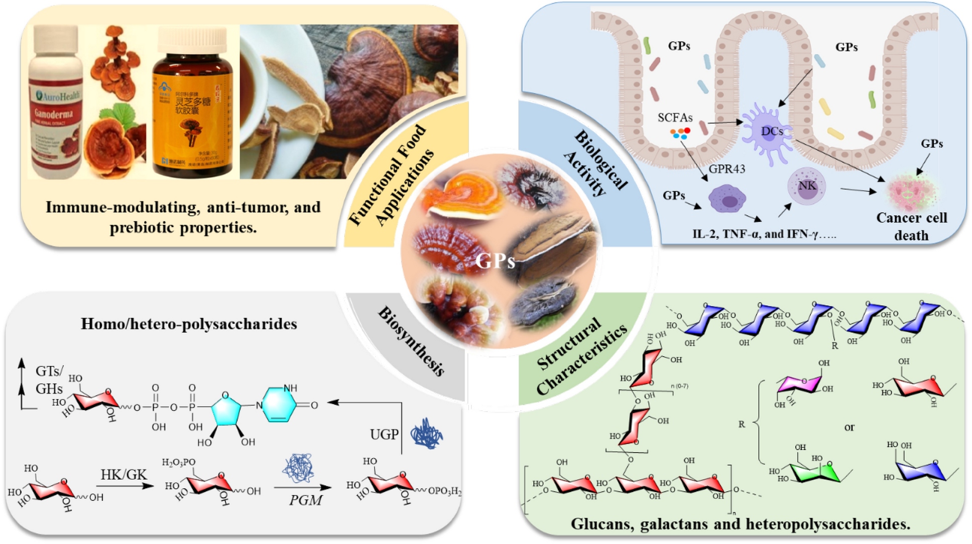
Remember me


BS11134 was classified as Bipolaris sorokiniana based on morphology and sequence of the internal transcribed spacer (ITS) region (GenBank accession number KU297882) [23]. We used the Hidden Markov Models file (Terpene_syn_C_2) classified in the Pfam database [24] to examine the BS11134 genome obtained in our previous study [23] for potential sesquiterpene cyclase (STC) genes, resulting in four homologous sequences of known STC proteins with DDXXD metal binding motifs. Further annotation of these four STC-encoding biosynthetic gene clusters using antiSMAS [25] and 2ndfinder online software revealed various post-modification enzymes, including cytochrome P450, acetyltransferase, decarboxylase, etc. (Fig. 1). Notably, the length of STC8 gene cluster was only 15 kb (Fig. 1), consisting of a three-gene cassette very similar to the recently identified seco-sativene gene cluste [26].
Fig. 1
Organization of the sesquiterpenoids biosynthetic gene cluster in BS11134
2.2 FBMN guided detection of sesquiterpenoidsWe used rice medium to ferment the fungus because this medium gave the most induced secondary metabolites compared with other six media used [23]. A previously reported seco-sativene type sesquiterpenoid helminthosporol (7) was characterized from the crude extract of BS11134 based on its UV spectrum and HR-MS data (Fig. 2B and Fig. S7a). Further analysis using GNPS FBMN identified new seco-sativene type sesquiterpenoids using helminthosporol (7) as the seed compound (Fig. 2A), the precursor m/z of 7 was found to be within Cluster I, comprising 97 nodes with similar MS/MS patterns.
Fig. 2
FBMN-guided characterization of new helminthosporol analogs from BS11134 crude extract. A Clustering of sesquiterpenoids analogs. Helminthosporol (7) was used as seed compound (yellow shaded square) in cluster analysis, and 26 analogs (Purple circles: compounds isolated in this study; Blue circles: other potential new sesquiterpenoids identified by HRMS annotation) were identified within the network. B HPLC–UV chromatogram of sesquiterpenoids fraction RD3 examined at 254 nm, and the UV spectrum of helminthosporol. C Structures of compounds 1–10 isolated from strain BS11134
Detailed HRESIMS data annotation (Table 1) revealed 27 potential sesquiterpenoids, ten of which showed similar UV spectra to helminthosporol in the HPLC chromatogram of subfraction RD3. Among these, five nodes exhibited previously unreported m/z values (291.1571, 291.1574, 321.1682, 301.1782, 253.1804), guiding the isolation of new sesquiterpenoids from several subfractions. Three new sesquiterpenoids (1–3) with a helminthoporene skeleton as well as seven known compounds (4–10) were identified (Fig. 2C).
Table 1 Detailed annotation for sesquiterpnoid related nodes in cluster I from the BS11134 featured based molecular networking2.3 Structure elucidation of isolated sesquiterpenoidsThe molecular formula of compound 1 was C15H24O4 confirmed by its HRESIMS data ([M+Na]+m/z 291.1567, calcd. for C15H24O4Na+ 291.1571) (Fig. S1a). Analysis of 1H NMR, 13C NMR, and HSQC spectra (Table 2 and Figs. S1b-S1 d) revealed a carboxyl cluster [δC 167.5], two olefinic carbons [δC 157.7, 128.5], four methylene (two of which were substituted with the hydroxyl groups [δC 55.6, 59.9]), four methine and three methyl groups, as well as one sp3 quaternary carbon, outlining a seco-sativene sesquiterpene skeleton. The 1H-1H COSY (Fig. S1e) revealed a continuous spin system extending from H2−4 to H3−10/H2−11. In addition, H-6 was further coupled with H-7, which also coupled with H-13, and H-13 coupled with H2−14 (Fig. 3 and Table S1). The two hydroxyl groups at C-12 (δC 55.6) and C-14 (δC 59.9) could be further determined by the HMBC (Fig. S1f) correlations of oxymethene protons at δH 4.54 (d, J = 13.2 Hz) and 4.12 (d, J = 13.2 Hz) with C-1 (δC 128.5), C-2 (δC 157.7) and C-3 (δC 49.2), and oxymethene protons at δH 3.44 (dd, J = 10.3, 4.9 Hz), and 3.05 (dd, J = 10.3, 9.4 Hz) with C-3, C-7 (δC 43.2) and C-13 (δC 62.0). A six-membered ring was formed on C-13 was connected to C-4 through C-3, according to the HMBC correlations between H3−8 and C-3, C-4, and C-13. The presence of a methyl group (Me-8) at C-3 was inferred as well. The olefinic carbons C-1 and C-2 were connected to C-7 and C-3, respectively, according to the HMBC correlations of H-7/C-15, H-7/C-1, H-7/C-2, H2−12/C-2, and H2−12/C-3. These observations suggested the structure of compound 1 closely resembled helminthosporic acid [27], with the notable substitution of a methyl group for a hydroxylmethylene group. The relative configurations of 1 could be established according to the ROESY (Fig. S1 g) correlations of H-7/H3−11, H-7/H-13, H3−8/H-13, and H-13/H-6, as well as comparison with literature [27] (Fig. 3). To figure out the absolute configurations of 1, the electronic circle dichromatography (ECD) calculation of two epimers 3R,6R,7S,13S-1/3S,6S,7R,13R-1 was performed, resulting in the 3R,6R,7S,13S configurations of 1 (Fig. 4A). Therefore, the structure of 1 was established (Fig. 2C) and it was named 12-hydroxyhelminthosporic acid.
Table 2 1H and 13C NMR Data of compounds 1–5Fig. 3
2D NMR correlations of compounds 1–5. A Key 1H-1H COSY and 1H-13C HMBC correlations. B Key ROESY correlations
Fig. 4
Experimental and computed ECD of compounds 1 (A), 2 (B), and 5 (C)
Compound 2 has a molecular formula of C15H24O4, as confirmed by HRESIMS data (Fig. S2a). Comparative analysis of 1H and 13C NMR data (Table 2 and Fig. S2b–d) between compounds 1 and 2 revealed that two secondary methyl groups [δC/δH 20.8/0.76 (d, J = 6.3 Hz), δC/δH 21.8/0.97 (d, J = 6.3 Hz)] were replaced by two methyl groups at lower field [δC/δH 28.0/1.13 (s), δC/δH 28.5/1.04 (s)]. The appearance of an oxygenated quaternary carbon (δC 70.5) suggested the presence of one hydroxyl group at C-9. HMBC (Fig. 3 and Supplementary Fig. S2e, f) further confirmed this hypothesis with correlations of H3−10/C-6, H3−10/C-9, H3−10/C-11. Resonances at δH 1.88 (s) showed correlations with carbons at δC 49.5(C-3), δC 127.6(C-1), and δC 156.1(C-2), establishing the methyl group location of C-12. The relative configurations of 2 were consistent with those of 1 by detailed analysis of their ROESY (correlations of H-7/H3−11, H-7/H-13, H3−8/H-13, and H-13/H-6) (Fig. 3, Fig. S2 g, and Table S2). The absolute configurations were assigned as 3R,6S,7R,13S through ECD calculation. Thus, the structure of 2 was established (Fig. 2C), and this compound was named 9-hydroxyhelminthosporic acid.
The molecular formula of compound 3 was deduced as C18H24O5 based on HRESIMS (Fig. S3a) (m/z 343.1514 [M+Na]+, calcd. for 343.1516) with 7 degrees of unsaturation. 1H, 13C and 2D NMR data (Figs. S3b–f) revealed a similar structural skeleton with compound 1, except for resonances of two additional carboxyls (δC 164.9, 166.8) and one olefinic bond (δC 145.6, 131.2). HMBC correlations from H-14 (δH 6.51 d, J = 10.9 Hz) to C-3, C-7, C-16, C17, and C-18 revealed that two carboxyl groups were connected with C-14 through non-protonated carbon C-16. The relative configurations of 3 were consistent with 1 based on key ROESY correlations of H-7/H3−11, H-7/H-13, H3−8/H-13, and H-13/H-6 (Fig. 3, Fig. S3 g, and Table S3). Given the shared biosynthetic pathway of compounds 1 and 3 through the seco-sativene sesquiterpene skeleton, the absolute configurations of 3 were deduced to align with that of 1, and was designated 3R,6R,7S,13S. This alignment was supported by its specific optical rotation ([α]−20) in contrast to compound 1 ([α]−38) and to bipolarisorokin H ([α]−137) [28]. Thus, 3 was a new compound (Fig. 2C) and named bipolarisorokin I.
HRESIMS of 4 displayed a molecular ion peak at m/z 301.1775 for [M+Na]+ (calcd. for 301.1774) (Fig. S4a) and indicating a molecular formula of C17H26O3. 1D and 2D NMR data (Figs. S4b–f) showed that compound 4 shared a structural framework with helminthosporol (7) [29], distinguished by an additional acetyl group [δC/δH 20.7/1.98 (s), δC 170.4]. HMBC correlations from H3−17 and H2−14 to C-16 confirmed compound 4 as a 14-OH acetylated analogue of 7. Through ROESY analysis, the crosspeaks of H-7/H3−11, H-7/H-13, H3−8/H-13, and H-13/H-6 revealed that the relative configurations of 4 were consistent with those of 1 (Fig. 3, Fig. S4 g, and Table S4). Its absolute configurations were deduced as 3R,6R,7S,13S considering its biosynthetic lineage from seco-sativene sesquiterpene, and comparison of specific rotation values (4, [α]+11.0) with literature (bipolenin H, [α]+17.5) [30]. Accordingly, compound 4 was definitively identified and named bipolarisorokin J, with a defined structure (Fig. 2C). The NMR data of compound 4 is reported here for the first time.
Compound 5 possessed a molecular formula of C15H24O3 (four degrees of unsaturation) determined by HRESIMS data (Fig S5a). NMR analysis (1H and 13C, Table 2 and Figs. S5b-S5 d) revealed a structural resemblance to helminthosporol (7), except that the methyl group (Me-11) was replaced by a hydroxylmethylene group. The presence of a hydroxyl group at C-11 could be assigned by HMBC (Fig. 3 and Fig. S5e, f) correlations of the downfield shifted signals at δH 3.87 (dd, J = 11.1, 3.9 Hz) and 3.62 (dd, J = 11.1, 4.7 Hz) with C-6, C-9 and C-10. Detailed analysis of ROESY afforded the 3R*,6S*,7S*,13S* relative configuration in 5 (Fig. 4B, Fig. S5 g, Table S5). The relative configurations between C-6 and C-9 in 5 was determined to be 6S*,9S* by 13C NMR calculations and DP4+ analyses (Fig. 5, Table S9) [31]. ECD calculations confirmed the absolute configurations of 5 as 3R,6S,7S,9S,13S, leading to the identification of compound 5 as 11-hydroxyhelminthosporol (Fig. 2) [32].
Fig. 5
13C NMR calculation results of two plausible isomers 5a/5b at the B3LYP/6–311++G(2 d,p) level. A Linear correlation plots of the calculated and experimental 13C NMR values. B Relative errors between the calculated and recorded 13C values. C DP4+ probability
Compounds 6–10 were identified as helminthosporic acid derivative (6) [33], helminthosporol (7) [29], helminthosporic acid (8) [27], sorokinianin (9) [34], and secolongifolene diol (10) [29], by comparing their spectroscopic data with those in previous literature.
2.4 Proposed biosynthetic pathway of the isolated sesquiterpenoidsSativene could be synthesized from FPP by sesquiterpene synthase (Fig. 6), by a mechanism involving ionization and successive rearrangement [35]. Compounds 1–9 belong to seco-sativene type irregular terpenoids, while compound 10 belongs to seco-longifolene type sesquiterpenoids [29]. The plausible biosynthetic routes of compounds 1–10 were described based on recent research [26, 36] (Fig. 6).
Fig. 6
Organization of the seco-sativene biosynthetic gene cluster in B. sorokiniana ND90Pr and BS11134 (A), and proposed biosynthetic pathway for isolated sesquiterpenoids 1–10 (B)
Intermediate IM2 was generated from sativene through successive oxidation by P450 and a spontaneous shift of the Δ2,12 olefinic bond. Subsequent reduction of the C-14 aldehyde, catalysed by a Aldo–keto reductase (AKR) yielded compound 7. Compounds 4, 5, and 8 can be derived from Compound 7 through acetylation at 14-OH, hydroxylation at C-11, and carboxylation of C-15, respectively. Hydroxylation at C-9 or C-12 of compound 8 yielded compounds 1 and 2, respectively. Compound 6 resulted from the acetylation at 14-OH of Compound 8. The γ-butyrolactone moiety in Compound 9 was proposed to derive from the TCA cycle intermediate 2-oxosuccinic acid via an aldol condensation reaction, as supported by previous isotope labelling studies [37]. A similar process occurred with compound 3, where the 2-methylenemalonic acid moiety was incorporated using malonic acid as intermediate through an aldol condensation reaction with C-14 aldehyde of IM2 (Fig. 6B). seco-Longifolene type sesquiterpenoid 10 was postulated to possess a divergent cyclization route, generating an end product with a seven-membered ring [27, 38].
2.5 In vitro anti-inflammatory assayAll isolated sesquiterpenoids from strain BS11134 were screened for their anti-inflammatory activity (Table 3) by evaluating inhibition effects on the NO production induced by LPS in RAW264.7, a mouse macrophage cell line. Compounds 1 and 9 exhibited anti-inflammatory effects in vitro, with inflammation inhibition rates of 28.0 ± 2.4% and 84.7 ± 1.7%, respectively, at a concentration of 10 μM, compared to the positive control, indomethacin, which showed a 51.2 ± 8.2% inhibition rate.
Table 3 NO inhibitory activities of compounds
Comments (0)