Neonatal ICHs include various types, such as IPH, IVH, SAH, SDH, and subpial hemorrhage. This retrospective study focuses on subpial hemorrhage, a subtype of ICH that has been underreported and not well understood until recently [1, 2, 8]. We investigate the demographic, pathophysiologic, clinical, and neuroimaging characteristics of subpial hemorrhage, emphasizing its distinct neuroimaging features that differentiate it from other extraaxial hemorrhages. Subpial hemorrhage may be associated with poor neurological outcomes, likely due to the extent of parenchymal injury [1]. Our results demonstrate a correlation between PMV and IPH, and introduce the HPm-sign as a promising indicator for differentiating subpial hemorrhage from other neonatal hemorrhage types in certain cases.
Our cohort of 28 patients with subpial hemorrhage included 5 preterm infants (two moderate-to-late preterm, one very preterm, and two extremely preterm). Of these, one survived without neurological sequelae, one developed quadriplegic cerebral palsy and epilepsy, and two died; follow-up was unavailable for one patient. These results are quite similar to those of Assis et al. [8], who reported a 60% mortality rate among premature infants, while our study found a 40% mortality rate. In contrast, Pinto et al. [9] did not identify worse outcomes for early preterm infants compared to others.
Hematologic abnormalities were found in 58% of our patients, which is lower than the rates reported by Cain et al. [7], but higher compared to Assis et al. [8], where only 1 of 16 patients had abnormal coagulation profiles. The most common abnormalities in our cohort were elevated D-dimer (23%), thrombocytopenia (15%), and increased activated protein C (15%). In contrast, Cain et al. [7] reported elevated D-dimer in 53% of patients, while another study found thrombocytopenia in 60% of neonates [9].
In our study, 86% of the lesions were unilateral, similar to the reported incidence by Zhuang et al. [10], but differing from Assis et al. [8], where all lesions were unilateral. Most lesions were located in the temporal lobes, consistent with other studies [8,9,10]. The occipital lobe had the lowest frequency of involvement, which agrees with previous studies [8, 9] but contrasts with Zhuang et al. [10], who reported it as the second most frequently affected region (29.4%). It has been postulated that the predominance of temporal lobe subpial hemorrhages may be related to skull deformation during birth at the confluence of sutures at the pterion, while occipital hemorrhages may be associated with the asterion, corresponding to the venous drainage territories of the superficial middle cerebral vein and the vein of Labbé [6, 11].
All patients in our study with subpial hemorrhage had underlying cortical infarcts, which is consistent with findings from other authors [7,8,9]. However, Zhuang et al. [10] reported 6 of 34 patients presenting with subpial hemorrhage only. Additionally, we found concurrent IPH in 86% of cases, similar to the 82% reported by Cain et al. [7], but higher than in other studies [8, 10] and lower than in Pinto et al. [9].
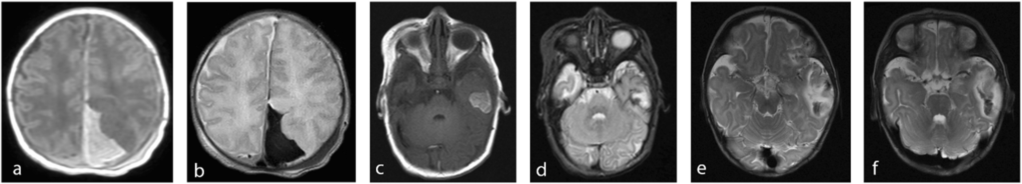
In 21 patients (75%), the distinct imaging pattern resembling the yin-yang symbol was observed, with a T2WI hypointense component corresponding to the subpial hemorrhage extending to the cerebral sulci and a T2WI hyperintense component reflecting the underlying infarcted cerebral cortex. Previous studies have reported the yin-yang sign in 12 of 34 patients [10], 9 of 10 patients [9] and all patients [8].
An interesting finding in our study was the visualization of a hyperintense line surrounding the external contour of the subpial hemorrhage on TOF-MRA in 12 of 18 patients, which we propose to call the hyperintense pia mater sign (HPm-sign). This is the first systematic report highlighting the value of TOF-MRA in diagnosing this condition; Barreto et al. [1] described only one case, suggesting that this finding indicates blood-stained pia mater. Similar to Barreto et al. [1], we believe that the HPm-sign could assist neuroradiologists in differentiating this type of hemorrhage from other intracranial hemorrhages in neonates in certain cases.
In our study, subpial hemorrhage was associated with another type of ICH in 96% of cases, most commonly parenchymal (86%) and subdural (64%) hemorrhages. Compared to previous studies, our incidence of IPH was higher (73%, 77%, and 64%) [2, 8, 10], but similar to that reported by Pinto et al. [9].
CBHs were identified in 36% of our patients, a significant finding considering that it has only been reported in one previous study, which identified bilateral cerebellar microbleeds in 60% of neonates [9]. Infants with CBH often have significantly lower gestational ages and higher rates of intubation at birth, hypotension, IVH, and sepsis [12]. In our cohort, all 5 premature patients had CBH, and 6 of 13 patients with IVH also presented with CBH. Furthermore, 4 of 10 patients with CBH required intubation, compared to 7 of 18 without cerebellar involvement.
It has been proposed that medullary venous congestion may play a critical role in the pathophysiology of subpial hemorrhage [2, 6,7,8]. In our study, prominence of the medullary veins, indicating congestion and/or thrombosis, was observed in 82%, consistent with findings from other authors [7, 9]. We also noted frequent occurrences of parenchymal and intraventricular hemorrhages, both of which are common in neonates with venous thrombosis [7, 13, 14]. Our findings indicate a significant correlation between PMV and IPH, supporting the theory that medullary vein congestion is central to the pathophysiology of subpial hemorrhage.
The subpial space is defined as the potential space bordered externally by the pia mater and internally by the external glial limiting membrane (glia limitans) [1, 3, 4, 15, 16]. Pial arteries run parallel to the cerebral cortex surface, encased by a single layer of pia mater that extends as a leptomeningeal sheath into the brain, separated from the glia limitans on the surface of the cortex by the subpial space. Veins in the subpial space may or may not be surrounded by pia [4, 16, 17], potentially increasing their rupture risk in this space [1,2,3]. Subsequent blood accumulation in the subpial space can compress the underlying cerebral parenchyma, leading to venous congestion, medullary vein thrombosis, and cortical infarction [1, 8]. Marín-Padilla in a neuropathological study found that subpial hemorrhages are invariable associated with focal disruptions of the external glial limiting membrane, observing disintegration of glial endfeet, rupture of perforating vessels, subpial hemorrhages, damaged capillaries with rupture of their wall and focal thrombosis in acute lesions [3].
On follow-up MR images, neither the subpial hemorrhage nor the underlying infarct progressed over time, consistent with findings by Assis et al. [8]. The average age at the most recent clinical follow-up was 15 months, ranging from 1 to 144 months. Two of 20 patients died (10%), but none due to subpial hemorrhage. Other studies reported mortality rates of 6% [7], 10% [9], and 19% [8]. In our study, 25% of surviving patients had neurological deficits, compared to 44% and 12.5% in two other studies [7, 8]. In our cohort, 10% exhibited motor delays, which is lower than the rates reported by Dabrowski et al. [2] and Pinto et al. [9], where 76% and 50% had motor delays, respectively. Only two patients (10%) developed remote symptomatic epilepsy, similar to previous studies that found rates of 6% and 10% [7, 9], but lower than Dabrowski et al. [2], who reported 24%.
This study has several limitations that should be considered, primarily related to its small sample size and retrospective design. Another limitation of our study is that outcomes are likely confounded by comorbid conditions and other concurrent neurological insults.


Comments (0)