89Zr was produced via the nuclear reaction of 89Y(p, n)89Zr using a medical cyclotron, CYPRIS HM-18 (Sumitomo Heavy Industries), fitted with an yttrium (89Y) foil, measuring 10 × 11 mm and with a thickness of 0.15 mm, into a gold disk as the target material. After irradiation, the 89Y foil was dissolved in 5 mL of 6 M HCl and passed through a hydroxamate-based resin (Zr resin; Triskem) preconditioned with 2.5 mL of 2 M HCl to purify 89Zr. After washing with 10 mL of 2 M HCl and 10 mL of water for injection, 89Zr was eluted from the Zr resin using 1 M oxalic acid.
[89Zr]Zr-EphA2 mAb (clone 230-1) was prepared by labeling a DFO-linked EphA2 mAb with 89Zr. Briefly, 200 µL of 1 M 4-(2-hydroxyethyl)piperazine-1-ethane-sulfonic acid (HEPES) buffer (pH 6.7) and 65 µL of 2 M Na2CO3 were added to 200 µL of 89Zr-oxalic acid (~ 80 MBq). Thereafter, 200 µL of EphA2 mAb coupled with the DFO chelate (497 µg) was dissolved in 0.5 M HEPES buffer (pH 6.7), reacted at 27 °C, and centrifuged at 550 rpm for 60 min. The [89Zr]Zr-EphA2 mAb was obtained with more than 95% radiochemical purity via radio-thin-layer chromatography (miniGITA; Raytest) analysis using iTLC-SG paper and 20 mM citric acid buffer (pH 5.0) after purifying the reaction mixture using a PD-10 column (Cytiva, Tokyo, Japan) and 0.25 M sodium acetate buffer (pH 5). The final solutions of the products contained 23.1 MBq/mL activity and 132 µg/mL antibody in 0.25 M sodium acetate buffer (pH 5). An average of 5.3 DFO chelate was coupled to an anti-EphA2 mAb, which was measured using MALDI-TOF.
1,4,7-triazacyclononane-N, N’,N"-triacetic acid (NOTA)-linked EphA2 mAb
EphA2 mAb (clone 230-1) (500 µg) was diluted in 0.1 M NaHCO3 (pH 9.5, 25 µL), added to a 20-fold molar excess of p-SCN-Bn-NOTA (38 µg) diluted in 0.1 M NaHCO3 (pH 9.5, 150 µL), and mixed at room temperature for 24 h. The reaction mixture was purified by gel filtration chromatography using a PD-10 column (GE Healthcare), and the appropriate fractions were collected and concentrated using a centrifugal filter (Amicon Ultra 0.5 mL, Ultracel-100 K; Merck). The reaction progress was observed using MALDI-TOF-MS (Bruker and Shimadzu Corporation). 3,5-Dimethoxy-4-hydrocinnamic acid and α-cyano-4-hydroxycinnamic acid were saturated with 0.1% trifluoroacetic acid solution/acetonitrile (1:1), respectively. EphA2 mAb was dissolved in this aqueous solution and measured.
Synthesis of [177Lu]EphA2 mAb
[177Lu]LuCl3 (lutetium chloride dissolved in 0.04 M HCl) was purchased from POLATOM and Eckert & Ziegler Radiopharm. Representatively, an EphA2 mAb (clone 230-1) coupled with NOTA chelates (0.5 mg) was dissolved in 0.3 mol/L sodium acetate (pH 6.5) and reacted with a [177Lu]LuCl3 solution (100 MBq) at 44 °C for 15 min. Then, the solution was added to a solution of 10% sodium ascorbate and reacted at 44 °C for 45 min. The obtained [177Lu]Lu-EphA2 mAb was analyzed by electrophoresis (strip, cellulose acetate; constant voltage,133 V; constant current, 1 mA/cm; eluant, 0.06 mol/L barbital buffer; pH 8.5; duration, 30 min) as a commonly used and validated method. Activity on the strip was measured using a Typhon FLA7000 bioimager (Cytiva). The radiochemical purity of the products ranged from 92.0 to 96.1% (average, 94.7%), and remained above 90% at 24 h after preparation. Concentrations of activity of the products were adjusted to 10 MBq/mL and 3 MBq/mL for treatment and biodistribution evaluation, respectively. Final solutions of the products contained 500 µg/mL antibody and 1% sodium ascorbate in 0.1 mol/L sodium acetate buffer solutions (pH 6.5). An average of 6.1 NOTA chelates was coupled to the EphA2 mAb, which was measured using MALDI-TOF.
In-vitro analysis using HT-1080 WT and EphA2-KO cells
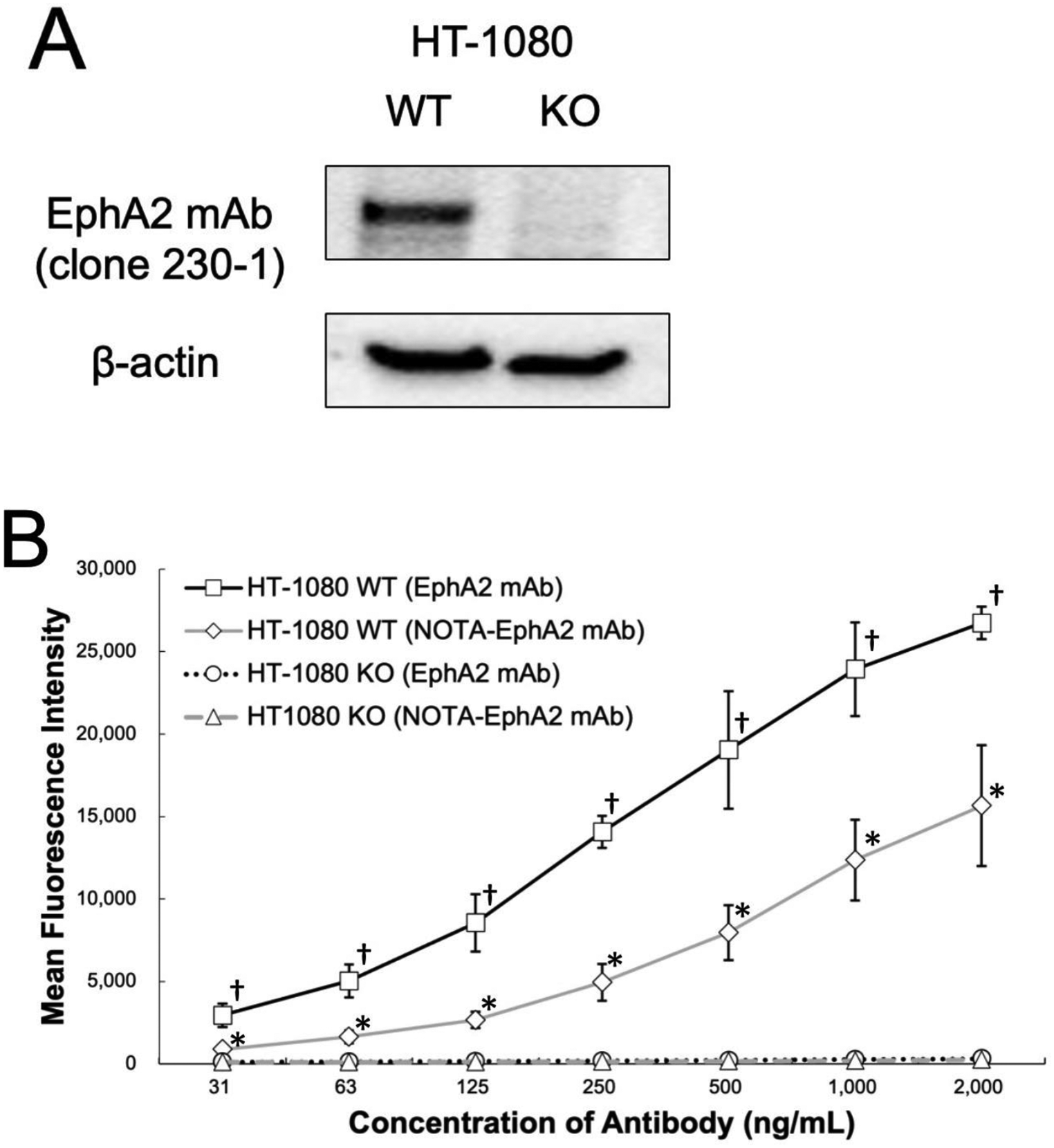
Target sequence identification for designing gRNA was performed using the online software CRISPRdirect (https://crispr.dbcls.jp/). The gRNA sequence GAGGGGCAGAAGTTGCGCGC (AGG) in exon 1 of EphA2 was cloned into a pCas-Guide-EF1a-GFP vector (OriGene Technologies). HT-1080 cells (EphA2-expressing human fibrosarcoma cells) were obtained from American Type Culture Collection. Cells were transfected with the CRISPR Cas9 plasmid using the Neon transfection system (Thermo Fisher Scientific). At 48 h post-transfection, the cells were trypsinized and sorted via fluorescence-activated cell sorting (FACS) to obtain green fluorescent protein–positive cells using FACSMelody (Becton Dickinson). Clonal selection of single green fluorescent protein–positive cells yielded several viable clones, which were screened for EphA2 gene knockouts (KO) by Sanger sequencing. EphA2 protein levels were then compared with those in wild-type (WT) cells by western blotting.
The reactivity of EphA2 mAb (clone 230-1) and NOTA–linked EphA2 mAb (clone 230-1) to EphA2 on cancer cell surfaces was determined using flow cytometry. Approximately 1 × 10⁵ HT-1080 WT or HT-1080 EphA2-KO cells were incubated with various concentrations (31.25–2,000 ng/mL) of mAb in 20 µL phosphate-buffered saline (PBS; pH 7.4) with 2% fetal bovine serum (FBS; staining buffer) for 60 min on ice. The cells were then washed and stained with the phycoerythrin-conjugated goat anti-mouse IgG secondary antibody (Invitrogen) for 30 min on ice. The cell suspension was washed three times with PBS and analyzed using a FACSymphony A1 flow cytometer (BD Immunocytometry Systems).
Preparation of xenograft models
HT-1080 cells were maintained in a culture medium (RPMI 1640 with l-glutamine and phenol red; Fujifilm Wako Pure Chemical) with 10% heat-inactivated FBS and 1% penicillin-streptomycin. Male nude mice (BALB/cSLC-nu/nu, origin: the Institute of Medical Science, the University of Tokyo, total n = 46) were purchased from Japan SLC, Inc. (Hamamatsu, Japan). The mice were housed under a 12 h light/12 h dark cycle with free access to food and water and were allowed to acclimate for at least one week. The housing chamber temperature was maintained at approximately 23 °C. Tumor xenograft models were established by the subcutaneous injection of 1 × 10⁶ HT-1080 cells suspended in 0.1 mL PBS in nude mice. The mice were evaluated 2 weeks after implantation, when the tumor size reached approximately 1 cm in diameter. The protocol was approved by the Animal Care and Use Committee of the Osaka University Graduate School of Medicine (approval number: 30-088-009). Euthanasia was performed under deep anesthesia using isoflurane inhalation when signs of intolerable suffering or a significant decrease in body weight (reduction of more than 30% compared to baseline) were observed. A tumor diameter of more than 2 cm (approximately 4,000 mm³) was also defined as the humane endpoint.
[89Zr]Zr-EphA2 mAb PET imaging and analysis
[89Zr]Zr-EphA2 mAb (clone 230-1) (1.30 ± 0.09 MBq, approximately 7.62 ± 0.52 µg of EphA2 mAb, adjusted to a volume of 0.1 mL) was intravenously administered to HT-1080 xenograft mice (9 weeks old, body weight = 23.4 ± 0.9 g, n = 6). Static PET/CT scanning was performed 1 and 5 days after administration, by PET scan durations of 10 and 20 min, respectively, under isoflurane anesthesia using a small-animal PET scanner (Siemens Inveon PET/CT). After the PET scan on day 5, the mice were euthanized, and the activity and weight of the major organs were determined using a well counter (BeWell; Molecular Imaging Laboratory). All PET data were reconstructed using 3-dimensional ordered-subset expectation maximization (16 subsets, 2 iterations), followed by maximum a posteriori (OSEM3D-MAP) with scatter and attenuation correction. The regional uptake of activity was decay-corrected for the injection time and expressed as a standardized uptake value (SUV). Regions of interest (ROIs) were manually defined on the tumor (ellipsoid-sphere ROI adjusted to the tumor size) and the muscle tissue (3 mm diameter spherical ROI in the right forelimb), referencing anatomical CT images using Osirix MD version 14.0 (Pixmeo SARL).
Evaluation of biodistribution of [177Lu]Lu-EphA2 mAb
HT-1080 xenograft mice (n = 6) were intravenously administered [177Lu]Lu-EphA2 mAb (clone 230-1) (3.12 ± 0.21 MBq, 50 µg) to evaluate biodistribution under isoflurane anesthesia. The volume of each injected solution was adjusted to 0.1 mL (as for the following experiments). At 24 and 72 h after administration, the mice were euthanized, and the tumors and major organs were collected. The activity and weight of the samples were measured using a well counter system. A blocking study was also performed by pre-administration of non-radiolabeled EphA2 mAb (500 µg) (5 min before the administration of [177Lu]Lu-EphA2 mAb (2.93 ±0.13 MBq)) to evaluate the uptake specificity of [177Lu]Lu-EphA2 in HT-1080 xenograft (n = 6). Uptake levels were compared 24 h post-administration between the blocking and non-blocking groups. The absorbed dose (Gy) in the tumor was calculated based on the S value (0.0233 mGy/MBq·s) reported in a previous publication [9]. The residence time (s) was determined using the trapezoidal method with biodistribution data collected at 24 and 72 h, assuming only physical decay occurs after 72 h.
Radioimmunotherapy using [177Lu]Lu-EphA2 mAb
The antitumor effect was evaluated by administering 10 MBq (9.7 ± 0.78 MBq, 46–150 µg, n = 9) or 3 MBq (2.84 ± 0.45 MBq, 12–50 µg, n = 9) of [177Lu]EphA2 mAb (clone 230-1) (10.36 ± 0.54 MBq), non-radiolabeled EphA2 mAb (clone 230-1) (50 µg, n = 6), [177Lu]LuCl3 solution (10 MBq, n = 6), or saline (control, n = 4) in HT-1080 xenograft mice (8 weeks old; body weight, 22.1 ± 1.24 g). Tumor size and body weight were monitored to evaluate treatment effects and systemic side effects. The tumor volume (mm3) was measured with a caliper and calculated using the elliptical sphere model equation. Preliminary blood tests were performed to evaluate hematological and kidney toxicity using iSTAT (Abbott).
Immunohistochemistry
Immunohistochemical staining was performed to confirm EphA2 expression in the tumor xenografts using an anti-EphA2 antibody. After the animals were euthanized, tumor xenografts were resected and fixed with 4% paraformaldehyde (overnight, 4 °C). The fixed tissues were immersed in 30% sucrose in PBS (overnight, 4 °C). The frozen sections (slice thickness: 10 μm) were incubated with an anti-EphA2(D4A2) rabbit mAb (CST #6997; Cell Signaling Technology, 100-fold dilution). Immunohistochemistry was performed using Dako EnVision + System– HRP Labeled Polymer Anti-Rabbit (K4003) (DAKO Corp.). The stained sections were analyzed by microscopy (Keyence BZ-9000). A total of five mice were used, with one section prepared per mouse. Two locations were selected per section, and each location was captured at four magnifications: ×40, ×100, ×200, and ×400.
Statistical analyses
Comparisons between the two groups were performed using the Mann–Whitney U test for tumor size and body weight, and the t-test, following normality assessment with the Shapiro-Wilk test, for flow cytometry analysis and blocking experiments. Statistical analyses were performed using SPSS version 25.0 (IBM Corp.), and differences were considered statistically significant at a p-value of < 0.05.


Comments (0)