Acquisition and analysis of public data resources
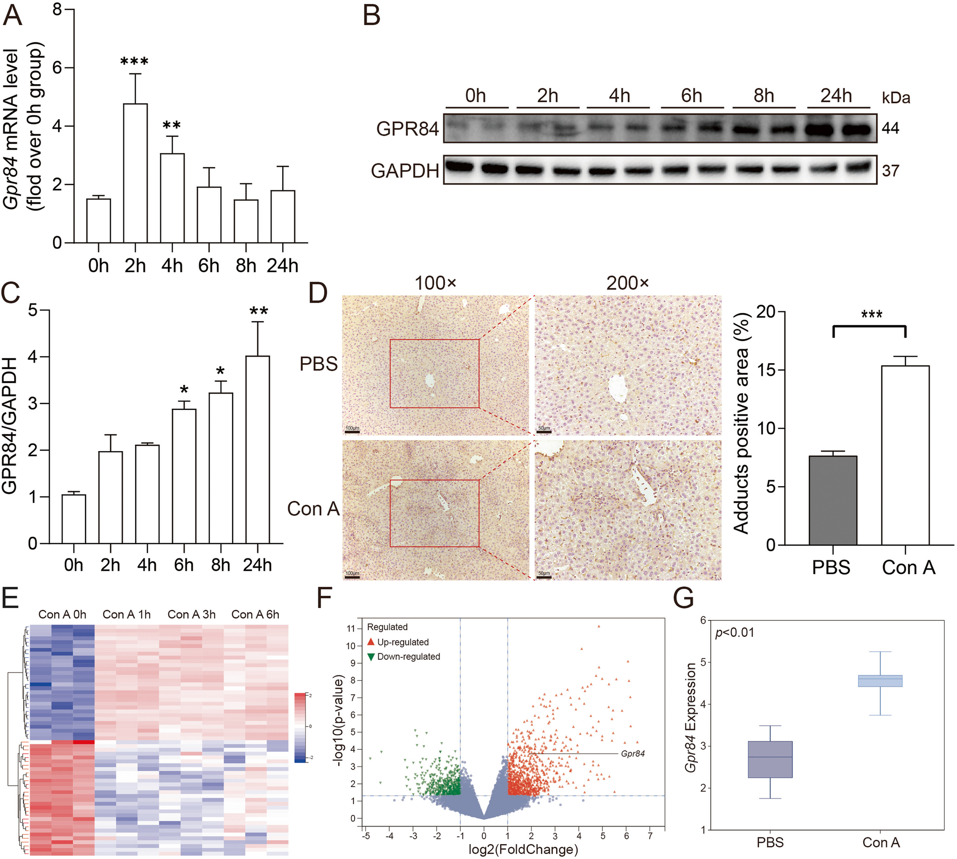
We retrieved the dataset for Con A-induced immune liver injury in mouse models (GSE17184, n = 12) from the Gene Expression Omnibus (GEO) database. Using the SangerBox platform (http://sangerbox.com), we identified differentially expressed genes (DEGs) and generated relevant visualizations.
Animals
The animal experiments were conducted using 6–8-week-old male C57BL/6 J wild-type (WT) mice, obtained from the Model Animal Research Center of Nanjing University. GPR84 knockout (KO) mice were provided by BRL Medicine Inc. Mice were housed under controlled temperature conditions with a standard 12-h light/dark cycle. All animal procedures were approved by the Animal Experimentation Center of Nanjing Medical University.
Induction of immune liver injury
WT and GPR84-deficient mice were subjected to immune liver injury by tail vein injection of Concanavalin A (Con A, 15 mg/kg, Sigma, #C2010) or phosphate-buffered saline (PBS). Mice were euthanized at different time points post-injection via CO₂ inhalation to collect liver tissues and blood samples for subsequent analyses.
Quantitative real-time PCR (qRT-PCR)
Total RNA was extracted from liver tissues using Trizol reagent, followed by reverse transcription into cDNA with HiScript III RT SuperMix (Vazyme, #R323). The cDNA was then amplified using ChamQ SYBR Color qPCR Master Mix (Vazyme, #Q421). Primers used in the PCR reactions were synthesized by Invitrogen, and their sequences are listed in Supplementary Table 1.
Immunohistochemistry (IHC)
Mouse liver tissues were fixed in 4% paraformaldehyde for 24 h, followed by paraffin embedding and sectioning. The sections were stained with H&E and subjected to immunohistochemical staining using a GPR84 antibody (Bioss, #bs-13507R). Stained sections were examined and photographed with an Olympus IX51 light microscope, and ImageJ software was used to analyze tissue morphology.
Western blotting
Total protein was extracted from mouse liver tissues or cell samples using RIPA lysis buffer. Protein samples were separated by SDS-PAGE and transferred to PVDF membranes. After blocking with 5% BSA for 2 h, membranes were incubated overnight at 4 °C with primary antibodies, followed by detection with HRP-conjugated secondary antibodies. The primary antibodies used included p-STAT3 (Cell Signaling Technology, #4074S), STAT3 (Cell Signaling Technology, #4904S), p-ERK (Cell Signaling Technology, #4370S), ERK (Cell Signaling Technology, #4695S), p-JNK (Cell Signaling Technology, #9251S), JNK (Cell Signaling Technology, #9252S), p-p38 (Cell Signaling Technology, #9211S), p38 (Cell Signaling Technology, #9212S), p-p65 (Cell Signaling Technology, #3033S), p65 (Cell Signaling Technology, #8242S), Cleaved Caspase-8 (Cell Signaling Technology, #8592S), Caspase-3/p17/p19 (Proteintech, #66,470–2-Ig), GPR84 (Bioss, #bs-13507R), and GAPDH (Proteintech, #10,494–1-AP). All antibodies were diluted according to the manufacturer’s instructions.
AST and ALT measurement
Blood samples were collected from the retro-orbital sinus of mice at 8 and 24 h after Con A injection. Serum was separated and analyzed using an automated biochemical analyzer (MODULAR EVO 4200) to measure AST and ALT levels.
TUNEL assay
To detect apoptosis, the One Step TUNEL Apoptosis Assay Kit (Beyotime, #C1089) was used according to the manufacturer's instructions for TUNEL staining.
Liver non-parenchymal cells (NPCs) isolation
The liver was perfused with HBSS containing EGTA to remove blood cells, followed by mechanical dissociation. The tissue was ground, filtered through a 100 µm cell strainer to obtain a single-cell suspension, and red blood cells were lysed using RBC lysis buffer. Liver NPCs were then isolated using 35% Percoll (Cytiva, #17,089,101). The resulting cells were resuspended in PBS with 2% FBS for subsequent staining.
Flow cytometry
Isolated liver NPCs were incubated with CD16/32 antibody for 30 min to block non-specific binding, followed by staining with fluorescence-conjugated antibodies. The antibodies used included: APC-conjugated anti-mouse F4/80 (BioLegend, #123,116), FITC-conjugated anti-mouse Ly6 C (BD Biosciences, #561,085), PE-Cyanine7 conjugated anti-mouse CD11b (BioLegend, #101,216), APC-Cyanine7 conjugated anti-mouse CD45 (BioLegend, #103,116), PE-conjugated anti-mouse Ly6G (BD Biosciences, #561,104), APC-eFluor™ 780-conjugated anti-mouse CD3 (eBioscience, #47–0032–80), APC-conjugated anti-mouse CD4 (eBioscience, #17–0042–83), and FITC-conjugated anti-mouse CD8a (eBioscience, #11–0081-81).
Isolation and culture of bone marrow-derived macrophages (BMDMs)
Mice were euthanized with CO₂, and bone marrow was flushed from femurs using RPMI 1640 medium. The cell suspension was passed through a 100 µm strainer, and red blood cells were removed with lysis buffer. The remaining cells were seeded in culture dishes with RPMI 1640 medium supplemented with 10 ng/mL macrophage colony-stimulating factor (M-CSF) and 10% FBS. After incubating at 37 °C with 5% CO₂ for 4 days, 4 mL of fresh medium containing 10 ng/mL M-CSF and 10% FBS was added. Cells were incubated for an additional 3 days before being used for further experiments.
Isolation of primary hepatocytes
Primary hepatocytes were isolated following the protocol (You et al. 2013). In brief, after euthanasia, the liver was first perfused with HBSS containing Ca2⁺ and Mg2⁺ for 5 min, followed by 2 min with HBSS without Ca2⁺ and Mg2⁺. Subsequently, 0.04% type IV collagenase (Sigma-Aldrich, #C4-BIOC) was perfused for 10 min. The liver was then gently stirred in Williams E medium and filtered through a 100 µm cell strainer. Hepatocytes were isolated by centrifugation at 40 × g for 3 min.
Bone marrow transplantation to create chimeric mice
Eight-week-old recipient mice were fed 0.2% neomycin for one week prior to X-ray irradiation (5 Gy for 2–3 min, repeated after 3 h). Donor bone marrow cells (1 × 10⁷ cells/mouse) were injected via the tail vein immediately after irradiation. The transplanted mice were housed in cages and maintained on 0.2% neomycin for two weeks to prevent infections. Eight weeks after transplantation, immune-mediated liver injury was induced by injecting Con A (15 mg/kg, Sigma, #C2010). To confirm bone marrow reconstitution, genomic DNA was extracted from peripheral blood and used for GPR84 genotyping (Fig. S1D).
Treatment
To induce immune-mediated liver injury, mice received an injection of Con A (15 mg/kg, Sigma, #C2010) or PBS. One hour later, 30 mg/kg of the GPR84 antagonist GLPG1205 (MCE, HY-135303) was administered via oral gavage. Mice were euthanized with CO₂, and liver tissues and blood samples were collected for further analysis. For cell experiments, cells were treated with 10 µM GLPG1205 and 10 µg/mL Con A, and samples were collected at various time points for downstream protein analysis.
Statistical analysis
All data are presented as the mean ± SEM. Statistical analyses were performed using GraphPad Prism software (version 8.0.2). Depending on the number of groups compared, either a two-tailed unpaired Student’s t-test or analysis of variance (ANOVA) was used to assess statistical significance. For comparisons between two groups, we utilized the Student's t-test, employing the Mann–Whitney U test for non-parametric data. For comparisons involving multiple groups, we conducted one-way or two-way analysis of variance (ANOVA), followed by post-hoc tests to identify specific differences among groups. Statistical significance was indicated by P values, with P < 0.05 considered significant. Levels of significance are denoted as P < 0.01 (**), P < 0.001 (***), and P < 0.0001 (****).


Comments (0)