We harvested cells during the logarithmic growth phase, seeded them, and replaced the standard culture medium 30 min before reaching approximately 70% confluence. Plasmids containing short hairpin RNA (sh-NC/sh-CKS2) and overexpression gene (oe-NC/oe-CKS2) were obtained from Fenghui Biotechnology (China). We transfect sh-NC/sh-CKS2 and oe-NC/oe-CKS2 plasmids into cells with the use of Ultra Fection 3.0. After incubating in a saturated humidity culture chamber at 37 ℃, 5% CO2 for 6 h, the culture medium was replaced with a fresh one for continued cultivation.
qRT-PCR
Total RNA was extracted from tumor tissues or cells using TRIzol (CWBIO, China). The extracted RNA was reverse transcribed into cDNA using Hifair® II 1st Strand cDNA Synthesis Kit (YESEN, China). qRT-PCR was performed by using uGreener Flex qPCR 2XMix (U&GBIO, China) on an ABI7500 real-time fluorescence quantitative PCR system (Applied Biosystems, USA). GAPDH was used as an internal control, and the calculation method was 2−ΔΔCt. The primer sequences are outlined in Table 1.
Table 1 Primer set for qRT-PCRWestern blot (WB)
The cells were lysed in lysis buffer (containing 1% phosphatase inhibitor and 1% protease inhibitor) on ice for 10 min at 4 ℃, 12,000 rpm. The supernatant was gathered after being centrifuged for 15 min and the total protein content was determined using the bicinchoninic acid (BCA) assay kit (Solarbio, China). Denatured proteins were separated by SDS-PAGE, transferred to the PVDF membrane, blocked with 5% skim milk, and then mixed with CKS2 (ABclonal A3791, China), LC3B (LC3I, LC3II ABclonal, China, A5618), p62 (ABclonal, China, A19700), GPX4 (AbclonalA11243, China), and SLC7A11 (AbclonalA13685, China) for overnight incubation at 4 ℃. The TBST washing lasted 3 times, 5 min for each time. Then the membrane was incubated with the secondary antibody (Beyotime A0208, China) at room temperature for 1.5 h. Following three rounds of TBST washing, lasting five minutes each, the protein bands were visualized using the ECL detection kit (Biosharp, China) and photographed with the ChemiScope 6200 imaging system (Clinx, China).
CCK-8 assay
Transfected CC cells were plated in a 96-well plate (density of 2000 cells/well), with cell viability detected at 0, 24, 48, and 72 h. 10 µl of CCK-8 reagent was introduced to each well, followed by 2 h of incubation at 37 ℃, with the absorbance optical density (OD) value of each well at 450 nm measured by a microplate reader.
We seeded 2 × 10⁴ cells in a 96-well plate and treated them with different concentrations of ferroptosis inducer Erastin (0/5/10/20/40/80 µM) or sora (0/2.5/5/10/20 µM). After 24 h, each well was supplemented with 10 µl of CCK-8 solution and kept at 37 ℃ for 2 h. OD and IC50 values were detected and calculated.
Colony formation assay
Cells were inoculated into a 12-well plate (200 cells/well) and cultivated for 7 days. Then, the plate was immobilized with 75% methanol, stained with 1% crystal violet for 30 min, rinsed with phosphate-buffered saline (PBS), dried at 37 ℃, and photographed with a Nikon D5600 camera (Nikon, Japan) to record the cell units in each well.
Transmission electron microscope (TEM)
Cells were inoculated into a T75 culture bottle and transfected with sh-NC/sh-CKS2 and oe-NC/oe-CKS2 plasmids respectively. When the cell density reached 90% or more, cells were gathered by trypsin digestion and washed with PBS, with cell precipitate fixed in 2.5% glutaraldehyde solution. Subsequently, the fixed cells were subjected to permeabilization and embedded overnight at 70 ℃. Embedded slices were stained with 1% uranyl acetate and 0.4% lead citrate, and then observed under the TEM.
Measurement of ROS and lipid ROS in cells
With the use of the ROS detection kit (Beyotime, Cat. No.: S0033S, China), the level of ROS in cells was determined. Subsequently, cells in each treatment group were seeded in 6-well plates and incubated for 12 h. We diluted the original DCFH-DA solution in the kit with the serum-free medium at a ratio of 1:1000, then added it to each well and cultivated it at 37 ℃ for 30 min. Cells were collected and rinsed three times with PBS solution to remove residual DCFH-DA dye. On an Agilent NovoCyte flow cytometer (Agilent, USA), we finished the analysis.
Next, cells or tumor tissue homogenate were resuspended in 500 µL PBS solution containing 2 µM BODIPY581/591 C11 (MCE, USA), and housed in a CO2 incubator at 37 ℃ for 30 min. Samples were analyzed using the Agilent NovoCyte (Agilent, USA).
Propidium iodide (PI) staining
The Hoechst 33,342/PI dual staining kit (Solarbio, Cat. No: CA1120, China) was employed to assess apoptosis. Cells from each treatment group were inoculated into a 6-well plate and maintained for 12 h. Subsequently, the cells were collected, and to each tube, 5 µL of PI reagent and 5 µL of Hoechst reagent were added. The tubes were then incubated on ice for 20 min and the analysis was performed using the Agilent NovoCyte flow cytometer (Agilent, USA).
Detection of GSH, glutamate, cysteine, and cystine intake levels and GSH/ oxidized GSH (GSSG) ratio
Relative GSH levels and GSH/GSSG ratios in cells or nude mouse tumor tissues were determined using GSH and GSSG detection kits (Beyotime, Cat. No.: S0053, China), with OD measured at 405 nm. The relative GSH level and GSH/GSSG ratio in the sample were calculated.
The glutamate colorimetric assay kit (Elabscience, Cat. No: E-BC-K903-M, China) was employed to determine glutamate concentration in cells or nude mouse tumor tissues, with OD measured at 450 nm. The glutamate concentration in samples was calculated.
Cysteine concentration in cells or nude mouse tumor tissues was assessed using a cysteine colorimetric test kit (Elabscience, Cat. No.: E-BC-K352-M, China), with OD measured at 600 nm and cysteine concentration calculated. The cystine uptake fluorescence assay kit (Elabscience, Cat. No: E-BC-F066, China) was applied in the measurement of cystine uptake level. The fluorescence values were measured using a SynergyH1 fluorescence microplate reader (Boteng, USA) at an excitation wavelength of 485 nm and an emission wavelength of 535 nm, with the cystine uptake in the samples calculated.
Immunofluorescence (IF)
Cells were immobilized in 75% alcohol for 30 min, followed by three times rinses with PBS (5 min for each), 10 min of permeabilization with 0.1% TritonX-100, as well as 1 h of block with 5% BSA at room temperature. The slides were incubated overnight at 4 ℃ with LC3B antibody (ABclonal, China, A5618) and then with the secondary antibody conjugated with AF555 (Bioss, China, bs-0295G-AF555) at room temperature for 1 h. After the addition of DAPI (10 ug/ml) (Solarbio, Cat. No: C0065, China) for nuclear staining, the slides were incubated for 5 min, washed with PBS, sealed, and observed under an inverted fluorescence microscope.
Immunohistochemistry (IHC)
4 µm-thick of CC tumor tissue sections were applied. In brief, we embedded the tissue in paraffin, then deparaffinized the sections and rehydrated them through graded alcohols, followed by 3 times washing in PBS, each for 5 min. Next, the sections were incubated overnight with primary antibodies GPX4 (A11243, abclonal, China) and SLC7A11 (A13685, abclonal, China). The sections were then incubated with secondary antibody goat anti-rabbit IgG at 37 ℃ for 30 min, stained with a working solution of 3, 3′-DAB for 3 min, and rinsed 3 times in PBS, each time for 5 min. Finally, the sections were counterstained with hematoxylin and sealed for microscopic examination. Three sections were taken from each tumor tissue for testing. Five independent fields were randomly selected for observation and photography by using a Leica microscope (×200).
Xenograft model
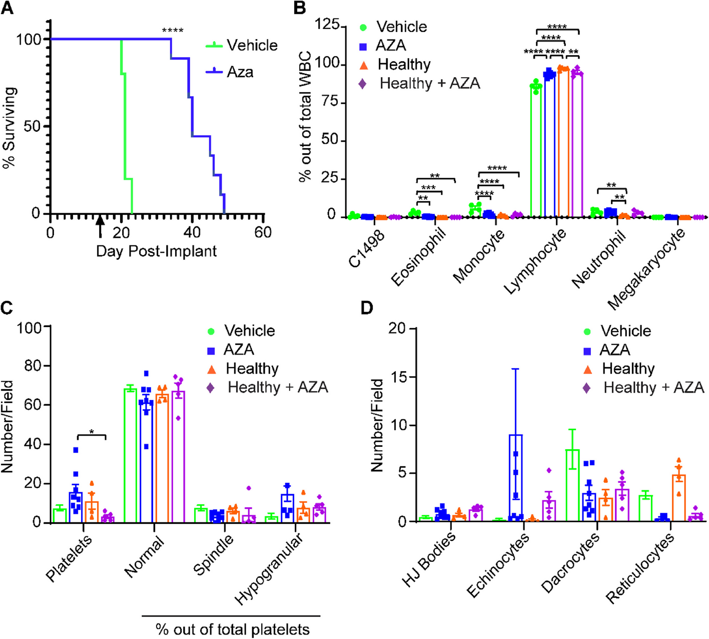
Twenty-four female BALB/c nude mice (4 weeks old, 18.0 ± 2.0 g) purchased from Shanghai SLAC Laboratory Animal Co., Ltd. were randomly divided into two groups for in vivo xenograft experiments. The mice were injected with 5 × 10⁶ CC cells with stable knockdown of CKS2 (sh-CKS2) or control vector (sh-NC). The cells were subcutaneously injected into the right axilla of the mice in both groups. When the tumor volume reached 100 mm (Yuan et al. 2021), the nude mice were intraperitoneally injected with sora (5 mg/kg) or PBS. Tumor size was measured every 5 days, and tumor volume was calculated as follows: tumor volume = (length × width2)/2. All animal experiments and procedures were guided and approved by Taizhou Hospital, Wenzhou Medical University Ethics Committee (approval number: tzy-2024084).
Data analysis
Three parallel groups were set up for each group, and the experiment was independently repeated three times. Data analysis was processed using SPSS 22.0 software. Quantitative data were expressed as mean ± standard deviation (x̅±s). Independent sample t-test was employed for comparison between two groups, while the one-way analysis of variance (ANOVA) was employed for comparison among multiple groups. A p-value < 0.05 referred to a significant difference. Statistical charts were drawn by utilizing GraphPad Prism 8.0 software.


Comments (0)