2.1 Ethics
This prospective, open-labelled, single-center study was approved by the ethics committee of the Medical University of Vienna (EC: 1348/2015) and the Austrian Agency for Health and Food Safety. It was registered at the European Union Drug Regulating Authorities Clinical Trials Database (EUDRACT 2015-001793-18) and conducted according to International Council for Harmonisation of Technical Requirements for Pharmaceuticals for Human Use good clinical practice guidelines and the Declaration of Helsinki. Since the participants were unconscious, consent was obtained from survivors once they could understand the study. The treating team informed patients’ relatives about their inclusion in the study, but the ethics committee did not mandate this as a requirement. The study procedures were conducted at the Department of Emergency Medicine (ED) and the ICU at the Medical University of Vienna from April 2016 to June 2022.
2.2 Study Population and Procedure
We included comatose, intubated patients aged ≥18 years who experienced witnessed CA of any rhythm, were eligible for hypothermic temperature control, and received AMO/CLAV or AMP/SULB for suspected or confirmed aspiration pneumonia. Exclusion criteria included pregnancy, unwitnessed CA, non-sustained ROSC, body mass index (BMI) outside the range of 18–40 kg/m2, significant coagulopathies or hemorrhagic conditions (platelet count < 50 G/L), a glomerular filtration rate < 50 mL/min/1.73 m2, advanced malignancies, terminal illnesses, or signs of impending clinical deterioration.
All patients underwent the same procedures during two study phases: the hypothermic phase and the subsequent normothermic phase. During each study period, plasma, microdialysate, and urine were collected at predetermined intervals after antibiotic steady state. In the AMP/SULB group, bronchial mucosa ELF was collected in both phases.
2.2.1 Hypothermic Temperature Control
Hypothermic temperature control procedures followed ED guidelines targeting 33 ± 1 °C for 24 h after sustained ROSC, followed by rewarming at 0.5 °C/h [20]. Normothermia (36.5 ± 1 °C) was maintained for at least 72 h using physical or pharmacological methods. Cooling was achieved noninvasively with Arctic Sun® 5000 (Bard Medical Division, C.R. Bard, Inc., Louisville, CO, USA) or external surface cooling pads (EMCOOL-Spad®, EMCOOLS AG, Pfaffstaetten, Austria, and TTcool-pad 302, Everynear GmbH, Baden, Austria) or invasively with Thermogard®XP (ZOLL Medical, Cologne; Germany). Body temperature was monitored with an esophageal temperature probe.
2.2.2 Study Medication
Patients received either 2 g amoxicillin/0.2 g clavulanate (Curam® 2.2 g powder for infusion solution, Sandoz GmbH, Vienna, Austria) or 2 g ampicillin/1 g sulbactam (Unasyn 3g Dry Vials®, Pfizer Corporation Austria Ges.m.b.H, Vienna, Austria). Antibiotics were prepared in 100 mL 0.9% saline solution (physiological saline solution “Fresenius”- Infusions Solution, Fresenius Kabi Austria GmbH., Graz, Austria) and administered via infusion pump intravenously over 15 min every 8 h. The line was flushed with 50 mL 0.9% saline after infusion. Antibiotics were given at least twice before sampling to ensure steady-state conditions.
2.2.3 Clinical Pneumonia Surveillance
C-reactive protein levels (normal: \(\le \hspace\)0.49 mg/dL) were measured daily. Chest X-rays or computed tomography scans were performed at the physician’s discretion, and pneumonia signs were evaluated by the attending radiologist.
2.3 Sampling2.3.1 Plasma Sampling
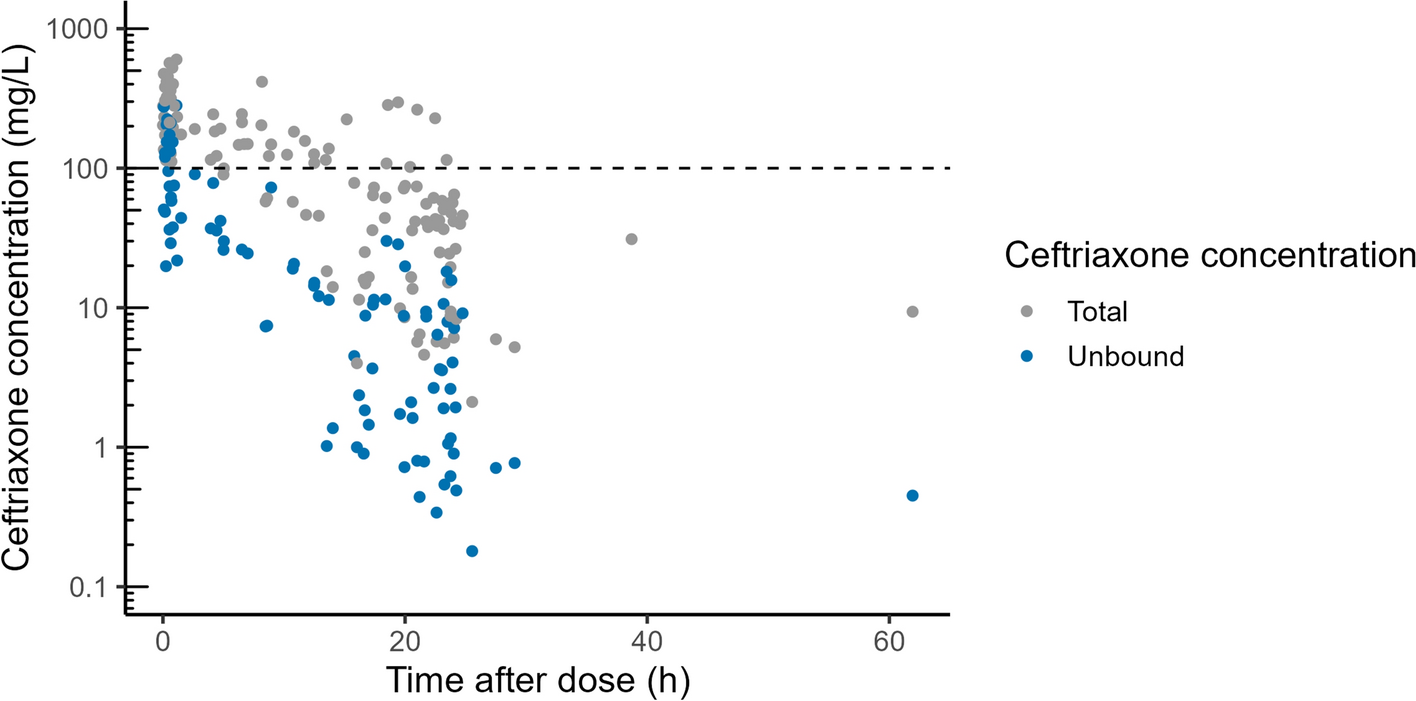
Blood samples (4 mL) were collected via an arterial catheter into lithium–heparin tubes. To measure total drug concentrations in plasma, samples were drawn at baseline (before the next antibiotic administration) and at 0.5, 1, 1.5, 2, 3, 4, and 6 h after start of drug infusion. The sample collection schedule was identical for both study periods. Samples were refrigerated immediately, centrifuged at + 4 °C, 2600g, 10 min, and the plasma was frozen at − 20 °C within 1 h.
2.3.2 Urine Sampling
Urine was collected at 0–2, 2–4, and 4–6 h after drug administration. About 5 mL per sample was refrigerated and frozen at − 20 °C within 1 h.
2.3.3 Bronchoalveolar Lavage, ELF Collection
To investigate antibiotic distribution in the bronchial mucosa, ELF was collected via bronchoscopy from endotracheally intubated patients. This procedure was conducted exclusively in the AMP/SULB group as part of an exploratory approach to assess target site antibiotic penetration. A single bronchoalveolar lavage (BAL) was performed at 2, 4, or 6 h after starting antibiotic administration during each study period. Up to three 20 mL saline aliquots were instilled and aspirated. BAL samples were mixed, filtered, and centrifuged at 2000g, 4 °C, 10 min, and the supernatant was frozen at − 20 °C within 1 h.
Bronchoscopies were performed using a flexible video bronchoscope (EB-1970K by Pentax©, HOYA Corporation, Tokyo, Japan). To determine the dilution factor of ELF in BAL, the urea method was applied as described previously [21].
2.3.4 Microdialysis
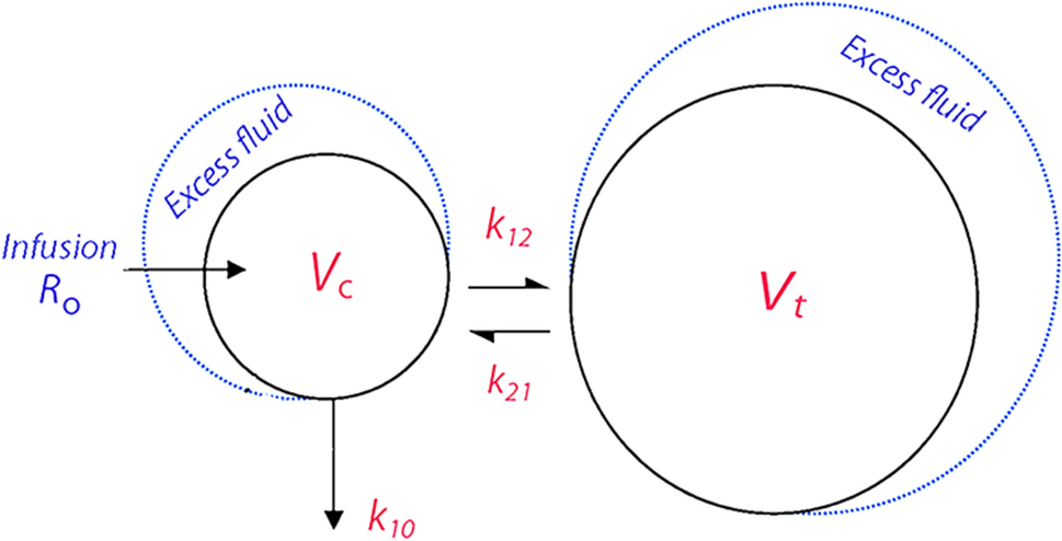
In the present study, two microdialysis probes were inserted into one of the patient’s thighs. The microdialysis catheters (63 Microdialysis Catheter 60/10, M Dialysis AB, Solna, Sweden) with a membrane length of 10 mm and a molecular weight cut-off of 20 kDa, were inserted into the subcutaneous adipose tissue and into the quadriceps muscle. Throughout the entire study (both sampling periods), microdialysis probes were perfused with 0.9% saline solution at a flow rate of 2 µL/min. Following an equilibration period of 30 min, microdialysis samples were collected at baseline (before antibiotic drug administration) and at 0–1, 1–2, 3–4, 4–5, and 5–6 h after drug administration. All microdialysis samples were immediately placed in a portable refrigerator box and frozen at − 20 °C within 1 h from collection.
After the last sample of each study day, each probe was calibrated using the retrodialysis method to calculate the concentration in the interstitial space fluid, as previously described [22]. For this part of the experiment, the perfusion medium for calibration contained a known concentration of the respective antibiotics and beta-lactamase inhibitor, and the relative loss rate was determined.
2.3.5 Sample Handling and Storage
At the end of the study day, plasma, microdialysate, ELF, and urine samples were transferred from − 20 °C to − 80 °C and stored at − 80 °C until analysis.
2.3.6 Sample Analysis: High-Performance Liquid Chromatography Method and Ultrafiltration
The high-performance liquid chromatography equipment consisted of a Shimadzu Prominence modular system with a three-channel degasser (DGU 20A3R), quaternary solvent pump (LC 20AD), autosampler (SIL 20AC HT, set to 6 °C), column oven (CTO 20AC, set to 40 °C), photodiode array detector (SPD M30A, detection wavelengths: AMP/SULB 205 nm, AMO 227 nm, CLAV 215 nm) equipped with cells of 10 mm or 85 mm optical path length, system controller CBM 20A, and LabSolution software (all from Shimadzu Europe, Duisburg, Germany). The flow rate was 0.4 mL/min, and the injection volume was 1–2 µL, for BAL 5 µL. Separation was performed isocratically using a Cortecs T3 2.7 µ 100 × 3 mm analytical column (Waters, Eschborn, Germany) preceded by a guard column (Nucleoshell RP18 2.7 µ 4 × 3 mm, Macherey-Nagel, Düren, Germany). The mobile phase consisted of 0.1 M sodium phosphate buffer/acetonitrile 90:10 (v/v), pH 3.1, for the determination of AMP/SULB, and 97:3 (v/v), pH 3.3, for the determination of AMO/CLAV. SULB and CLAV were eluted after 2.1 min, AMP and AMO after 4.2 min.
Total drug concentrations were analyzed according to a published protocol [23]. In brief, serum (100 µL) was buffered with 25 mM sodium phosphate buffer (pH 6.0, 200 µL) and deproteinized with acetonitrile (500 µL). The precipitated protein was separated by centrifugation, the acetonitrile was extracted into dichloromethane (1.5 mL), and an aliquot of the aqueous layer was injected. The free concentrations were measured using a published ultrafiltration method [24]. In brief, plasma (300 µL) was mixed with 10 µL potassium phosphate (3M, pH 7.43 ± 0.02) in a Vivafree™ 500 30 kD Hydrosart® centrifugal ultrafiltration device (Vivaproducts Inc., Littleton, MA, USA) before ultrafiltration at 37 °C. Microdialysate or BAL were injected directly, and urine after dilution 1:50 with 10 mM sodium phosphate buffer, pH 6.0.
The lower limit of quantification (LLOQ) for AMP/SULB and AMO/CLAV was 0.3/0.5 and 0.3/0.2 mg/L, respectively, in plasma, and 0.03/0.05 and 0.03/0.02 mg/L, respectively, in saline, which was used as a surrogate for the other matrices. Based on in-process quality controls (high/low), the relative standard deviation (SD) for precision was < 3%/≤ 7% for AMP/SULB and < 3%/< 5% for AMO/CLAV, respectively; the inaccuracy was < 3% for all substances. The estimated LLOQ (signal-to-noise ratio = 5) in BAL (injection volume 5 µL) was 0.02/0.01 for AMP/SULB and 0.01/0.01 mg/L for AMO/CLAV; the LLOQ in urine was estimated at < 30/50 and < 30/20 mg/L, respectively. The accuracy of the determination of free drug in plasma cannot be specified, as the extent of protein binding in a particular sample is not known. The precision was assessed by analyzing spiked pooled plasma of healthy subjects; in these samples, the mean ± SD unbound fraction of AMP/SULB and AMO/CLAV was 80.6 ± 6.9%/91.7 ± 6.9% and 87.3 ± 1.9%/81.7 ± 3.4%, respectively.
2.4 Pharmacokinetic Analysis
The pharmacokinetic data were analyzed using Phoenix® WinNonlin® Build 8.0 (Certara USA, Inc., Princeton, NJ, USA). We employed a non-compartmental analysis for this study to determine the pharmacokinetic parameters.
2.4.1 Plasma Protein Binding
Plasma protein binding was assessed for each subject at three distinct timepoints using ultrafiltration. Mean plasma protein binding per subject was used to calculate unbound drug concentrations, which were used for pharmacokinetic analysis.
2.4.2 Pharmacokinetic Parameters
We determined key plasma pharmacokinetic parameters, including maximum plasma concentration, elimination half-life, apparent volume of distribution, total body clearance, and area under the concentration–time curve from 0 to 6 h.
We also calculated the duration that unbound plasma concentrations exceeded the minimal inhibitory concentration [MIC] (fT>MIC) for AMO and AMP, using MIC values of 4 mg/L, 8 mg/L, and 16 mg/L, representing the upper range for relevant pathogens per European Committee on Antimicrobial Susceptibility Testing MIC distributions [25]. Calculations of the fT>MIC were based on the individual concentrations from each study subject.
2.4.3 Exposure Comparison
To compare drug exposure between the normothermic and hypothermic states, we calculated concentration ratios (individual concentration at each timepoint in normothermia divided by the concentration at the same time point in hypothermia) for plasma, muscle, and subcutaneous tissue.
2.5 Data Analysis2.5.1 Urine Excretion
The amount of drug excreted in urine within the first 6 h after infusion initiation at the targeted temperature (hypo- or normothermia) was quantified based on concentration measurements at various urine sampling intervals. This was expressed as a percentage of the administered drug dose for one dosing interval.
2.6 Statistical Methods
Data presentation included mean values ± SDs or geometric means with 95% confidence intervals, as appropriate. A paired, parametric t test was used to test for statistical significance of differences, at a two-sided p value of < 0.05.


Comments (0)