High-pressure injection injuries are a rare type of injury and predominantly seen in the extremities. Due to the limited external damage, the severity of this type of injury is often underestimated. The prognosis of the injury is determined by several factors:
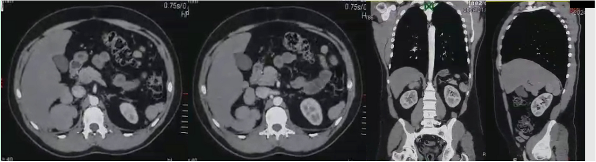
the nature and volume of the injected material, injection pressure, injection site and the delay in presentation to the ED [1,2,3]. The nature of the injected material is the most important prognostic factor and therefore also leading in treatment choices [4]. Injection of materials such as air and water generally have a better prognosis compared to oil and paint. Paint particularly contains many toxic elements that induce a more severe inflammatory response. However, injection with water or air should not be underestimated, as these materials can also lead to severe infections and compartment syndrome. Bacteria that are present on the skin or in the equipment are injected into deep tissues with a high pressure. In severe cases, this can result in necrotizing fasciitis. A pressure of 7 bar is sufficient to penetrate the skin, even without direct contact between the tool and the skin [5, 6]. Our case describes air injection into the eye with a pressure of 8 bar. In contrast to the skin, there is no known minimum pressure for conjunctival penetration. However, in 1986, an ocular injection injury was described with a minimal pressure of 3.4 bar (50 psi) [7]. Current literature mainly describes patients with injection injuries to the extremities. Due to the anatomical differences between the face and the extremities, different treatment options and potential complications should be considered. Regardless of the location, treatment of this type of injury always involves prevention of infection through tetanus immunization and adequate antibiotic therapy. Our patient had extensive subcutaneous emphysema and emphysema present in the orbit, intracranially and in the parapharyngeal spaces. It is important to be aware that bacteria, present on the conjunctival surface and in the pressure cleaner itself, were spread to these locations and that these bacteria can cause severe infections.
Since intracranial emphysema was not detected in the cerebral spinal fluid or subarachnoid space and the patient did not have any signs of CSF leakage, it was less probable that the patient would develop post-traumatic meningitis [8]. In our case, antibiotic therapy consisted of intravenous Amoxicillin-clavulanate on the first day, followed by oral Amoxicillin-clavulanate treatment for one week. This antibiotic was chosen for its adequate coverage of the most common skin flora and anaerobic oral and pharyngeal flora. The patient was participating in the Dutch National Immunization Program, thus no additional tetanus immunization was necessary.
Subsequently, it should be determined whether acute surgical intervention is necessary. For injection of paint and oily substances, emergent decompression and debridement is required in the majority of the cases due to the severe chemical reaction that occurs. For non-toxic materials, such as water or air, a conservative treatment with careful observation can be sufficient in certain cases. In these cases, it is important to consider not only the ratio between the size of the affected body part and the amount of injected material, but also the injection pressure. Compartment syndrome is a feared complication in injection injuries and requires emergent decompression. While compartment syndrome is predominantly seen in the extremities, it can also develop in the orbit. Orbital compartment syndrome (OCS) is a rare condition, developing in less than 0.1% of all facial traumas [9] and in 3.6% of patients with orbital injuries [10]. Any condition that causes an increase in intraorbital mass and subsequently an increase in intraorbital and intraocular pressure can potentially lead to OCS. Normal range of IOP in adults is 10-20mmHg [11]. In children IOP increases with age, approaching adult levels at 12 years. Sihota et al. [12] described an average IOP of 12.02 ± 3.74 mmHg in children aged 0–12 years. The most common cause of OCS is facial or ocular trauma that results in retrobulbar hemorrhage [13]. The rapid increase in intraorbital pressure causes compression of the optic nerve. Simultaneously the increase in intraocular pressure causes compression of the central retinal artery resulting in retinal ischemia. Symptoms of OCS include severe pain, swelling of the eyelids, proptosis, chemosis, impaired vision, relative afferent pupillary defect (RAPD) and elevated IOP [13]. OCS is a clinical diagnosis based on the history and the findings at physical examination. Treatment of OCS consists of orbital decompression which can be achieved by an emergent lateral canthotomy and cantholysis (LCC) [13]. Delay in decompression can result in complete vision loss. Our patient had an increased risk of developing OCS based on the trauma mechanism, the elevated IOP of 28mmHg, the swelling of the eyelids, chemosis, slight proptosis and slight reduction in visual acuity. However, the proptosis was not severe (Fig. 2.), pain was adequately controlled with only Paracetamol, there were no signs of RAPD, ocular movements were uncompromised and IOP was below the threshold of 40mmHg which is an indication for acute LCC [14]. It was decided not to perform any type of emergent surgical intervention. An important factor in this decision was the non-toxic nature of the injected material. As earlier described, air does not cause the same severe chemical reaction as toxic materials and is naturally absorbed over time by the surrounding tissues. It was not expected that the injected air would cause further swelling of the involved tissues or that IOP would continue to increase at this stage. However, the patient was admitted to the pediatric ward for close observation and was kept nil per os (NPO) for potential emergent surgery. During admission, the patient did not develop any signs of a compromised airway despite the parapharyngeal emphysema. Swelling of the eye, proptosis and pain did not progress. Visual acuity remained stable and the patient did not develop any additional abnormalities of the eye. Due to the favorable clinical course, there was no indication for a surgical intervention or repeated imaging. After discharge, the patient was followed up at the out-patient clinic. After two weeks visual acuity was completely recovered, subcutaneous emphysema had almost completely resolved and there were no signs of complications. In 2018, Bagheri et al. [15] published a case report describing a similar injection injury with air in a child’s eye. In both cases, patients were treated with prophylactic systemic and local antibiotics. However, in the case that Bagheri et al. described, patient underwent aspiration of the subconjunctival air in addition to the antibiotic treatment. In both cases, the patient’s visual acuity fully recovered.
In conclusion, high-pressure injection injuries to the face are rare and demand a different approach compared to the most common high-pressure injection injuries to the extremities. The nature of the injected material is paramount in choosing the appropriate treatment. This case illustrates that a high-pressure injection injury with air in the facial region, leading to extensive emphysema, can be managed conservatively with antibiotic therapy and inpatient observation.


Comments (0)