
Remember me


Forty male albino Wistar rats were sourced from the Animal House Colony of the National Research Centre (NRC) in Giza, Egypt, weighing between 150 and 200 g. The animals were given a week to acclimate before the study began. The animals were placed in appropriate cages (5 rats/cage) in an air-conditioned room with alternating 12-h day/night cycles at 23 ± 2 °C. The animals received an unlimited water supply and standardized food pellets. Throughout the experiment, all attempts were made for the animals to reduce the degree of pain or suffering that may have been caused. The study was ethically approved by the Research Ethics Committee (REC) of the Faculty of Pharmacy, Ain Shams University, Cairo, Egypt (ACUC-FP-ASU REC#180, 2023), ensuring compliance with institutional and international standards.
Chemicals and DrugsDoxorubicin (Doxorubicin®) was purchased from Ebewepharma (Unterach, Austria). In addition, DAPA, or dapagliflozin (Forxiga®), was purchased from AstraZeneca (Giza, Egypt). All highest purity grade chemicals and solvents were stored at temperatures between 2 and 8 °C.
Experimental ProtocolAll rats were distributed randomly into four groups (10/group) (Fig. 1). Group 1: Injected with normal saline intraperitoneally for 28 days and considered as the normal control group. Group 2 (DAPA group): Animals were administered dapagliflozin (2 mg/kg/day, p.o.) starting on the first day of the experiment for 28 days [14, 15]. Group 3: Animals were given doxorubicin (DOX; 2 mg/kg, i.p.) once every week (on experiment days 1, 7, 14, and 21) [16, 17]. Group 4 (DOX + DAPA): Animals were administered doxorubicin (2 mg/kg, i.p.) once every week for 28 days (on experiment days 1, 7, 14, and 21) with dapagliflozin (2 mg/kg/day, p.o.) starting on the 1st day of the experiment for 28 consecutive days. DOX and DAPA were freshly prepared every day and dissolved in a saline solution.
Fig. 1
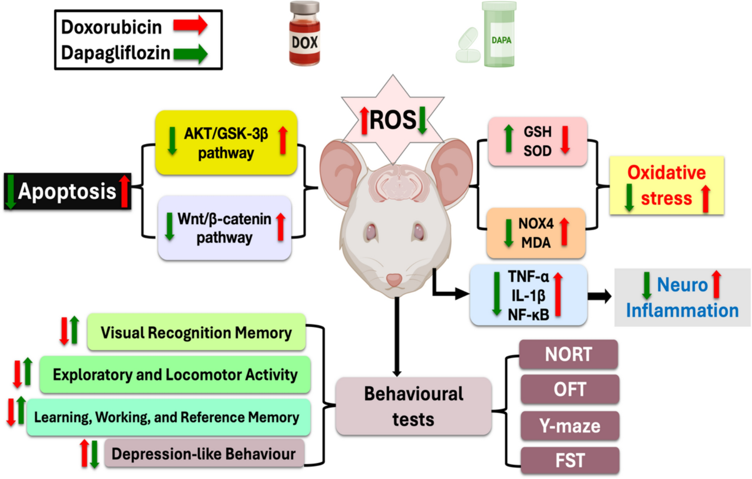
Experimental protocol. Animals were given DOX (2 mg/kg, i.p.) once every week for 28 days (on the experiment’s days 1, 7, 14, and 21). DAPA in a dose of 2 mg/kg, p.o., was given daily for 28 days beginning on the first day of the study schedule, and behavioral tests were carried out from day 29 to 33; then, on the next day, animals were euthanized. Brain tissues were separated for biochemical, histopathological, and immunohistochemical analyses. Where AKT protein kinase B, DAPA Dapagliflozin, DOX Doxorubicin, FST Forced Swimming test, GSH Reduced glutathione, GSK-3β Glycogen synthase kinase-3beta, i.p Intraperitoneal, IL-1β Interleukin-1β, MDA Malondialdehyde, NF-κB Nuclear factor kappa-B, NORT Novel Object Recognition test, NOX4 NADPH oxidase 4, OFT Open Field test, p.o Orally, SOD1 Superoxide dismutase 1, TNF-α Tumor necrosis factor-alpha, Wnt Wingless-related integration site, and β-Catenin beta-Catenin
Cognitive behavioral tests were conducted 24 h following the last DAPA dose. The Novel Object Recognition test (NORT) was applied across three days, on experiment days 29, 30, and 31. The Open Field test (OFT) was carried out on experiment day 30, and the Y-maze test was applied on experiment day 31. Days 32 and 33 of the experiment were implemented for the Forced Swimming Test (FST).
After the behavioral assessments on day 34, thiopental sodium (50 mg/kg) was given to euthanize the animals in each group, and they were killed via cervical dislocation. After that, brain tissues were quickly separated, collected, and washed with ice-cold normal saline. Brain samples were split and isolated into two distinct sets. Tissue samples from the first set (n = 6) were kept for enzyme-linked immunosorbent assay (ELISA) and Western blot analyses. For the second set, 10% formalin was used to preserve the tissue samples (n = 3) for histopathological and immunohistochemical analyses. Then, all animals’ cadavers were frozen till incineration.
Cognitive Behavioral TestsNovel Object Recognition Test (NORT)NORT was completed over three days (on experiment days 29 to 31): habituation day (day 29), training day (day 30), and testing day (day 31). Rats were allowed to investigate two identical objects during the training day. On the test day, one of the training objects was substituted with a new object. Rats exhibit an inherent preference for novel stimuli; thus, when presented with a familiar object, they allocate the majority of their time to the novel object [18]. The ANY-maze video tracking system (Version 7.36, Wood Dale, USA) was used to analyze the animal’s behavior by tracking the number of entries to the familiar object, the number of entries to the novel object, the familiar object exploration time, and the novel object exploration time. The NORT assesses visual recognition memory, which enables comparing previously stored information with presented stimuli [19].
The Open Field Test (OFT)OFT was applied on the 30th day of the experiment. It is a widely used indicator of behavior and general activity in an open field over time (5 min) [20]. The animal’s behavioral analysis was conducted using the ANY-maze video tracking system (Version 7.36, Wood Dale, USA) by tracking the distance traveled, line crossing, number of corner zone entries, and mean speed. It is employed in the assessment of rats’ emotionality, general locomotor activity, and exploratory behavior [21].
The Y-maze TestOn day 31 of the experiment, the rats’ willingness to explore novel settings was evaluated through the Y-maze test. Usually, rats would rather explore a new maze arm than go back to one they have already explored. Testing takes place in a Y-shaped maze with three opaque arms spaced 120° apart. After an introduction to the maze’s center, the animal was allotted 8 min to freely explore the three arms. This test is used for the evaluation of novel medications for their impact on cognition [22]. The behavioral analysis was carried out using the ANY-maze video tracking system (Version 7.36, Wood Dale, USA) by tracking the total distance, number of zone A entries, number of zone B entries, and number of zone C entries. Rats’ spatial reference memory can be evaluated well with the Y-maze test. It investigates working memory, reference memory, and discriminative learning [23].
Forced Swimming Test (FST)The FST is a behavioral test utilized to evaluate behavioral despair and “depressive-like” states in rodents [24]. On experiment days 32 and 33, the rats were kept for 5 min at a height of 20 cm in a plastic cylinder that measured 30 cm in height and 20 cm in width, containing water at 24 ± 1 °C. The animals were exposed to an identical cylinder for 5 min in the second trial, which was carried out 24 h after the first trial [24]. The animal’s behavioral analysis was performed manually by keeping track of the latency time, swimming time, immobility time, and struggling time.
Histopathological ExaminationAfter being stored in 10% neutral buffer formalin, the brain tissues were carefully dissected, cleaned with water, dehydrated in increasing ethyl alcohol grades, cleared with xylene, and then embedded in paraffin. Thin Sections (4–6 µm) were processed and stained with hematoxylin & eosin [25].
Immunohistochemistry (IHC) ExaminationParaffin slices were fixed onto positively charged slides. Immunohistochemical staining was conducted utilizing the Avidin–Biotin-Peroxidase Complex (ABC) method. Two polyclonal antibodies were used: rabbit NF-κB (p65) (1:100; Cat#E-EL-R0674, Elabscience, USA) and rabbit total caspase-3 (1:100; Cat#E-AB-63602, Elabscience, USA) (Supplementary File). After incubation with the primary antibodies, ABC reagents from the Vectastain kit (Vectastain ABC-HRP kit, Vector Labs, Newark, USA) were applied per the manufacturer’s instructions. Diaminobenzidine (DAB) (DAB; Sigma-Aldrich, USA) was utilized for visualization of the antigen–antibody complexes, which produced a brown precipitate indicating positive immunoreactivity. Negative controls were included by replacing the primary or secondary antibody with non-immune serum. IHC-stained sections were observed using an Olympus microscope (BX-53, Olympus Corporation, Tokyo, Japan).
Quantitative Assessment of Biochemical Markers in the Brain Tissue Homogenates by ELISA and Colorimetric TechniquesUsing a polytron homogenizer at 4 °C, 10% of the brain tissues were homogenized in 0.05 mol/L of phosphate buffer (pH 7). The homogenate was centrifuged for 20 min at 10,000 rpm to eliminate mitochondria, erythrocytes, nuclei, intact cells, and cell debris. Rat NOX4 ELISA kit (Cat#NBP2-76792, Novus Biological, USA), rat MDA ELISA assay kit (Cat#MBS268427, MyBioSource, San Diego, CA, USA), and SOD1 Rat ELISA kit (Cat#E4584-100, Biovision, Milpitas, CA, USA) were utilized for the determination of oxidative stress biomarkers within the brain samples. Furthermore, rat IL-1β ELISA kit (Cat#E0119Ra, Bioassay Technology Lab, Shanghai, China), rat TNF-α ELISA kit (Cat#438206, Biolegend, Sandiego, CA, USA), and rat NF-κB ELISA kit (Cat#E-EL-R0674, Elabscience, USA) were used for the assessment of the inflammatory markers within the brain samples. A sandwich ELISA format was used, where each kit contained pre-coated antibody plates, biotin-conjugated detection antibodies, enzyme reagents, and chromogenic substrates. All reagents, working standards, controls, and samples were prepared and brought to room temperature before use. A biotin-conjugated detection antibody specific to the collected antigen was then added to bind it. Next, avidin-horseradish peroxidase (HRP) was applied to bind to the biotin, the tetramethylbenzidine (TMB) substrate binds to the HRP enzyme to form a blue color, and sulfuric acid stop solution was used to stop the reaction, producing a yellow color, and the optical density (O.D.) of the well was measured at 450 nm, with corrections at 540 nm or 570 nm using a filter-based multi-mode microplate reader (Stat Fax 2200, Awareness Technologies, Florida, USA). Sample concentrations were calculated using a standard curve by interpolating their optical densities. In addition, GSH was assayed colorimetrically using a reduced glutathione colorimetric assay kit (Cat#K464-100, Biovision, Cairo, Egypt). All procedures followed the kit manufacturer’s instructions.
Western Blotting (WB)Following the manufacturer’s instructions, the ReadyPrep™ Protein Extraction Kit (Cat# 163-2086, Bio-Rad, Hercules, CA, USA) was applied to extract the total protein from the brain tissue samples. Protein concentrations in each sample were measured using the Bradford Protein Assay Kit (Cat# SK3041, Bio Basic, Markham, Ontario, Canada) [26]. An equivalent amounts of protein (20 µg) were mixed with the same volume of 2 × Laemmli sample buffer containing 4% sodium dodecyl sulfate (SDS), 10% 2-mercaptoethanol, 20% glycerol, 0.004% bromophenol blue, and 0.125 M Tris–HCl (pH 6.8), then boiled at 95 °C for 5 min to ensure complete denaturation. Sodium dodecyl sulfate–polyacrylamide gel electrophoresis (SDS–PAGE) was used for protein separation by using the TGX Stain-Free™ FastCast™ Acrylamide Kit (Cat# 161-0181, Bio-Rad, CA, USA). Proteins were then transferred to polyvinylidene difluoride (PVDF) membranes using the Trans-Blot® Turbo™ Transfer System (Cat# 1704150, Bio-Rad, CA, USA) at 25 V for 7 min in a transfer buffer composed of 25 mM Tris, 190 mM glycine, and 20% methanol. Membranes were blocked with 3% BSA in TBS-T (20 mM Tris, 150 mM NaCl, 0.1% Tween-20, pH 7.5) for an hour at room temperature. Protein Kinase B (AKT) (1:1000; Cat# 9272, Cell Signaling Technology, MA, USA), Glycogen Synthase Kinase-3 beta (GSK-3β) (1:1000; Cat# 9315, Cell Signaling, Danvers, MA, USA), Wingless-related integration site (Wnt) (1:100; Cat# sc-514531, Santa Cruz Biotechnology, CA, USA), and Beta-Catenin (β-Catenin) (1:200; Cat# sc-7963, Santa Cruz Biotechnology, CA, USA) antibodies were used. After washing 3–5 times (5 min each) with Tris-Buffered Saline with Tween 20 (TBST), membranes were incubated for 1 h at room temperature with Horseradish Peroxidase–conjugated (HRP-conjugated) secondary antibody (goat anti-rabbit IgG-HRP, 1:1000; Novus Biologicals, USA), then washed again in TBST. For signal detection, a chemiluminescent substrate (Cat# 1705060, Bio-Rad, Hercules, CA, USA) was applied by mixing equal volumes of Clarity™ Western Luminol/Enhancer and Peroxidase Solutions. A charge-coupled device (CCD) camera-based imaging system (ChemiDoc™ MP, Cat# 12003154, Bio-Rad, CA, USA) was utilized for visualizing the protein bands. Using image analysis software, band intensities were measured, and as a housekeeping control, each target protein’s expression was normalized against β-actin.
Statistical AnalysisData were presented as means ± S.D. Groups comparison was performed by one-way analysis of variance (ANOVA) followed by Tukey’s test. GraphPad Prism (version 5.0, GraphPad Software, Inc., San Diego, USA) was used for all statistical analysis and graphs. P < 0.05 was considered statistically significant.
Comments (0)