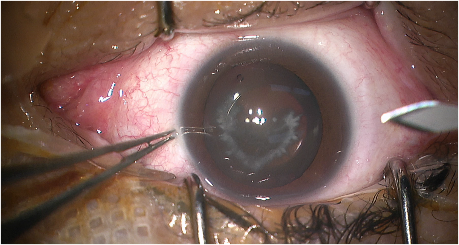
With the development of science and technology, MIGS has gradually been integrated into the diagnosis and treatment methods of ophthalmology. The term ‘MIGS’ was coined by Dr. Ike Ahmed in 2009. It is a type of ultra-minimally invasive surgery that operates from a small corneal limbus incision through the anterior chamber without damaging the bulbar conjunctiva and sclera. Its characteristics include minimal wound and tissue destruction, high safety, and easy combination with cataract surgery. Its postoperative characteristics include short recovery time, good preservation of residual vision, relatively good curative effect, and IOP can usually be dropped to the normal range. There are many classifications of MIGS, including outflow through the trabecular meshwork/Schlemm canal, suprachoroidal outflow, subconjunctival filtration and reduction of aqueous humor production, including Trabectome, Kahook Dual Blade, XEN [28, 29]. It is mainly used in patients with early POAG, mild to moderate POAG, secondary glaucoma, and patients with both cataract and glaucoma.
NTG is a specialized type of POAG. Its characteristics are that the IOP is within the normal range (10-21mmHg), the difference between the two eyes is not > 5mmHg, and the 24-hour IOP range is not > 8mmHg. Such patients may have visual field defects, including paracentral scotoma in the early stages. As the disease progresses, the visual field may gradually become restricted, and finally a tubular visual field (central visual field) may remain. The optic disc will have corresponding depressions, and the optic nerve will also have corresponding damage, which is often accompanied by some vascular diseases, such as migraine, ischemic disease, hypotension, etc [30].
Although the pathogenesis of NTG is unclear, several hypotheses have been proposed. Some studies suggest that blood flow and vascular factors lead to vascular ischemia, insufficient perfusion pressure, and subsequent optic nerve damage. Others indicate that elevated plasma endothelin-1 (ET-1), a potent vasoconstrictor that regulates ocular blood flow, may play a role. Additionally, impaired autoregulation of ocular blood flow, increased vascular resistance, and nocturnal hypotension have been implicated. Mitochondrial dysfunction and oxidative stress may also contribute to retinal ganglion cell apoptosis. Furthermore, some researchers have proposed that abnormalities in cerebrospinal fluid pressure across the lamina cribrosa could lead to optic nerve damage. Collectively, these mechanisms highlight the complex and multifactorial nature of NTG pathogenesis [31]. On the one hand, MIGS can increase aqueous humor outflow by decreasing the resistance to outflow in the trabecular meshwork and Schlemm’s canal pathways [32]. On the other hand, it can shunt aqueous humor to the suprachoroidal or subconjunctival space [33, 34]. In conclusion, because of the different mechanisms of blood pressure lowering by MIGS surgery, the specific MIGS procedure applicable to NTG needs to be studied in more detail. In addition, NTG’s optic nerve damage mechanism also includes blood flow and vascular theory [35]. The pathogenesis of NTG is not yet fully understood, and some scholars believe that it is related to the blood flow theory. The mechanisms underlying the blood flow abnormalities in NTG patients are unknown, but oxidative stress, vasospasm, and endothelial dysfunction appear to be risk factors for glaucomatous optic neuropathy. Thus, lowering IOP alone for the treatment of glaucoma is not enough. Treatment strategies should also include optic nerve protection, of which improving ocular blood flow(OBF) is key [36]. Of course, some scholars believe that it is the cerebrospinal fluid dynamics theory.
European Glaucoma Society terminology and guidelines for glaucoma state that the target IOP reduction percentage (i.e., 20%, 30%, 40%) depends primarily on the VF defect at diagnosis and the rate of progression [37]. A Collaborative Normal Tension Glaucoma Study (CNTGS) shows that reducing IOP by 30% from baseline is a therapeutic goal [38]. So is the IOP-lowering effect of MIGS surgery still applicable to NTG patients? Some scholars believe that traditional trabeculectomy can achieve the effect of reducing NTG IOP, but the postoperative follow-up time is required to be at least 3 months, and postoperative complications need to be treated through massage and adjustment of adjustable sutures. The psychological and financial burdens on patients are heavy, so most patients prefer emerging MIGS surgeries with less invasive risks, shorter operation times, and less postoperative follow-up time [39]. Some scholars believe that each method should be provided for patients with different situations, different indications, and different clinical situations to obtain different results [39]. In contrast, traditional trabeculectomy is supported by a large amount of literature and data from ophthalmology colleagues, while MIGS, as a new technology, currently lacks a large amount of experience and long-term results. All in all, NTG patients tend to choose minimally invasive surgery, but some scholars are skeptical about the specific efficacy of MIGS for NTG patients.
However, many current studies have proven that MIGS is effective in controlling IOP and reducing medications in NTG patients. One study showed that NTG patients with a baseline mean IOP of 14.8 ± 2.3 mmHg who underwent combined EX-PRESS implantation and phacoemulsification had significantly lower IOPs at all postoperative time points compared with baseline (P < 0.0001) [22]. From the immediate postoperative period to 1 month postoperatively, the IOP was stable. From months 1 to 12, the mean IOP ranged from 9.4 to 10.0 mmHg; the mean falling IOP increased from 4.9 mmHg to 5.4 mmHg; and the percentage reduction in intraocular pressure decreased from 21.1 to 35.4%. At 12 months, mean IOP was 10.0 ± 3.1 mmHg, a decrease of 4.9 mmHg (31.1%) from baseline (P < 0.0001) [17]. And while achieving these IOP reductions, almost all NTG patients no longer need to take IOP-lowering medications [38]. Another prospective, single-center case series of all NTG Asian eyes undergoing combined iStent injection implantation and phacoemulsification showed significant and sustained reductions in IOP and glaucoma medications 12 months postoperatively [17]. And in a real clinical population with NTG, significant and sustained IOP stabilization and reduced or even no medication use were achieved 1–3 years after MIGS surgery, with a good safety profile [19, 20, 22, 24, 25]. Due to variations in baseline IOP and the number of IOP-lowering medications across studies, direct comparisons of outcomes at different follow-up time points may not be meaningful.
Potential bias may arise from the lack of detailed classification of MIGS procedures across studies, with some failing to distinguish between standalone MIGS and MIGS combined with phacoemulsification. Additionally, baseline differences in IOP and medication use, especially among NTG patients, could affect treatment outcomes. Variations in IOP measurement methods and devices also contribute to inter-study heterogeneity. These factors may influence the overall effect estimates and should be considered when interpreting the results.
The results of this study show that MIGS combined with phacoemulsification can well reduce postoperative IOP in patients, which also contributes to the IOP-lowering medications of NTG patients. While MIGS combined with phacoemulsification reduces IOP, the incremental IOP-lowering effect attributable to phacoemulsification alone appears minimal. However, we cannot ignore the role of phacoemulsification surgery. Studies have shown that MIGS combined with phacoemulsification surgery can improve the VA and IOP of patients with glaucoma and cataract, and is definitely a better surgical option for eligible NTG patients [40]. MIGS combined with phacoemulsification surgery can also reduce the risk of secondary surgery for patients due to postoperative cataract aggravation [41]. Therefore, the choice of whether to combine phacoemulsification for NTG patients requires clinicians to make a careful decision based on the patient’s condition. Several epidemiologic studies have shown that NTG accounts for the largest proportion of POAG in Asian populations, which is also consistent with the racial proportions of this study [42, 43]. Therefore, it is hoped that in the future, Asian countries will increase the number of studies related to surgery in NTG patients.
At present, only postoperative IOP and medications indicators are included, and not all studies include detection data of optic nerve structure and function. Although reducing IOP is the most obvious and effective way to protect glaucoma optic nerve, it is not always possible in real clinical diagnosis and treatment. NTGs with well-controlled IOP (target IOP reduction of 20% or more) or low basic IOP may still experience decreased Retinal Nerve Fiber Layer (RNFL) and Visual Field Index (VFI) after MIGS. Therefore, it is recommended that follow-up studies pay more attention to the optic nerve and visual field, and comprehensively evaluate the RNLF thickness and VFI in the mid- and long-term follow-up of NTG patients after surgery. It is also recommended that follow-up studies further refine the surgical methods and indication groups, compare intraocular pressure control between high-tension glaucoma (HTG) and NTG patients, and examine patients at different glaucoma stages after different MIGS surgeries. Additionally, these studies should clarify the best indications for various MIGS procedures based on individualized factors such as disease stage and patient age. A multi-center, large-sample, prospective controlled study would be ideal to address these questions. MIGS is a relatively new surgical approach in glaucoma management, especially for NTG patients, where high-quality evidence remains limited. This study was initiated to address that gap. Previous meta-analyses confirmed IOP reduction with MIGS with Phaco in NTG but lacked subgroup analysis and evaluation of medication reduction.
One of the major limitations of this meta-analysis is the presence of substantial heterogeneity in several pooled estimates. This can be partially attributed to differences in baseline IOP values across studies, which directly affects the postoperative IOP reduction. Furthermore, variations in measurement methods, devices used, follow-up durations, and the number and type of preoperative glaucoma medications contribute to this heterogeneity. While subgroup and meta-regression analyses were performed to explore these sources, residual heterogeneity remained. Nonetheless, the overall direction of effect was consistent, suggesting a generally favorable efficacy profile for MIGS interventions in NTG patients.


Comments (0)