
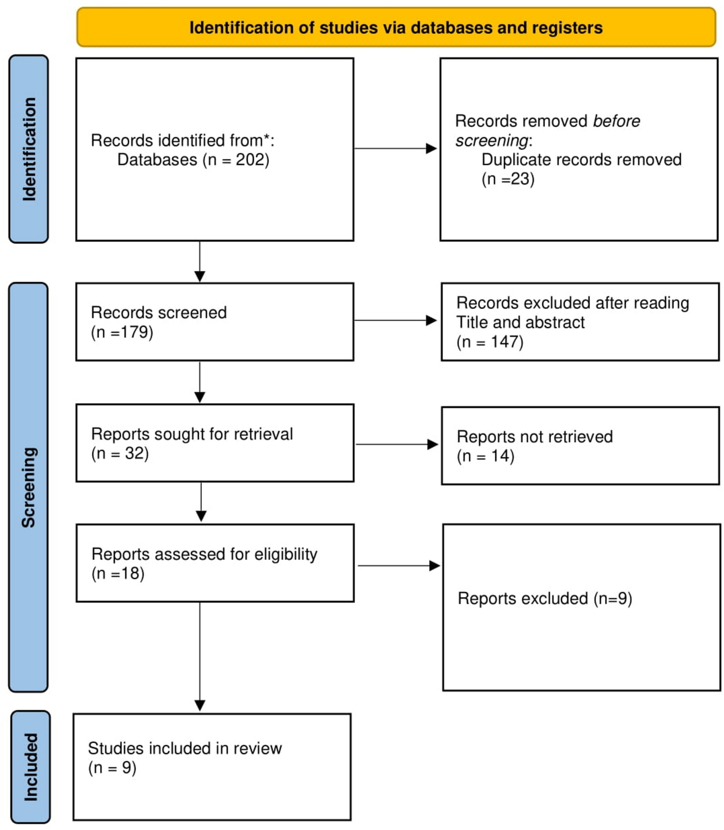
Remember me


Traumatic brain injury (TBI) occurs when an external mechanical force is exerted upon the head, resulting in temporary or permanent impairments in brain function (Jamjoom et al. 2021). Depending on the underlying pathophysiological processes involved, TBIs can be classified as primary or secondary cerebral injuries. Brain injuries result from the complex processes initiated by the initial impact, characterized by various neuropathological processes including ischemia, oxidative stress, excitotoxicity, apoptosis, necrosis, and neuroinflammation (Yang et al. 2023a). The initial inflammatory response following TBI serves to protect the site of injury from invading pathogens and tissue debris. However, the activation of microglia and astrocytes constitutes a primary factor in secondary cell death subsequent to TBI (Ni et al. 2019). When infected or injured, microglial cells and astrocytes will be momentarily activated and increase inflammation (Zhang et al. 2023). Activated astrocytes and microglial cells trigger inflammatory responses by producing various chemokines and cytokines (Zhang et al. 2023). MiRNAs have emerged as a promising therapeutic approach for the diagnosis and treatment of TBI, as they regulate microglial polarization and specifically promote the transition of microglia from the M1 to the M2 phenotype after TBI. This specific transition is crucial for minimizing the detrimental effects of excessive neuroinflammation and facilitating the repair and regeneration processes associated with TBI.
Specifically, a controlled cortical impact (CCI) model was used to induce TBI by mechanically contusing the cortex through an open-head procedure (Cai et al. 2022). In short, after administering anesthesia, mice underwent craniotomy while preserving the intact dura mater above the cortex. A contusion injury was then inflicted perpendicular to the brain's surface using a CCI device. Comprehensive neurological assessments were conducted on mice with nerve injuries before and after miRNA therapy to evaluate the treatment efficacy. This includes assessments such as the Neurological Severity Score (NSS), modified NSS (mNSS), Morris Water Maze (MWM), Rotarod test, Foot Fault test, and Adhesive Removal test. The NSS evaluates the severity of neurological damage, encompassing analyses of motor function, alertness levels, and physiological behavior (Shao et al. 2020). The mNSS score is a modified version of NSS used to better assess animal models or specific research objectives (Huang et al. 2018). The Morris Water Maze (MWM) was used to study the functional assessment of brain regions associated with spatial learning and memory (Li et al. 2019). The Rotarod test is a classic behavioral experiment used to assess mice's coordination, balance, and motor endurance (Ge et al. 2018). The Adhesive Removal test can identify sensory motor deficits and motor asymmetry (Zhang et al. 2021). The Foot Fault Test can detect and assess bilateral and unilateral motor deficits in rodent models of central nervous system diseases (Xin et al. 2017).
miR-124Yang et al. used lipofectamine-2000 to transfect miR-124 into rat bone marrow MSC (BMMSC) and then separated Exo-miR-124 from the culture medium supernatant (Yang et al. 2019). By intravenous injection of 100 μg/mL Exo-miR-124, a significant increase in miR-124 was detected in hippocampal microglia, which promoted M2 polarization of microglia and the production of anti-inflammatory factors IL-4, IL-10, and TGF-β after TBI stimulation. After treatment with Exo-miR-124, the NSS scores of TBI rats significantly decreased. In the MWM, the platform crossing time and target quadrant route of TBI rats significantly increased compared to the control group rats. RT-PCR results indicate that miR-124 promotes M2 polarization of microglia by inhibiting the TLR4 signaling pathway. This study elucidated the mechanism by which Exo-miR-124 facilitates the M2 polarization of microglia through the inhibition of the TLR4 pathway, thereby affecting hippocampal neurogenesis and the recovery of neurological behavior and cognition after TBI. These findings suggest that Exo-miR-124 may represent a novel therapeutic strategy for improving outcomes in TBI.
miR-873a-5pLong et al. transfected mouse astrocytes with miR-873a-5p mimic and then separated Exo-miR-873a-5p from the culture medium supernatant (Long et al. 2020). By injecting 5 nM Exo-miR-873a-5p into the lateral ventricle, a significant reduction in brain injury area and degree of brain edema was observed, while also increasing the mNSS score. Through qRT-PCR analysis, it was found that the expression of markers for M1-type astrocytes, including INOS, CD32, and IL-1β, significantly decreased, while the expression of markers for M2-type astrocytes, such as CD206, IL-4, and Arg1, significantly increased. Immunofluorescence studies further confirmed that overexpression of miR-873a-5p significantly decreased the expression of the M1-type astrocyte marker INOS in the damaged cortex, while increasing the number of cells marked with the M2-type astrocyte marker Arg1. Western blot analysis further revealed that miR-873a-5p effectively converts the activation of M1-type microglial cells into that of M2-type microglial cells by inhibiting the ERK phosphorylation and NF-κB signaling pathways. The study reveals that Exo-miR-873a-5p fosters M2 polarization of microglia by suppressing the ERK phosphorylation and NF-κB, thereby improving neural function following brain injury. These findings suggest that miR-873a-5p could serve as a promising therapeutic target for enhancing recovery and improving neurological function after TBI.
miR-124-3pHuang et al. transfected miR-124-3p mimic into mouse microglial cells using lipofectamine-3000 and then separated Exo-miR-124-3p from the culture supernatant (Huang et al. 2018). After intravenous injection of 30 μg/mL Exo-miR-124-3p, the escape latency of TBI mice in the Morris water maze test was significantly shortened compared to the control group mice, and the time spent in the target quadrant was significantly increased compared to the control group mice. The results of luciferase reporter gene assays indicated that miR-124-3p targets PDE4B, regulating the mTOR signaling pathway. The main finding of this study is that miR-124-3p inhibits neuronal inflammation and promotes neurite outgrowth by targeting PDE4B to suppress mTOR signaling activity.
Using the same method, Li et al. transfected and isolated exosomes and then administered 30 μg/mL Exo-miR-124-3p via tail vein injection. TBI mice showed significantly higher mNSS scores than the control group at 7, 14, and 21 days, along with significant improvements in the rotarod test performance (Li et al. 2019). In the MWM test assessing spatial learning ability, TBI mice exhibited significantly shortened escape latencies compared to control group mice, along with increased time spent in the target quadrant. Further validation through luciferase reporter gene analysis confirmed the miR-124-3p-specific inhibition of FIP200 translation, aiding in the regulation of excessive neuronal autophagy. This study demonstrates that Exo-miR-124-3p suppresses neuronal autophagy mediated by FIP200, thus exerting a protective effect against neuronal damage post-TBI. Consequently, miR-124-3p may offer a novel therapeutic strategy for managing neuronal inflammation following TBI.
miR-17-92Zhang et al. transfected human MSC (HMSC) with miR-17-92 and then isolated Exo-miR-17-92 from the culture supernatant (Zhang et al. 2021). After tail vein injection of 10 μg/mL Exo-miR-17-92, there was a significant improvement in the mNSS scores of TBI rats. Compared to the control group rats, treatment with Exo-miR-17-92 promoted sensory and motor recovery in TBI rats, as evidenced by the adhesive removal test and foot fault tests of the right forelimb. Additionally, performance in the MWM test indicated enhanced spatial learning and memory abilities. Furthermore, immunofluorescence staining revealed a significant increase in vascular regeneration in the dentate gyrus and neurogenesis at the border of the lesion, accompanied by reduced inflammation. This study demonstrates that Exo-miR-17-92 reduces neuroinflammation and neuronal cell loss, increases neurogenesis and angiogenesis, and significantly improves the therapeutic efficacy of functional recovery after TBI. MiR-17-92 may thus serve as a promising alternative to stem cell therapy for treating nerve damage or diseases.
Ischemic StrokeIschemic stroke (IS), caused by a clot or other obstruction blocking a blood vessel, results in significant physiological changes in brain tissue. Around 70% of all IS cases are caused by the occlusion of a major cerebral artery, typically the middle cerebral artery (Feigin et al. 2018). Most IS cases are thromboembolic in nature, with the primary causes of embolism being large artery atherosclerosis and cardiac conditions, particularly atrial fibrillation. Following an IS, various mechanisms such as excitotoxicity, inflammation, impaired mitochondrial function, free radical generation, and accumulation of misfolded proteins lead to neuronal death (Kadir et al. 2022). Increasing evidence demonstrates that modifications in the microenvironments of cerebral tissues, specifically the inflammatory response, play a significant role in post-ischemia cerebral injury (Stoll and Nieswandt 2019). Reperfusion therapy is the primary treatment for cerebral thrombosis. However, secondary brain damage caused by ischemia–reperfusion injury exacerbates neuronal death and leads to poor prognosis (Lan et al. 2024). The imbalance between M1 and M2 phenotypes in microglial cells exacerbates neuroinflammatory responses and neuronal death following IS. Controlling the transition of microglial cells from M1 to M2 phenotype can alleviate various forms of neuronal death after cerebral ischemia–reperfusion injury (Lan et al. 2024).
To establish a mouse model of cerebral ischemia–reperfusion, middle cerebral artery occlusion (MCAO) was utilized (Deng et al. 2019). In brief, mice were anesthetized and the common carotid artery and the proximal portion of the carotid artery were ligated. Distal to the ligature of the carotid artery, a filament was inserted and secured to induce MCAO at the bifurcation of the carotid artery. Reperfusion commenced following the induction of localized ischemia. Animals in the control group underwent a midline neck incision, isolation, and direct suture of the right common carotid artery. Before and after miRNA treatment of mice with neural injury, a comprehensive assessment of their neurological function was conducted to evaluate the therapeutic effects. This included assessments such as NSS, mNSS, Neurological Deficit Score (NDS), MWM, corner test, adhesive removal test, and foot fault test. NDS evaluates the severity of neurological symptoms based on sensory, motor, visual, and consciousness functions (Chen et al. 2014). The corner test can detect sensory impairments caused by striatal damage (Wang et al. 2020).
miR-17-92Xin et al. infected rat BMMSC with miR-17-92 lentivirus and isolated Exo-miR-17-92 from the culture supernatant (Xin et al. 2021). After tail vein injection of Exo-miR-17-92 at a concentration of 3 × 10¹¹ particles/mL, the minimum threshold for cortical microstimulation-induced forelimb motor impairment significantly decreased. Compared to control rats, rats treated with Exo-miR-17-92 showed a significant reduction in the time taken for adhesive removal in the adhesive removal test and a significant decrease in mNSS scores. Double immunofluorescence staining results indicated that miR-17-92 enhanced neurological function recovery in stroke patients by mediated through the downregulation of PTEN and subsequent activation of the PI3K/Akt/mTOR pathway. This study suggests that Exo-miR-17-92 may promote neuro-functional recovery after IS by enhancing axon-myelin remodeling through the PI3K/Akt/mTOR pathway. This provides a positive strategy for the treatment of IS.
miR-126Wang et al.’s study demonstrated that the infarct area increases in diabetic mice with ischemic stroke, and administration of endothelial progenitor cells can reduce the infarct area. They transfected mouse endothelial progenitor cells with a miR-126 mimic and isolated Exo-miR-126 from the culture supernatant (Wang et al. 2020). Diabetic mice underwent MCAO surgery to induce cerebral thrombosis. Following tail vein injection of 50 μg/mL Exo-miR-126, significant improvements in NDS were observed on the 2nd and 14th days compared to the control group rats. Sensory and motor functions significantly recovered on the 2nd and 14th days, as evidenced by the corner test results compared to the control group rats. Adhesive removal test results showed significantly reduced contact time and tape removal time on the 2nd and 14th days compared to the control group rats. WB analysis indicated that miR-126 promotes vascular regeneration and neurogenesis in the chronic phase by upregulating VEGFR2. This study suggests that Exo-miR-126 protects the brain from acute injury and promotes neurological recovery by upregulating VEGFR2, thereby enhancing angiogenesis and neurogenesis. It provides a promising strategy for the treatment of IS in diabetes.
miR-133a-3pYang et al. transfected mouse BMMSC with miR-133a-3p mimics using lipofectamine-2000 and isolated Exo-miR-133a-3p from the culture supernatant (Yang et al. 2023b). Following intraventricular injection of Exo-miR-133a-3p, compared to the control group rats, MCAO rats showed significantly reduced NSS, and MWM results indicated a significant decrease in escape latency. Luciferase reporter gene assays revealed that upregulation of miR-133a-3p significantly downregulated the expression levels of DAPK2, upregulated the Akt signaling pathway, and inhibited progression at the injury site. This study demonstrates that Exo-miR-133a-3p alleviates neuronal damage by targeting DAPK2. Consequently, this research provides a theoretical foundation for exploring new targets against neuronal injury.
Intracerebral HemorrhageIntracerebral hemorrhage (ICH) is a localized bleeding within the brain tissue caused by the rupture of blood vessels. Primary risk factors associated with ICH development include hypertension, amyloidosis, vasculitis, substance abuse, coagulation dysfunction, and genetic factors (Zhou et al. 2022). Cerebral hemorrhage can cause both primary and secondary brain injuries. The rupture of blood vessels and the formation of hematoma can compress normal brain tissue, resulting in primary brain injury (Zhou et al. 2022). Hematoma not only causes mechanical compression but also leads to the formation of edema, inflammation, and cell death around the hematoma, further damaging the nervous system and resulting in secondary brain injury (Zhou et al. 2022). Microglia, as innate immune cells, act as protectors of neurons and respond to various acute brain injuries, including ICH. Research indicates that proper regulation of M1 and M2 neuroglial cells plays a significant role in the development of neuroinflammatory responses following ICH, making it a crucial target for inhibiting neuroinflammation (Lei et al. 2023). After ICH, reducing the brain's infiltration of white blood cells and lowering the levels of inflammatory cytokines within the brain can inhibit inflammation (Guo et al. 2023).
The method for establishing the ICH model involves first anesthetizing the mice, then slowly inserting a needle into the right basal ganglia region of the brain and infusing bacterial collagenase at a constant rate to damage the blood vessel walls and cause blood extravasation. As a control group, an equal volume of saline solution is infused at a constant rate. Before and after administering miRNA treatment to the mice with nerve injury, their neurological function is comprehensively examined to assess the treatment efficacy. This includes the beam walking test, motor deficit score, mNSS, corner test, and foot fault test. The beam walking test is used to assess motor function, limb strength, balance, and body coordination (Duan et al. 2020). The motor deficit score assesses sensory, motor, and other aspects to evaluate motor deficits (Duan et al. 2020).
miR-146a-5pDuan et al. transfected miR-146a-5p into rat BMMSC using a lentiviral vector and isolated Exo-miR-146a-5p from the culture medium supernatant (Duan et al. 2020). After tail vein injection of 100 μg/mL Exo-miR-146a-5p, significant enhancements in the beam walking test and motor deficit score of ICH mice were observed on days 7 and 28 compared to the control group rats. Luciferase reporter gene assays indicated that reduced expression of IRAK1 and NFAT5 could inhibit M1 polarization in microglial cells. This study demonstrates that Exo-miR-146a-5p reduces neuronal apoptosis and inflammation associated with microglial M1 polarization by downregulating IRAK1 and NFAT5, thereby exerting neuroprotective effects and improving functional outcomes after ICH.
miR-124Guo et al. transfected BV2 microglial cells with a lentiviral vector containing miR-124 and isolated Exo-miR-124 from the culture medium supernatant (Guo et al. 2023). On days 1 and 3, intranasal instillation of 2.11 × 10⁹ ± 1.04 × 10⁷ particles/mL was performed. Through mNSS, foot fault tests, and corner tests, significant improvement in neurological function was observed compared to the control group rats. Flow cytometry analysis revealed that Exo-miR-124 limited leukocyte infiltration into the brain to exert its function. The results of this study indicate that intranasal administration of Exo-miR-124 reduces the infiltration of immune cells into the brain after ICH, significantly alleviating hemorrhagic brain injury and neuroinflammation.
Optic Nerve InjuryOptic nerve injury is a significant cause of visual impairment worldwide and can ultimately result in blindness (Li et al. 2018b). This type of injury hinders the regeneration capability of optic nerve axons and leads to the formation of glial scars primarily composed of reactive astrocytes (Li et al. 2018b). Therefore, research efforts focused on optic nerve protection should prioritize the preservation of retinal ganglion cells (RGCs) and the promotion of optic nerve axon regeneration (Zhou et al. 2023). Glaucoma is a contributing factor to RGC damage and mortality, and a key pathological mechanism underlying its development is retinal IRI (Yu et al. 2022). Retinal microglia serve as resident phagocytes in the retina, carrying out innate immune functions and maintaining tissue homeostasis. They are involved in almost all retinal pathological processes, and in the context of glaucoma, they are closely associated with RGC damage and mortality (Yu et al. 2022).
To create a mouse model of optic nerve injury, the following steps were followed: an incision was made along the upper edge of the unilateral eyelid, the conjunctiva was incised from the lateral side of the cornea, and the optic nerve was exposed beneath the extraocular muscles by blunt dissection and retraction of the eyeball. The optic nerve was then clamped 2 mm posterior to the globe and held for 9 s. Following the surgery, a temporal conjunctival incision at the corneal margin was made to facilitate clearer observation of the sclera under ophthalmic microscopy. A tangential scleral incision was made between two vortex veins outside the corneal margin, approximately 1 mm apart. Similar surgical procedures were performed on control group mice, but without clamping of the optic nerve. Immediately after modeling, intravitreal injection of Exo-miRNA was administered. Another method to create a mouse model of retinal IRI involves inducing and maintaining intraocular pressure at 100 mmHg for 50 min using a syringe filled with sterile saline solution. Subsequently, the syringe is removed, allowing blood to reperfuse the retina. Twelve hours prior to the IRI surgery, Exo-miRNA is injected intravitreally. The contralateral eye, without elevated intraocular pressure, serves as the control group. To assess the functional responses of the retina and optic nerve, Flash Visual Evoked Potentials (F-VEP) are measured following optic nerve crush injury, and retinal function is analyzed through pattern electroretinography (PERG) and visual acuity (Li et al. 2018b).
miR-21-inhibitorLi et al. transfected miR-21 inhibitor (miR-21-in) into rat astrocytes using lipofectamine-2000 and then isolated Exo-miR-21-in from the culture medium supernatant (Li et al. 2018b). After intravitreal injection of miR-21-in, the peak latency of F-VEP N1, P1, and N2 waves was significantly reduced compared to the control group rats. Through double-labeled immunofluorescence and Western blot detection, Exo-miR-21-in alleviated F-VEP functional impairment by targeting the EGFR/PI3K/AKT/mTOR pathway. The results of this study indicate that miR-21 mediated the activation of the EGFR/PI3K/Akt pathway, improving astrocyte hyperactivation and glial scar progression, and promoting axonal regeneration and functional recovery after optic nerve crush. This study suggests that miR-21 may be a therapeutic target for optic nerve injury.
miR-21-5pYu et al. transfected miR-21-5p mimic antibody into human gingival MSC and then isolated Exo-miR-21-5p from the culture medium supernatant (Yu et al. 2022). Intravitreal injection of 500 ng/mL Exo-miR-21-5p significantly reduces the thickness of the inner nuclear layer induced by IRI. Results from pattern electroretinography and visual response indicate that Exo-miR-21-5p exhibits stronger anti-apoptotic effects. Dual-luciferase reporter gene assays demonstrate that miR-21-5p functions through binding with PDCD4. This study demonstrates that Exo-miR-21-5p protects retinal cells from inflammation and cell death by binding to PDCD4, providing a new therapeutic approach against IRI.
Spinal Cord InjurySpinal cord injury (SCI), a serious condition, can result in temporary or permanent changes to the spinal cord, leading to partial or complete loss of autonomic, sensory, and motor functions (Sykova et al. 2021). Primary injury is directly caused by external forces, such as spinal cord contusion, compression, transection, or stretching, resulting in mechanical damage to the spinal cord (Sun et al. 2024). Within minutes after the initial injury, a secondary SCI occurs, characterized by localized vascular damage, progressive hemorrhage, ischemia, edema, thrombosis, ionic changes, oxidative stress, and lipid peroxidation due to the release of free radicals. These factors ultimately lead to cell death (Sykova et al. 2021). Inflammatory reactions and gliosis hyperplasia can lead to the formation of inhibitory environments and scars, which hinder axonal regeneration and limit therapeutic potential (Kwiecien et al. 2020). Additionally, arachnoiditis, characterized by granulomatous infiltration surrounding the damaged spinal cord, contributes to the formation of a fully developed scar lacking astrocytes or other glial cells (Kwiecien et al. 2020). Astrocytes play a significant role in SCIs as they can both impede and facilitate recovery from central nervous system disorders, including SCI (Gao et al. 2023).
To establish a rat spinal cord injury (SCI) model, rats were first anesthetized, and then a laminectomy was performed at thoracic vertebrae 9–11 (T9–T11). Subsequently, instruments were used on the dura mater exposed at the T10 level to induce moderate contusion. In the control group, only laminectomy was performed without inducing SCI. Before and after miRNA treatment of nerve-injured mice, comprehensive assessments of their neurological functions were conducted to evaluate the therapeutic effects. These assessments included the Basso–Beattie–Bresnahan method (BBB), Basso Mouse Scale (BMS) scoring, footprint analysis, and horizontal ladder walking testing. The BBB is used to assess the recovery of forelimb and hindlimb motor function following mild to moderate injuries (Li et al. 2018a). The BMS takes into account the potential inconsistencies in hindlimb function caused by spinal cord impact (Liu et al. 2020). Footprint analysis can reflect changes in animal gait (Liu et al. 2020). The horizontal ladder walking testing evaluates aspects such as limb placement, stepping, and coordination by observing the gait of rats on a horizontal rung ladder (Huang et al. 2020).
miR-133bLi et al. used lipofectamine-3000 to transfect miR-133b mimics into rat BMMSC, and the supernatant was collected to isolate Exo-miR-133b (Li et al. 2018a). Five days after tail vein injection of 100 μg/mL Exo-miR-133b, significant recovery of hind limb motor function in SCI rats was observed compared to the control group, as assessed by the BBB locomotor rating scale. HE staining demonstrated a significant reduction in lesion area after Exo-miR-133b injection. Neuronal staining targeting neurons in the injured spinal cord showed a significant increase in the number of mature neurons in injured rats receiving Exo-miR-133b. Using Western blotting, miR-133b was found through the activation of the ERK1/2, STAT3, and CREB pathways, as well as the suppression of RhoA expression. The results of this study indicate that injecting Exo-miR-133b promotes axonal regeneration in neuronal cells, partially through the activation of ERK1/2, STAT3, and CREB, as well as the inhibition of RhoA expression. This suggests a new therapeutic approach for SCI.
miR-29bYu et al. transfected rat BMMSC with a recombinant lentiviral vector containing miR-29b and isolated Exo-miR-29b from the supernatant of the culture medium (Yu et al. 2019). Four weeks after tail vein injection of 200 μg/mL Exo-miR-29b, a significant decrease in the number of contractile nerve cells was observed in SCI rats compared to the control group rats. At 2, 4, and 8 weeks post-injection, SCI rats showed significantly higher BBB scores compared to the control group rats. Immunohistochemical staining results showed that miR-29b alleviated rat SCI by increasing NF200 and GAP-43-positive neurons while decreasing GFAP-positive neurons, thereby promoting neuronal regeneration. In summary, injecting Exo-miR-29b may be associated with regulating proteins i
Comments (0)