Mouse model
All experimental processes comply with East China Normal University (ECNU) animal ethics (ethical approval number m20230206). ECNU Animal Center provides 8–12 week old or 18 months old WT C57BL/6J mice. It was established that all mice were maintained over a period of 12 h under a complete dark-light cycle (with a white illumination level of 200 lux) as well as a constant temperature and humidity level (20–26 °C), unless otherwise indicated.
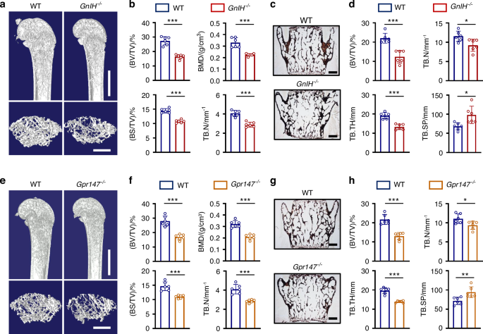
GnIH−/− mice and Gpr147−/− mice were generated by Cyagen using the CRISPR/Cas9 system in the C57BL/6N mouse strain. To create a GnIH knockout mouse, Exons 1 to 3 were targeted, encompassing a 567 bp coding sequence within the region. Specifically, gRNA1 (GATATTCTATACACGCTAGCTGG) targets exon 1, and gRNA2 (CTTCTCCAGACCTAGTGAACAGG) targets exon 3. For the creation of a Gpr147 knockout mouse, Exons 2 to 3 were targeted, covering a 415 bp coding sequence. Notably, gRNA3 (AGCCCAAGCACTTTCGAAGGTGG) targets exon 2, while gRNA4 (CATGCAGACGGAGTAAAGCCAGG) targets exon 3.
Genotype identification of mice was performed using the following primer sequences: GnIH−/−-F: CATTTGCCAAATTAGACCCTTAGGG, GnIH−/−-R: AAATGCAACCCAGGGTTGATGTC, GnIH−/−-He/Wt-F: AGCCCGACTTCAAGAGGCTAC; Gpr147−/−-F: GTGGACAGTAATAAGTGGGCTTAGGGT, Gpr147−/−-R: AGCTAAACAACAGTCTCCTGCATG, Gpr147−/−-He/Wt-F: GTAATTCTGGGACTGGCACGC.
Cell culture and osteoclast differentiation
Refer to our previous research reports,6,55,56 bone marrow macrophages (BMMs) were obtained from the femurs and tibias of 8-week-old WT, GnIH−/− or Gpr147−/− mouse. They were then cultured in α-MEM medium (Gibco) with the addition of 10 ng/mL M-CSF (R&D), 10% FBS (Gibco), and 1% penicillin-streptomycin (HyClone). For osteoclast differentiation assay, BMMs were plated in 96-well plates at a density of 1 × 10⁴ cells per well. Subsequently, they were stimulated with 50 ng/mL of RANKL (R&D) and 10 ng/mL of M-CSF (R&D). After 5–7 days, The TRAP staining kit (Sigma-Aldrich) was used to identify TRAP+ osteoclasts (five or more nuclei) from osteoclasts fixed in 4% Paraformaldehyde (PFA). For LPS-induced osteoclast differentiation assay, BMMs were plated in 96-well plates at a density of 1 × 10⁴ cells per well. The cells were incubated with 50 ng/mL of RANKL (R&D), 10 ng/mL of M-CSF (R&D), 50 ng/mL of LPS (Beyotime) and 10 μmol/L GnIH for 5 days. After fixed in 4% PFA, the cells were stained for TRAP assay.57,58,59
Osteoblast-Osteoclast co-culture
Isolated preosteoblastic cells from the calvaria of 3–5-day-old neonatal mice. Co-culturing was carried out in 96-well plates with BMMs (1 × 10⁴ cells per well) and preosteoblastic cells (1.5 ×10³ cells per well) in α-MEM (Gibco) (10% FBS), 100 ng/mL 1 alpha, 25-dihydropyrene (HyClone), 1% penicillin-streptomycin. One week later, TRAP staining was performed, and the number of osteoclasts exhibiting 5 or more nuclei and positive for TRAP staining was quantified.
CCK-8 assay
After seeding 96-wells with a combination of 100 μL α-MEM and 20 ng/mL M-CSF-containing BMMs (1.2 ×10⁴ cells per well) for two days, then supplemented with CCK-8 solution (10 μL per well) for 90 min and read the absorbance at 450 nm.
Cell migration assay
For BMM migration, GnIH at dosages of 0, 0.01, 0.1, 1, 10 μmol/L was added in the lower chamber while BMMs were seeded in the upper chamber (1 × 10⁵ cells per well) of transwell inserts. After one day, the migrated BMMs were fixed in 4% PFA for 0.5 h, then used water wash 4 times and stained them for 2 h with 0.1% crystal violet. In total, four fields of view were photographed per insert and then quantified using Media Cybernetics’ Image-Pro Plus 6.0 (Media Cybernetics).
Treatment with GnIH peptide in vivo
OVX-induced and LPS-induced (LPS was injected intraperitoneally at a dose of 5 mg/kg body weight on day 0 and day 4) bone loss mouse models were used as previously described.5,6,60 Mouse GnIH peptide with the sequence Phe-Pro-Ser-Leu-Pro-Gln-Arg-Phe-NH2 was intraperitoneally injected daily,61 at the dosage of 0.1 mg/kg in 200 μL PBS.62,63 OVX mice and aging mice were treated with GnIH for one month, LPS-induced mice were treated with GnIH for seven days. GnIH peptide was synthesized by Shanghai Bootech BioScience & Technology.
Green light exposure
The green LED light source (Shanghai wence) was fixed on the top of a special customized cabinet. OVX mice were subjected to green LED light exposure within a custom-designed cabinet, while being provided unrestricted access to food and water. Green light (520–525 nm, 400 Lux) therapy exposure for 8 h (8:00 am-16:00 pm) daily for 60 consecutive days38,64 was applied to treated mice. Following daily green light therapy, the mice were subsequently returned to their standard animal housing facility.
A total of five healthy adult males between the ages of 20 and 40, who do not have any visual impairment or eye diseases, have not taken any hormonal or osteoporosis medications within the past three months, and do not have a history of chronic diseases or other medical conditions, were selected for this study. For human green light or regular light exposure experiment, the subjects were initially exposed to regular light for 7 days as control, followed by green light for another 7 days after a two-week rest period. LED green light source emitting wavelengths between 520–525 nm or regular light (6000 K LED light) were utilized. The experiment took place in their designated room from 9:00 am to 11:00 am daily. The LED green light or regular light source was positioned at a distance of 1-2 m from the subjects’ eyes, and the light intensity ranged from 400 to 1 000 lux. To ensure individual comfort, subjects had the flexibility to adjust the distance between the light source and their eyes within the specified range. During the experiment sessions, subjects were specifically instructed to remain awake and maintain a normal blink rate without directly staring at the light source. When exposed to green light or regular light, each subject wore a hospital gown made of the same material. They were encouraged to engage in activities that did not require additional sources of light including conversation and listening to music, while watching television or using screen devices, were forbidden. Subjects were collected for peripheral venous blood sampling before the experment and after one week of green light or regular light therapy. The experiment underwent thorough review and was approved by the Medical Ethics Committee of Yangzhi Rehabilitation Hospital (Approval No. YZ 2023-070).
Human peripheral blood mononuclear cells differentiation
Obtaining blood from healthy adult individuals. After ficoll centrifugation, human PBMCs were selected and were plated in 96-well plates at a density of 1.5 ×10⁴ cells per well. Subsequently, they were stimulated with 60 ng/mL of RANKL (R&D) and 20 ng/mL of M-CSF (R&D), while treated with the 10 μmol/L human GnIH with the sequence Val-Pro-Asn-Leu-Pro-Gln-Arg-Phe-NH2. After 7 days, The TRAP staining kit (Sigma-Aldrich) was used to identify TRAP+ osteoclasts (five or more nuclei) from osteoclasts fixed in 4% Paraformaldehyde (PFA). Human GnIH peptide was synthesized by Shanghai Bootech BioScience & Technology.
Western blotting analysis
Following the treatment of cells with GnIH (10 μmol/L), proceed to lyse the cells using RIPA buffer in order to extract proteins. The protein concentration can then be quantified using the BCA assay (Thermo). Electrophoresis and transfer to nitrocellulose filter membranes (Beyotime) were followed by 3 h of treatment with 5% bovine serum albumin (Beyotime), and incubated with specific antibodies: GAPDH antibody (CST), p-PI3K (Tyr467/199) antibody (Abmart), p-AKT (Ser473) antibody (Abmart), p-p65 (Ser536) antibody (Abmart), p-IκB (Ser32/Ser36) antibody (Abmart), p-p38 (Thr180) antibody (Abmart), p-JNK (Tyr185) antibody (Abmart), p-ERK (Thr202/Tyr204) antibody (Abmart), p65 antibody (Abmart), IκB antibody (Abmart), p38 antibody (Abmart), JNK antibody (Abmart), ERK antibody (Abmart), AKT antibody (CST), PI3K antibody (CST), Nfatc1 antibody (SANTA CRUZ). A secondary antibody (Licor) was added to the membranes after overnight incubation at 4 °C. Images were captured using the Odyssey Infrared Imaging System. Image-Pro Plus 6.0 software was used to quantify the bands. First, all readings were normalized to the corresponding band of GAPDH. Next, to calculate the fold changes, p-AKT was normalized to AKT samples, p-PI3K was normalized to PI3K samples, p-p65 was normalized to p65 samples, p-IκB was normalized to IκB samples, p-p38 was normalized to p38 samples, p-JNK was normalized to JNK samples and p-ERK was normalized to ERK samples.
Immunofluorescence staining
For paraffin sections, after treatment with gradient of dehydration and 20 mg/mL proteinase K for 20 min, paraffin sections were fixed in 2% BSA and 0.1% Triton X-100 buffers for 1 h, they were incubated overnight at 4 °C with first antibody Trap (Novus), then with secondary antibody for 1 h, followed by DAPI staining for nuclei (Sigma).65
RNA and RT-qPCR
RNA and cDNA were extracted separately using Trizol (Invitrogen, USA) and 2× Hifair® II. SuperMix (Yeasen, China). Subsequently, the quantitative real-time PCR (RT-qPCR) reaction was performed using the Hieff® qPCR SYBR® Green Master Mix (YEASEN). Hypothalamic tissue was isolated from the brains of mice treated with green light, and the bone tissue was derived from the femur of the mouse after stripping the muscle tissue. The tissues were snap-frozen using liquid nitrogen and subsequently pulverized. BMMs and BMSC were isolated from 8-week-old WT mice. Osteoblasts and chondrocytes were differentiated from BMSCs, while osteoclasts were differentiated from BMMs.
The PCR primer sequences are as follows: Nfatc1-F: CCCGTCACATTCTGGTCCAT, Nfatc1-R: CAAGTAACCGTGTAGCTGCACAA; Ctsk-F: ATGTGGGTGTTCAAGTTTCTGC, Ctsk-R: CCACAAGATTCTGGGGACTC; Trap-F: CAGCTCCCTAGAAGA TGGATTCAT, Trap-R: GTCAGGAGTGGGAGCCATATG; Actin-F: GTACGCCAACACAGTGCTG, Actin-R: CGTCATACTCCTGCTTGCTG; GnIH-F: CAAGACACCCGCTGATTTGC, GnIH-R: TTCGCTTTCCACCAGGACTC; Gpr147-F: CCGAGTCTGAACGAGAGTGA, Gpr147-R: CGGTTCTTAAGCACGATGAA;
RNA-sequencing analysis
BMMs were isolated from 8-week-old WT and Gpr147−/− mouse and then stimulated with RANKL (50 ng/mL) and M-CSF (10 ng/mL) for 48 h. Cells were lysed using Trozil and sent to Shanghai Origin-gene Biological Company for RNA extraction, sequencing and analysis. Genes exhibiting a false discovery rate (FDR) < 0.05 were classified as differentially expressed.
Micro-CT
Femurs of mice were fixed in 4% PFA for 48 h and washed with water 4 times, then transferred into 75% alcohol for preservation. Skyscan-1272 micro-CT (Bruker micro-CT, Belgium) was used to examine the bone micro-architecture related parameters including bone mineral density (BMD), bone volume density (BV/TV), bone area density (BS/TV), trabecular number (Tb.N) and trabecular thickness (Tb.Th). The following software was used: Skyscan NRecon software (Bruker), CT Analyser software (Bruker), CT Voxsoftware (Bruker), and scanning parameters and analysis methods were as previously described.6,7
TRAP staining
After treatment with a dehydration gradient, the paraffin sections were treated with 0.1% Triton X-100 for 30 min and washed with PBS 2 times, and then stained with TRAP staining kit (Sigma-Aldrich) at 37 °C for 1 h. The OsteoMeasure Analysis System (Osteometrics) was used to analyze the number, surface area and eroded surface area of osteoclast. For calvarias TRAP staining, calvarias were isolated and fixed in 4% PFA for 24 h, then treated wtih 0.1% Triton X-100 for 1 h and washed with PBS 5 times. After stained with TRAP staining kit at 37 °C for 4 h, the positive area were determined using the Image-Pro Plus 6.0 software.
Calcein labeling
WT, GnIH−/− and Gpr147−/− mice were injected with calcein (30 mg/kg) on postnatal day 55 and postnatal day 65, and euthanized on postnatal day 72. Vertebrae were fixed in 4% PFA for 24 h, processed through an alcohol dehydration gradient, then embedded with methyl methacrylate. 5 µm sections were cut for calcein double labeling analysis and Goldner’s staining. For Goldner’s staining, the sections were stained with hematoxylin, Ponceau Acid, Orange G and light green solution. The OsteoMeasure Analysis System (Osteometrics) were used to measure bone formation rate per bone surface, mineral apposition rate, osteoblast number, osteoblast surface area, and osteoids per bone surface.
Elisa assay
Analysis of GnIH levels in mouse serum by using the KL-GnIH-Mu kit (Kanglang). Analysis of TNF-α, IL-1β, IL-6 levels in mouse serum by using the ELISA Kit (Jingmei). Analysis of GnIH, TRACP, CTX, OCN levels in human serum by using the ELISA Kit (Jingmei).
Statistical analysis
All data are reported as means ± SD. GraphPad Prism 8.0 was employed to evaluate significant differences in the data. One-way ANOVA followed by Tukey’s t tests or two-way ANOVA followed by Tukey’s t tests was used for multiple comparisons. When comparing only two groups, paired or unpaired Student’s t test was used as appropriate. Statistical significance was P < 0.05.


Comments (0)