CFP purification
Polysaccharides from C. fragile were extracted following the procedure described by Tabarsa et al. [25]. Briefly, dried samples were immersed in 90% ethanol, followed by hot water extraction at 65 °C once the ethanol had completely evaporated. The water-soluble crude polysaccharides were precipitated using ethanol treatment. Additionally, the Sevag technique was employed to remove the unbound proteins from the polysaccharides. To further enhance the purification, the sample was loaded onto a diethylaminoethyl Sepharose fast flow column (17–0709-01, GE Healthcare Bio-Science AB, Uppsala, Sweden). Three active fractions, CFP-F1, CFP-F2, and CFP-F3 were collected. Among these fractions, CFP-F2 demonstrated an excellent immune response and was therefore selected for further studies [15,16,17, 25, 26].
CC-ICG synthesis
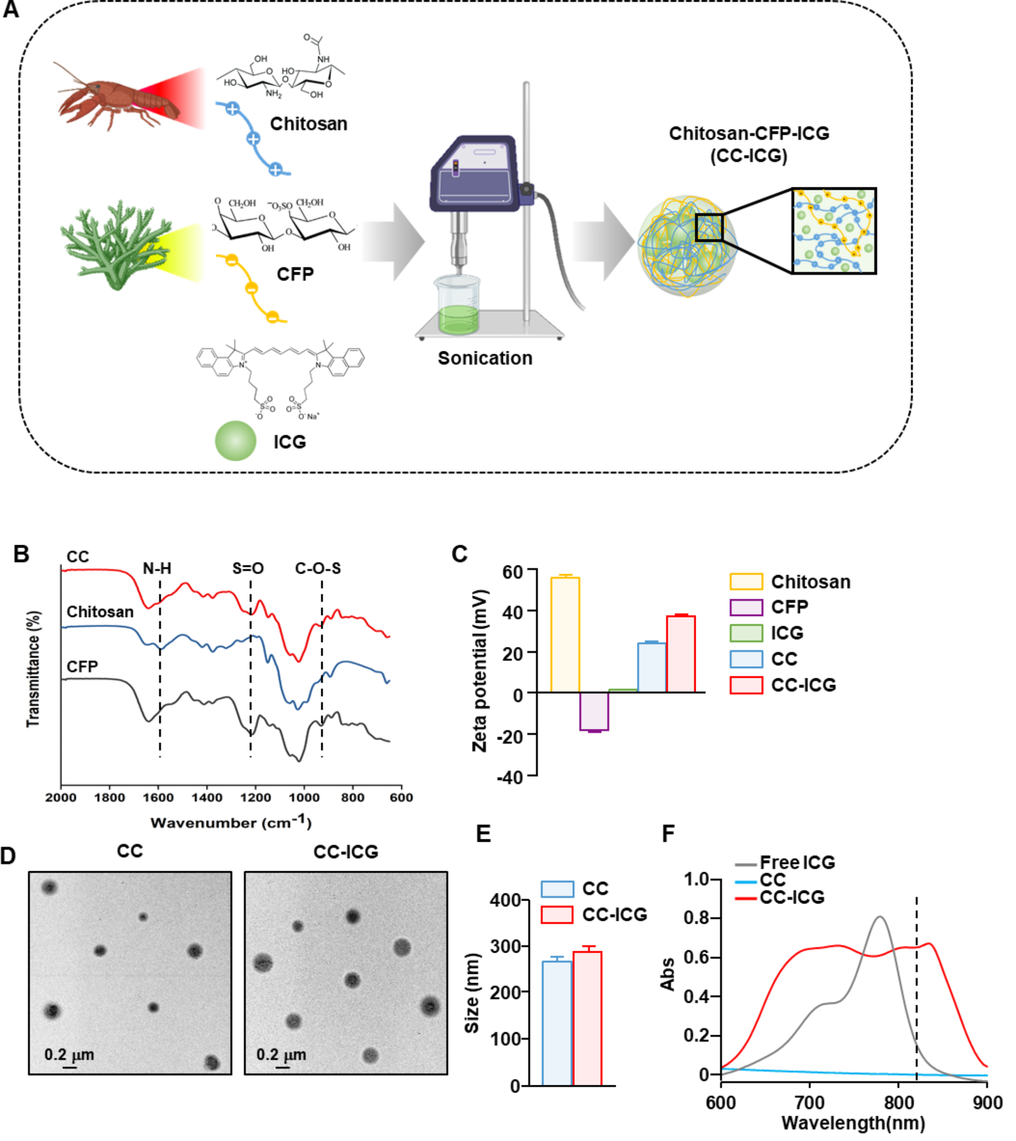
CC-ICG nanoparticles were synthesized using ultrasonication at room temperature. CFP was dissolved in distilled water, and chitosan (Daejung, Korea) was dissolved in a 1% (v/v) acetic acid solution. The two polysaccharides were mixed in the following ratios: 1:1, 1:2, and 1:3. Ultrasonication was then performed to form nanoparticles through material entanglement on ice under the following conditions: pulse-on 3 s, pulse-off 7 s, for a total of 3 min at 40% amplitude. The total volume of the mixture was fixed at 6 mL, and 2 mg of ICG was added (Tokyo Chemical Industry, Tokyo, Japan). The nanoparticles were harvested using centrifugation at 6200 × g for 10 min, and the pellet was resuspended in distilled water.
CC-ICG characterization
CFP, chitosan, and chitosan-CFP (CC) levels were measured using a Fourier Transform Infrared Spectrometer (JASCO, FT-4100, Tokyo, Japan). The zeta potential and nanoparticle size were determined using a Dynamic Light Scattering Particle Analyzer (Litesizer DLS 500; Anton Paar, Graz, Austria). Transmission electron microscopy (TEM) images were obtained using an H-7600 transmission electron microscope (Hitachi, Tokyo, Japan). The encapsulated ICG amount was quantified using a UV–vis spectrophotometer (Cary 100 Bio; Varian, Palo Alto, CA, USA). PTT was performed using a fiber-coupled continuous-wave diode laser (808 nm; Changchun New Industries Optoelectronics Technology Co., Ltd., Jilin, China), and temperature changes and thermal images were obtained using a MobIR Air Mobile Phone Thermal Imaging Camera (ZIYOUHU, China).
Cell line and cell culture
The murine colon carcinoma cell line CT-26 (ATCC, CRL-2638; Korean Cell Line Bank, Seoul, Korea) was cultured in RPMI-1640 medium with 1% penicillin/streptomycin and 10% fetal bovine serum (FBS). CT-26WT-iRFP-Neo cells (CT-26-iRFP; Imanis Life Sciences, CL091, Rochester, USA) were cultured in an RPMI-1640 medium with 1% penicillin/streptomycin, 10% FBS, and 0.4 mg/mL G418. Both cell lines were maintained in a 5% CO2 incubator at 37 °C.
Annexin V/DAPI staining
CT-26 cells (1 × 10⁵ cells/well) were seeded in 24-well plates (SPL Life Sciences Co., Ltd., Korea) and treated with phosphate-buffered saline (PBS), chitosan, CFP, CC, and CC-ICG. One hour after treatment, cells were irradiated with an NIR laser at 1, 1.5, and 2 W/cm2 for 5 min. Twenty-four hours post-irradiation, cells were harvested and stained with annexin V- fluorescein isothiocyanate (FITC) (BioLegend, San Diego, CA, USA) and 4′,6-diamidino-2-phenylindole (DAPI; Sigma-Aldrich) at room temperature in the dark for 15 min. The apoptotic/necrotic cells were analyzed using flow cytometry (NovoCyte; ACEA Biosciences, San Diego, CA, USA).
Mice
Five- to six-week-old female C57BL/6 and BALB/c mice (20 ± 0.2 g) were purchased from Orient Bio (Gyeonggi, Korea). Mice were raised in pathogen-free conditions at the Laboratory Animal Center of Asan Medical Center. All animal experiments were approved and conducted according to the guidelines of the National Research Council’s Guide for the Care and Use of Laboratory Animals and the Institutional Animal Care and Use Committee at Asan Medical Center (Protocol number: 2023-20-260).
Antibodies
Fluorescently labeled Brilliant Violet 785™ anti-mouse cluster of differentiation (CD)11c, FITC anti-mouse CD3, FITC anti-mouse CD90.1, FITC anti-mouse Gr-1, FITC anti-mouse CD49b, FITC anti-mouse TER-119, allophycocyanin (APC) anti-mouse CD40, Brilliant Violet 605™ anti-mouse CD80, phycoerythrin (PE)/Cyanine7 anti-mouse CD86, peridinin-chlorophyll-protein (PerCP) anti-mouse I-A/I-E, PerCP/Cyanine5.5 anti-mouse H-2 Kb, PE/Cyanine7 anti-mouse T cell receptor (TCR)-β, PerCP5.5 anti-mouse CD4, Brilliant Violet 785™ anti-mouse CD8, and APC anti-mouse CD44 were purchased from BioLegend (San Diego, CA, USA).
Generation of bone marrow-derived dendrite cells (BMDC)
Bone marrow (BM) was harvested from the hind limbs of six-week-old C57BL/6 mice. The collected BM was suspended in red blood cell lysis buffer (Thermo Fisher, Waltham, MA, USA) and washed with PBS. BM cells (1 × 10⁶ cells/mL of culture medium) were seeded in 24-well plates. The cells were incubated with 100 ng/mL of recombinant murine interleukin-4 and 100 ng/mL of recombinant granulocyte marcrophage colony stimulating factor. The differentiation of BMDCs was confirmed by CD11c expression on day 6 of culture using flow cytometry (NovoCyte, ACEA Biosciences, Inc.).
Analysis of BMDC activation
CT-26 cells (1 × 10⁵ cells/mL) were seeded in 24-well plates and treated with PBS, chitosan, CFP, CC, and CC-ICG, followed by irradiated with an NIR laser for 5 min at 1.5 W/cm2. Twenty-four hours after treatment, the culture medium was collected. On day 6, the BMDC culture medium was removed and replaced with the collected CT-26 culture medium. Twenty-four hours after incubation, BMDC morphology was observed using a microscope (EVOS M5000; Thermo fisher), and co-stimulatory and major histocompatibility complex (MHC) molecule expression were analyzed using flow cytometry (ACEA Biosciences Inc.)
Analysis of splenic DC activation
To analyze splenic DC activation, C57BL/6 mice were injected intravenously (i.v.) with PBS, chitosan, CFP, CC, and CC-ICG. Six hours post-injection, the mice were sacrificed, and their spleens were harvested. The spleens were sectioned into small pieces using curved scissors and digested with collagenase IV and DNase-containing culture medium at 37 °C for 20 min. Aggregated and undigested tissues were removed using a 100-nm nylon mesh and washed with PBS. The pellet was resuspended in 5 mL of Histopaque-1077 (Sigma-Aldrich) and layered with 5 mL of fresh Histopaque-1077. FBS was then added to the upper layer of the cell suspension. The cells were centrifuged at 600 × g for 10 min without a break. The fraction that was denser than 1.077 g/cm3 was harvested as leukocytes. The splenocytes were then stained with lineage and DC activation markers, as shown in previous studies [12, 27, 28]. The stained cells were analyzed using flow cytometry (NovoCyte, ACEA Biosciences, Inc.).
PTT of CT-26 tumor
CT-26 cells (5 × 10⁵/100 µL of PBS) were subcutaneously injected into BALB/c mice. On day 7, the mice were randomly divided into five groups: PBS, chitosan, CFP, CC, and CC-ICG. The aforementioned compounds were injected intratumorally and were then irradiated with an NIR laser at 1.5 W/cm2 for 5 min. Temperature changes and thermal images were obtained using a MobIR Air Mobile Phone Thermal Imaging Camera (ZIYOUHU, China). Tumor growth and survival rates were monitored.
Rechallenge of lung metastatic CT-26 cancer
For the rechallenge of lung metastatic CT-26 cancer, on day 38 after primary tumor inoculation, mice that were cured by CC-ICG and laser irradiation were i.v.-injected with CT-26-iRFP cells (5 × 10⁵ cells/100 µL of PBS). The PBS-, chitosan-, CFP-, and CC-treated mice were also inoculated i.v. with CT-26-iRFP cells as control groups for the rechallenge. The survival of the mice was monitored for 30 days post-injection (day 62 of the primary CT-26 challenge). On days 7, 10, and 14 after the rechallenge of lung metastatic CT-26 cancer, near-infrared fluorescent protein (iRFP) fluorescence was imaged using the Xenogen In Vivo Imaging System 200 BLI system (Caliper Life Sciences). Survival rates of mice were monitored for 20 days after the rechallenge of 4T1-iRFP. The detailed schedule of tumor challenge is shown in Fig. S8.
Hematoxylin and eosin (H&E) staining
On day 21 of the CT-26 rechallenge, mice were sacrificed, and 1 mL of 3.7% formaldehyde was injected intratracheally. The lungs were harvested and fixed with 3.7% formaldehyde at 4 °C overnight, dehydrated, and embedded in paraffin. The embedded lung tissue was sectioned into 5-µm thick slices and placed on glass slides. The sections were stained with H&E (Sigma-Aldrich), and images were obtained using an EVOS M5000 microscope (Thermo Fisher Scientific).
Analysis of memory T cells
Splenocytes were harvested on day 21 post-rechallenge of lung metastatic CT-26 cells. The splenocytes were stained with anti-CD8, anti-CD4, anti-TCR-β, and anti-CD44 for 20 min at 4 °C. Cells were washed with PBS and suspended in PBS containing DAPI (Sigma-Aldrich). The cells were analyzed using Novocyte (ACEA Biosciences, Inc.).
Live imaging for CTL activity
CD8 T cells were isolated from the splenocytes of CT-26 rechallenged mice using a CD8 T cell isolation kit (Miltenyi Biotec, Bergisch Gladbach, North Rhine-Westphalia, Germany). Isolated CD8 T cells (1 × 10⁵) were cultured with CT-26-iRFP cells (1 × 10⁴) in 24-well plates. Live images of CTL activity were obtained using a fluorescence microscope (EVOS M5000, Thermo Fisher Scientific) and live cell instruments (LCIbio, Gyeonggi-do, Korea) for 16 h.
Statistical analysis
Data are presented as the mean ± standard error of the mean. One-way or two-way Tukey multiple comparison tests were used to analyze the dataset. Statistical significance was set at p < 0.05.


Comments (0)