
Remember me


From June 2020 to August 2022, 23 patients were assessed for eligibility and 22 patients were enrolled (detailed clinical information was listed in Supplementary Table 1). One patient was excluded due to central nervous system involvement (Fig. 1b). Four patients discontinued the induction treatment owing to disease progression. One patient achieved partial response (PR) at interim evaluation but withdrew consent at 5 months for personal reasons, who was confirmed as responder in the intention-to-treat (ITT) analysis. Seventeen patients completed the induction treatment and 13 patients achieved CR, 1 achieved PR and 3 documented as progressive disease (PD) at final evaluation. Among the 14 CR/PR patients, 11 continued maintenance treatment of sintilimab and 8 of them completed the maintenance treatment, one patient received autologous hematopoietic stem cell transplantation (auto-HSCT), two patients did not proceed to maintenance treatment due to pseudoprogression and mucositis during induction treatment, respectively.
Fig. 1
Trial profile. a Study schema. Patients received pegaspargase 2500 IU/m2 intramuscularly on day 1 and sintilimab 200 mg intravenously on day 2 for 6 cycles of induction treatment, as well as sintilimab 200 mg for 28 cycles of maintenance treatment. Treatment was continued until disease progression, unacceptable levels of adverse events, or withdrawal of consent occurred. b Flow diagram. Among 22 patients enrolled, 17 completed the induction treatment and 11 continued the maintenance treatment
Baseline characteristics are listed in Table 1. The median age was 51 years (range, 24–74) with 23% of patients more than 60 years old. 68% had Ann Arbor stage IV disease, 59% had elevated serum lactate dehydrogenase (LDH) levels, and 45% had remote lymph node involvement. Risk stratification showed that 82 and 55% of the patients were categorized into a high-risk group according to the prognostic index for NKTCL (PINK) and PINK in combination with circulating EBV DNA (PINK-E), respectively.
Table 1 Characteristics of the patientsEfficacyIn the first stage with 7 patients enrolled, 5 achieved CR and 2 achieved PR. In the ITT analysis, the overall response rate (ORR) was 68% (95%CI, 47–84%) after 6 cycles of induction treatment, with the CR and PR rate as 59 and 9%, respectively. Representative PET-CT images from a CR patient are shown in Fig. 2a. Tumor burden decreased by ≥ 50% in most patients (71%, Fig. 2b). One patient, with the appearance of new lesions in the mediastinum, was diagnosed as pseudoprogression by biopsies (supplementary Fig. 1). Circulating EBV DNA levels were significantly decreased after 3 cycles of induction treatment in responders, as compared to those of non-responders, and turned negative after 6 cycles of induction treatment (Fig. 2c). With a median follow-up of 30 (range, 3–48) months for PFS and 30 (range, 6–50) months for OS, the 2 year PFS and OS rates for the chemo-free cohort were 68% (95%CI, 45–83%) and 86% (95%CI, 63–95%), respectively (Fig. 2d). The median duration of remission (DOR) was not reached. Of note, responders showed better PFS and OS than those of non-responders (Fig. 2e). Considering that upfront auto-HSCT showed no survival benefit,21,22 consolidation with auto-HSCT was not generally performed in this trial and only one responder received auto-HSCT. The 13 CR patients remained alive and disease-free under maintenance treatment (n = 10) or follow-up evaluation (n = 3). Of note, none of them experience recurrence after discontinuation of treatment (median 5 months, range, 0–34 months). One PR patient experienced disease progression after 20 cycles of sintilimab maintenance and remained alive after second-line treatment. The 7 non-responders received ≥2 L chemotherapies (Table 1). with 2 of them achieving CR. One treated with selinexor, sintilimab and P-GEMOX, another treated with sintilimab and MESA.
Fig. 2
Response and outcomes. a Representative PET-CT image of a CR patient before and after induction treatment. b Percentage decline of metabolic tumor volume (MTV) during the induction treatment in 21 patients with available PET-CT evaluation. The decline in MTV was measured with interim evaluation of PET-CT in 4 of 22 cases (marked with an asterisk), 3 patients discontinued the treatment, and another withdrew after interim evaluation. The decline in MTV could not be measured in 1 case, who discontinued the treatment before interim evaluation. c Dynamical documentation of circulating EBV DNA copies during induction treatment in responders and non-responders. d Kaplan-Meier plot showing PFS and OS. The numbers of patients who could be evaluated for each endpoint are shown. Tick marks indicate censored data. e Kaplan-Meier plot showing PFS and OS of responders and non-responders in the chemo-free cohort. P values in (c) were compared between responders and non-responders using student’s t test. P values in (e) were compared between responders and non-responders using log-rank test
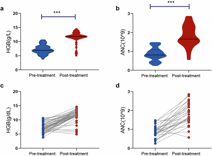
SafetyAmong 22 eligible patients treated with protocol therapy, 91% (n = 20) experienced at least one AE, most of which were mild and moderate. Grade 3/4 AEs were observed in 10 (45%) patients, including neutropenia (32%, n = 7), hypofibrinogenemia (18%, n = 4), leukopenia (9%, n = 2), anemia (5%, n = 1), thrombocytopenia (5%, n = 1), hypoalbuminemia (5%, n = 1), aspartate aminotransferase (AST)/alanine aminotransferase (ALT) elevation (5%, n = 1), and heart failure (5%, n = 1), which were manageable and led to no discontinuation of treatment. No hypersensitive reaction to pegaspargase occurred and no premedication of corticosteroids was administrated. No bleeding or thromboembolic events occurred. One patient received implantable cardioverter defibrillator surgery due to paroxysmal ventricular tachycardia 6 months before being diagnosed as NKTCL and experienced heart failure after the first cycle of induction treatment. The patient continued the treatment and achieved CR after induction treatment. Therefore, this is considered treatment unrelated.
The most frequent grade 1/2 hematological AEs were anemia (64%, n = 14), leukopenia (55%, n = 12) and neutropenia (32%, n = 7), while non-hematological AEs were hyperbilirubinemia (73%, n = 16), hypoalbuminemia (68%, n = 15), hypofibrinogenemia (55%, n = 12), AST elevation (50%, n = 11), and hyponatremia (50%, n = 11).
Eight patients had documented grade 1/2 hypothyroidism (36%), which were asymptomatic and need no medications. No grade 3/4 hypothyroidism was observed. Other immune-related AEs, such as pneumonitis, adrenal insufficiency, hepatitis, vitiligo, etc.,23 were not observed. All grade AEs that occurred during induction treatment are listed in Table 2.
Table 2 Treatment-related AEsComprehensive characterization according to treatment responseNone of the clinical characteristics was associated with the response to pegaspargase plus sintilimab treatment, such as Ann Arbor stage, extra nasal type, remote lymph node involvement, circulating EBV DNA, PINK, and PINK-E, etc. (Table 1, supplementary Table 2 and Fig. 2a, b). Tumor EBV gene expression before treatment showed no difference between responders and non-responders (supplementary Fig. 2c). PD-L1 expression is detected by PD-L1 immunohistochemical assay and quantified by tumor proportion score (TPS), which is defined by the percentage of stained tumor cells over total tumor cells.24 PD-L1 TPS of pre-treatment tumor tissues in CR patients was significantly higher than that in PD patients (Fig. 3a), while PD-1 expression on TIL correlated with poor response to treatment (Fig. 3b).
Fig. 3
Molecular signatures according to treatment response. a PD-L1 expression by tumor proportion score (TPS) according to treatment response. b PD-1 expression on tumor infiltrating lymphocytes according to treatment response. c Gene mutations identified in responders and non-responders. d Pathway enriched with differentially expressed genes in responders and non-responders. e Heatmap of genes involved in cytokine interactome and lipid metabolism highly expressed in non-responders. f Dynamical documentation of indicated lipoproteins in circulation upon induction treatment in responders and non-responders. Data in (a) and (f) were represented as mean ± SEM. P value in (a) was compared between CR and PD patients using student’s t test. P value in (b) was compared between patients with PD-1+ and PD-1- TILs using chi-square test. P values in (f) were compared between responders and non-responders using student’s t test
For exploratory biomarker analysis, we performed targeted DNA sequencing (n = 22) and RNA sequencing (RNA-seq, n = 17) on pre-treatment tumor samples of the patients. No significant difference in the mutation pattern was shown between responders and non-responders (Fig. 3c and supplementary Table 3). Mutation pattern in newly diagnosed and paired recurrent tumor biopsies was shown in supplementary Fig. 3a. We next compared differentially expressed genes using RNA-seq data and identified 303 upregulated and 519 downregulated genes in non-responders (supplementary Table 4), among which cytokine-cytokine receptor interaction, arachidonic acid, alpha-linolenic acid and glycerophospholipid metabolism were significantly activated in non-responders (Fig. 3d). Similar results were observed by comparing the RNA-seq data of newly diagnosed and paired recurrent tumor biopsies derived from one of the non-responders (supplementary Fig. 3b). Further revealed in Fig. 3e, genes upregulated in non-responders included CCL8, CXCR1, IL1B, and XCL1 involving recruitment, migration, expansion and function of immunosuppressive Treg cells,25,26,27,28 as well as ACHE, PLA2G4D, PLA2G4E, PLA2G2F, ALOXE3 and ALOX15B, contributing to phosphatidylcholine catabolism and high-density lipoprotein (HDL) homeostasis.29 Accordingly, peripheral blood HDL cholesterol (HDL-C) and apolipoprotein A-I (apoA-I), the main protein component of HDL, were elevated, whereas free fatty acids were decreased upon induction treatment in responders (Fig. 3f). Other blood lipids indicators, including triglyceride, cholesterol, low-density lipoprotein cholesterol (LDL-C), apoB, apoE, and lipoprotein A (LPA), showed no difference between responders and non-responders (Fig. 3f). It was worth noting that none of the patients took oral lipid-lowering drugs before treatment and all patients were on a low-fat diet during induction treatment due to administration of pegaspargase.
Inspired by recent studies characterizing the peripheral blood immune signature with response to checkpoint blockade in Hodgkin lymphoma,20 we further applied single-cell proteomic analysis (mass cytometry by time-of-flight, CyTOF) to investigate peripheral blood immune signature (n = 8, with qualified blood samples) using 40 immune cell markers designating 34 immune cell clusters (supplementary Table 5). As revealed by self-organizing map (SOM)-based gene clustering and visualization, 2 protein clusters (C1 and C2, Fig. 4a) were identified, exerting different expression levels in responders (n = 5) and non-responders (n = 3). Proteins in C1 (such as HLA-DR, CD141, CD11b, CD11c, CD14, CD16, and CD107a), mainly expressed on monocytes, DCs and B cells, were upregulated in responders, while proteins in C2 (such as FOXP3, CD25, CD127, CD62L, and CD197), mainly expressed on Treg and Naïve T cells, were upregulated in non-responders (Fig. 4b and Supplementary Fig. 4a). Next, we analyzed the protein expression of immune checkpoints and found that PD-1 was increased on T cell clusters in responders, while CTLA-4 was increased on monocytes, DC, and Treg cells in non-responders (Fig. 4c). The proportions of peripheral blood immune cell clusters in NKTCL significantly differed from those of healthy volunteers (n = 10, supplementary Fig. 4b, c). Sharing similar features of downregulated naïve CD4+ and CD8+ T cells in responders and non-responders, Treg cells were downregulated in responders, but upregulated in non-responders (Fig. 4d). Furthermore, peripheral immune cell subsets of all 22 enrolled patients before treatment were assessed by flow cytometry (FCM), confirming a significantly increased CD4+CD25+CD127low Treg cell subset in non-responders than that of responders (Supplementary Fig. 4d). Immune-suppressive Treg cells were positively correlated with triglyceride, apoE and FFA, while immune-activated cytotoxic T cells were positively correlated with lipid proteins, such as HDL-C, apoA-I, apoB, cholesterol, LDL-C and LPA (Supplementary Fig. 4e).
Fig. 4
Peripheral immune signatures according to treatment response. SOM plot showing the expression pattern of different cell markers (a) upon combination treatment in responders and non-responders (b). c Heatmap plot of PD-1 and CTLA-4 expression across indicated cell subsets in responders and non-responders. d Percentage of distinct cell subsets across healthy volunteers (n = 10), responders (n = 5) and non-responders (n = 3). Data in (d) were represented as mean ± SEM. P values in (d) were compared using student’s t test
Comments (0)