In this paper, we analyzed the timing, frequency and type of relevant IHT related complications in patients with aSAH who underwent an MRI during the NCCU-stay. Additionally, we evaluated the changes in medical decisions/management and the need of further interventions based on the MRI-findings. As a key finding, the rate of IHT complications in our population is relatively low. Moreover, the new information obtained from MRI induced changes in treatment management in one third of the patients, a proportion that reflects a relevant diagnostic and therapeutic impact in this critically ill population.
The relatively low frequency of IHT-related complications suggests that well-structured workflows and highly trained personnel seem to be essential for safe transport. The first guidelines for IHT were published in 1993 [26] and were last updated in 2004 [27]. Given the increasing use of advanced diagnostic modalities in NCCUs [28], the current guidelines may no longer adequately reflect present-day clinical demands. In response, checklists have been introduced to enhance patient safety during IHT [27, 29,30,31]. Many of the reported complications of the study population (56%) were systemic, primarily involving an increase in blood pressure, which required administration of sedatives and/or antihypertensive therapy and could therefore be treated promptly.
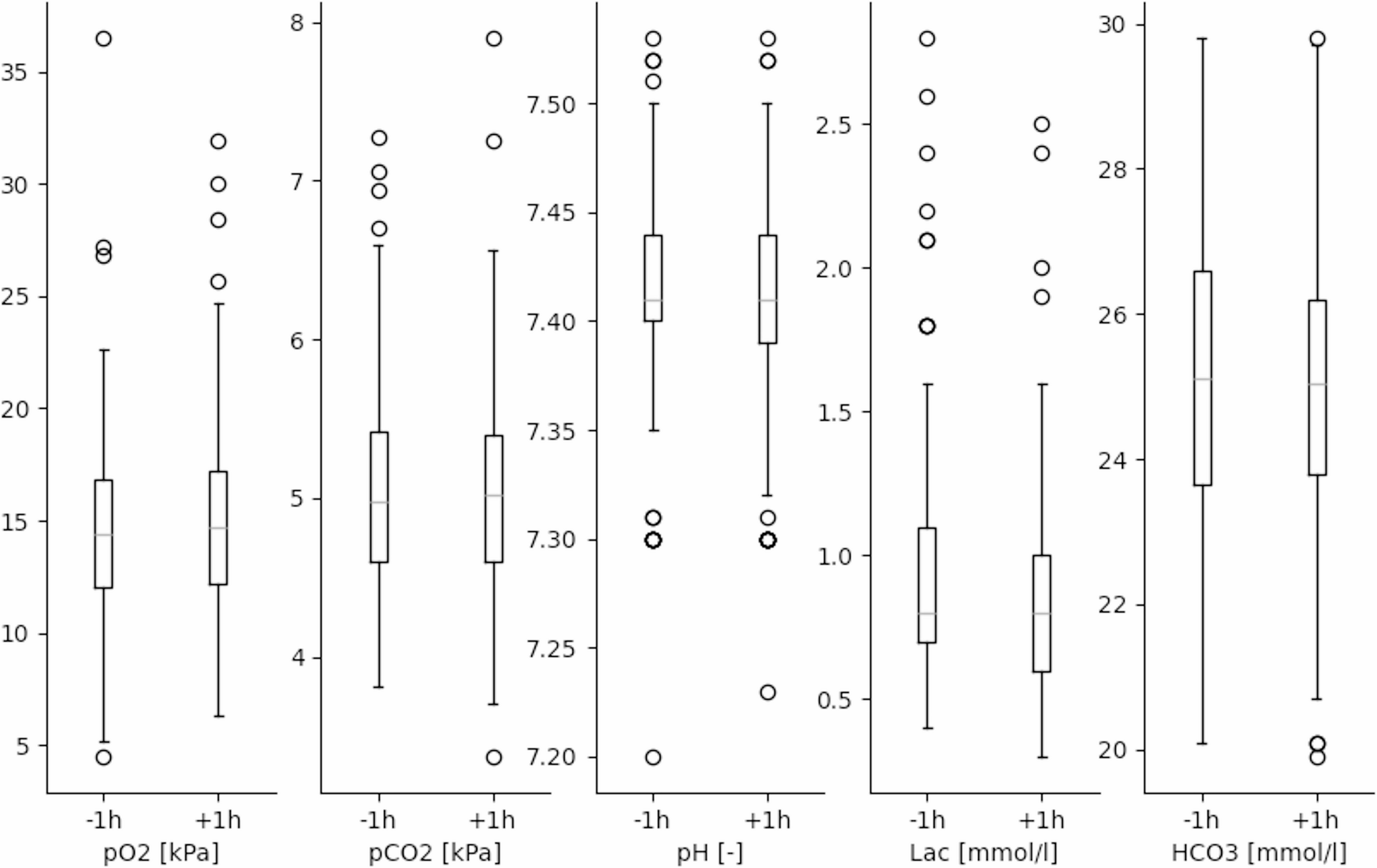
Ensuring patient safety during IHT for MRI is critical, with several studies identifying key risk factors and monitoring parameters to maintain physiological stability. Schmidbauer et al. were the first to propose specific thresholds based on expert opinion and retrospective data, though these have not yet been prospectively validated [32]. When applying their criteria to our cohort, we observed only 6% of cases exceeding neurological thresholds (ICP > 22 mmHg), while we argue that systolic BP > 180 mmHg should not be classified as a complication in aSAH patients, given its role in preventing vasospasm and DCI [33]. In our study, vital signs, blood gas values, and ICP remained largely stable before and after transport, likely reflecting the high level of neurocritical care expertise and the focus on MRI-specific IHTs. While Schmidbauer’s safety limits remain unvalidated [32], elevated ICP is widely acknowledged as a risk factor for reduced CPP and DCI. Prior studies have demonstrated that IHT can provoke ICP elevations, particularly in patients with already elevated baseline levels [3, 4, 13, 34], and that use of elevators or ramps may further exacerbate this and alter cerebral metabolism [4]. Additionally, the recommended 30° head-up positioning for optimal venous drainage and ICP reduction cannot be maintained during MRI [35].
Pinggera et al. investigated IHT for early MRI (median day 6) in a cohort of ventilated patients with acute, severe traumatic brain injury [36]. Their monitoring relied solely on end-tidal CO₂—a known surrogate with limitations compared to direct PaCO₂ measurement [37]—as well as MAP and ICP. While they reported stable ICP and MAP before and after transport, a significant decrease in end-tidal CO₂ up to 45 min after transport was recorded. Although they argue that these findings lack clinical relevance, the true impact remains rather uncertain—particularly with regard to potential hypocapnia-induced cerebral vasoconstriction and its effect on cerebral perfusion [38, 39], which was not directly measured. However, they highlight how important the coordination of involved teams (e.g. radiology and anesthesia) is along with the requirement for patients to be hemodynamically stable for four hours.
These observations further underscore the critical importance of evidence-based standard operating procedures (SOPs) in the management of high-risk patients. The study by Cuschieri et al. demonstrated that the implementation of SOPs in severely injured patients led to a significant reduction in mortality from 22 to 11% [40], highlighting that even partial adherence can improve outcomes—despite the inherent challenges in full protocol compliance. Although their cohort did not include patients with traumatic brain injury, the findings emphasize the broader value of structured protocols. An early example for this was proven Weg et al. in 1989, who concluded that manual ventilation during intrahospital transport was as safe when performed by trained personnel with defined protocols as mechanical ventilation [41]. However, this study also did not assess intracranial parameters, which are particularly sensitive in neurocritical care patients. Taken together, these findings [40, 41] highlight that clinical safety cannot rely solely on advanced technology; rather, it depends on well-trained staff capable of managing both equipment and unexpected complications in adherence to validated clinical standards.
Interestingly, nearly half of Pinggera et al.’s cohort underwent more than one MRI [36] whereas we count 36%. In our cohort, repeated MRIs were primarily used to improve diagnosis compared to CT (to investigate abscesses, partial thrombosis, residual perfusion of an aneurysm, or artifacts from endovascular coils). Their implications—such as suggested prolonged intubation for better functional outcome—were not evaluated [36].
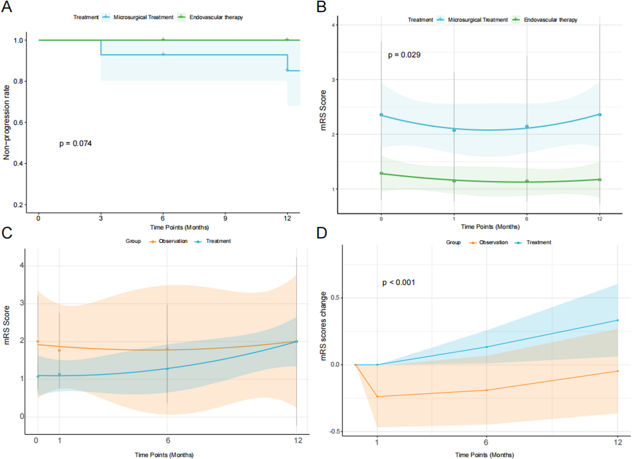
Following MRI and interdisciplinary review, there was a change in our medical management in one third of the patients, comparable to the data reported by Schmidbauer et al., where the authors specifically analyzed IHT in SAH patients who underwent a head-CT scan and they reported that imaging findings directly influenced clinical decisions in 40% of cases. However, “no consequence” may still imply that ongoing therapy was continued and therefore had a clinical impact, which is significantly different from “no consequence at all” in clinical decision-making. In 5% of our cases the interdisciplinary decision agreed to change therapy goal based on the MRI results with most of them having a poor grade WFNS classification.
While CT remains the first-line modality in acute neurocritical care, MRI provides significantly greater sensitivity for detecting early ischemic changes, small infarcts, and posterior fossa lesions - areas where CT often underperforms due to beam-hardening artifacts and limited resolution due to anatomical complexity [42, 45]. In our cohort, MRI findings frequently led to specific and clinically relevant management changes that would not have been initiated based on CT alone. For example, early DCI-related infarcts detected on DWI [46] prompted the implantation of multimodal neuromonitoring; confirmation of a cerebral abscess following aneurysm clipping led to revision surgery [47]; and vessel wall imaging revealed residual perfusion or incomplete exclusion of aneurysms, prompting further surgical or endovascular treatment [48]. Particularly in posterior fossa strokes—where CT sensitivity is notably low—MRI proved essential for accurate diagnosis and therapeutic guidance, especially using DWI, which outperforms CT and CT perfusion in this region [43, 44]. Therefore, while MRI is not universally required, it provided critical diagnostic and therapeutic value in selected high-risk patients.
Martin et al. demonstrated a significant correlation between adverse events during IHT and prolonged NCCU stays in patients with traumatic brain injury undergoing IHT for CT imaging [13]. Technical complications during IHT have been reported at rates up to 46% [49, 50]. This frequency was lower in our population, but this is probably due to the lack of a standardized definition for technical failures and challenges in retrospective data collection, which makes direct comparisons difficult.
While literature reports 17–42% of IHTs as emergencies [4, 14, 32], in our study, MRI was mostly performed electively, usually on the same day of indication, after interdisciplinary discussion which allowed for safe preparation of the IHT. In emergency cases, CT was preferred for rapid decision-making. Given the diagnostic value of MRI, portable ultra-low-field MRI may offer a promising alternative, avoiding IHT-related risks [28, 51]. However, its lower image resolution, increased artifact susceptibility, and potential interference with life-sustaining NCCU equipment remain important limitations [52]. Due to the limitations of portable MRIs, we believe the focus should be on selecting patients for a safe transport and for whom the result of the imaging would impact the clinical management.
This study has several limitations. First, its retrospective and single-center design without a control group limits generalizability. Furthermore, all MRIs were performed outside the acute phase of aSAH, which may have influenced both complication rates and diagnostic yield. The most significant methodological limitation is that only single time-point values for intracranial and blood pressure parameters were analyzed, rather than averaged values over defined intervals (e.g., 5-minute means), which may have reduced the sensitivity for detecting transient but clinically relevant fluctuations. Another key limitation lies in the potential for selection bias. However, it is important to note that our cohort includes all consecutive patients who received MRI, without further selection, thereby capturing a broad spectrum of clinical conditions. While the potential for systematic bias remains an inherent limitation in this non-randomized study, we believe our findings reflect real-world clinical practice and offer relevant insights into the safety profile and practical applicability of MRI in aSAH patients under intensive care.

Comments (0)