
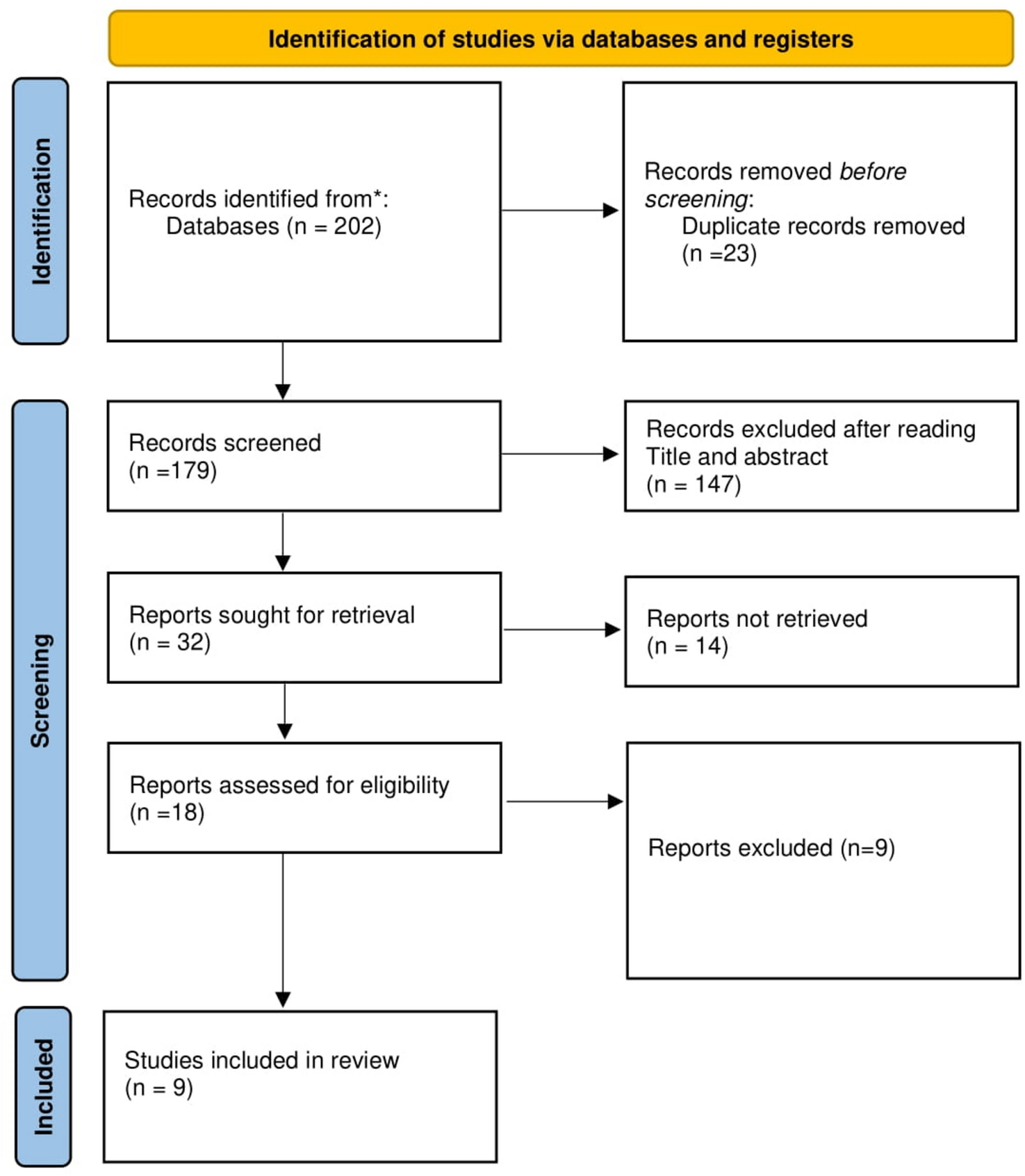
Remember me


Recent developments in neuromodulation technology provide hopeful treatment possibilities for many neurological and behavioral disorders. These developments have improved our understanding of the neural circuitry controlling brain activity and highlighted the potential of using these technologies to treat many mental health diseases. The study of neural networks linked to response inhibition is critical since it is vital for processes of self-control and decision-making. Developing neuromodulatory treatments meant for diseases characterized by poor inhibitory control, including attention deficit hyperactivity disorder (ADHD), impulse control issues, and many neuropsychiatric disorders, requires an understanding of the brain’s response inhibition networks.
Focusing on the prefrontal cortex and insula, two important regions involved in controlling impulsive behavior, Osada et al. (2024a, b) conducted a thorough study of the brain circuits linked to response inhibition. Using sophisticated neuroimaging techniques such as functional magnetic resonance imaging (fMRI) and transcranial magnetic stimulation (TMS), the researchers identified the brain areas involved in suppressing prepotent responses and clarified the neural circuits supporting self-control. From perception to execution, the study showed that response inhibition triggers particular insular-prefrontal networks at various stages. The results show that the prefrontal cortex and insula interact dynamically, each performing a different function in different stages of the inhibition process. This thorough knowledge of the neural networks linked to response inhibition would significantly influence the development of focused neuromodulatory medicines. Activating certain brain areas with TMS or other neuromodulation methods, for example, might improve inhibitory control and operate as a therapeutic intervention for people with ADHD or other disorders marked by weakened self-regulation (Osada et al. 2024a, b).
Recent developments in neuromodulation technologies, including deep brain stimulation (DBS) and transcranial direct current stimulation (tDCS), are increasing treatment options for conditions linked with compromised neural circuits. For example, while tDCS has shown promise in altering cortical excitability and improving cognitive performance in people with depression and other mood disorders, DBS has successfully treated movement disorders like PD by concentrating on specific areas of the basal ganglia. These approaches offer non-invasive or slightly invasive ways to affect brain activity and restore neuronal circuits damaged by injury, disease, or developmental problems.
Furthermore, optogenetics—a novel technique allowing exact control of brain activity by light—has dramatically improved our knowledge of the neural networks linked to complicated behaviors. Optogenetics has been used to study response inhibition in animal models, clarifying the causal links between brain networks and inhibitory control (Warden et al. 2024). Careful activation or suppression of specific neurons allows researchers to track the real-time effects on behavior, hence offering a level of control and accuracy that was previously unattainable with more traditional methods. This has opened doors for therapeutic approaches directly targeting brain circuits linked to cognitive and behavioral issues.
Apart from neuromodulation’s potential therapeutic applications, current research has underlined the need for circuit-specific treatments in understanding and treating psychiatric diseases. Sadeh et al. (2024) found that improving specific brain circuits with focused neuromodulation could increase cognitive flexibility and reduce anxiety and depression symptoms. Their results suggest that tailored neuromodulatory treatments, designed to the particular brain circuits linked to every patient’s condition, might offer a more effective substitute for traditional uniform therapy.
Recent developments in neuromodulation technologies have produced new knowledge on brain networks linked to response inhibition and other complex behaviors. Neuroimaging, optogenetics, and neuromodulatory methods have helped scientists map the brain areas and networks controlling inhibitory control, hence opening new paths for therapeutic interventions. Neuromodulation is anticipated to become a more important tool in treating mental and neurological disorders as our understanding of these circuits develops, hence offering customized and focused therapy to improve patient outcomes.
Neuromodulation in Mental Health TreatmentThe role of glucagon-like peptide-1 (GLP-1) receptor agonists in mental health therapy has drawn significant interest because of their putative therapeutic advantages outside their usual use in the control of type 2 diabetes and obesity. Recent studies have highlighted the possible benefits of GLP-1 receptor agonists in treating many mental health disorders, including neurocognitive issues, depression, and substance addiction. The rising prevalence of neurodegenerative diseases like AD and other psychiatric illnesses that still challenge traditional therapies amplifies the relevance of these consequences.
Chiefly responsible for the control of glucose metabolism and insulin generation, GLP-1 is a peptide hormone. Besides their metabolic functions, recent research suggests GLP-1 and its receptor greatly affect brain activity and mental wellness. Emphasizing their ability to reduce the frequency of substance misuse, psychotic diseases, and seizures, Au et al. (2023) undertook comprehensive research on the neuropsychiatric benefits of GLP-1 receptor agonists. Their study underlined the possible neuroprotective and antidepressant qualities of these medications, which appear to be linked to their ability to control neuroinflammation, improve synaptic plasticity, and promote neurogenesis in important brain areas connected to mood control and cognition (Au et al. 2023, 2024).
Among the most fascinating aspects of GLP-1 receptor agonists in mental health is their neuroprotective qualities, particularly in neurodegenerative diseases like AD. Recent studies show that GLP-1 receptor activation could reduce the pathogenic processes linked to AD, including amyloid plaque formation, tau tangles, and synaptic dysfunction (Bäckman et al. 2023). According to preclinical studies, GLP-1 receptor agonists may increase cognitive function, reduce neuronal loss, and improve behavioral symptoms in animal models of AD, thus providing a hopeful, creative approach for treating AD (Sweeney et al. 2023). Human clinical studies have shown that GLP-1 receptor agonists may slow down disease progression in those with moderate cognitive impairment (MCI) or early-stage AD and have cognitive benefits (Li et al. 2023a, b).
GLP-1 receptor agonists also show antidepressant-like effects as they alter important brain regions linked to mood control, including the hippocampus and prefrontal cortex. Studies have indicated that in areas usually damaged in people with depression, GLP-1 receptor activation can enhance neuroplasticity and promote neuronal survival (Liu et al. 2023). The results suggest that, especially for those unresponsive to traditional antidepressant drugs, GLP-1 receptor agonists may be a viable therapy option for depression.
Apart from their apparent effects on cognitive and mood control, GLP-1 receptor agonists have been connected to reduced drug-seeking behavior, hence suggesting them as a potential therapy option for handling substance use disorders. Research shows that stimulation of GLP-1 receptors might influence the reward circuits of the brain, which are usually compromised in people with addiction. By changing the action of vital neurotransmitters, including dopamine, GLP-1 receptor agonists could reduce cravings and prevent relapses in people with substance use disorders (Zhang et al. 2023a, b, c).
These results encourage more research on the development of GLP-1 receptor agonists as treatment choices for several neurodegenerative and mental diseases. The neuroprotective and antidepressant effects shown in preclinical and clinical studies highlight the possibility of these drugs to provide new, unusual approaches for mental health therapy. Research still indicates that GLP-1 receptor agonists might be essential in managing addiction, neurological diseases like AD, and depression, so offering fresh possibilities for those with limited choices in traditional treatments.
Aromatherapy and Sleep IllnessesInitially used in aromatherapy for its calming and sleep-promoting qualities, lavender essential oil (LEO) has recently attracted much attention for its neuronal pathways that support improved sleep quality. Modern research has started identifying the specific brain areas and pathways involved in LEO’s therapeutic effects, offering a deeper understanding of how this natural medicine could treat sleep problems and related conditions. Using polysomnographic recordings to evaluate changes in brain activity during sleep, Ren et al. (2023) recently looked at how LEO affected sleep patterns in freely roaming C57BL/6J mice.
The study found that breathing LEO during the light (inactive) phase significantly lowered the latency to start non-rapid eye movement (NREM) sleep, extended the total duration of NREM sleep, and increased cortical electroencephalographic (EEG) slow-wave activity, a marker suggestive of restorative sleep. Two compounds known for their sedative qualities, linalool and d-limonene, were found to be the main components of LEO that appeared to affect these outcomes. The researchers also found that the GABAergic system in the central amygdala, a brain region crucial for emotional control and stress responses, helped promote LEO’s sleep-inducing effects.
Pharmacogenetic reduction of GABAergic neurons in the central amygdala, or disruption of the olfactory system, completely neutralized LEO’s effects on sleep, emphasizing the vital relevance of these systems in enabling its therapeutic efficacy. The central amygdala regulates sleep, anxiety, and stress responses, which is therefore quite important (Zhao et al. 2022). The participation of the olfactory system emphasizes the complex interaction between sensory input and the brain’s internal processes controlling physiological activities like sleep (Chen et al. 2023a, b). These results confirm the efficacy of lavender essential oil in treating sleep problems and provide a neurological basis for its historical use in conventional medicine.
This work enables the study of how specific brain circuits are modulated by aromatic compounds and how these circuits interact to influence behaviors, including sleep. Recent developments in chemogenetics and optogenetics have enabled more precise mapping of neural circuits linked to sleep control; future studies might apply these techniques to explore the particular pathways via which LEO exerts its effects. For example, the optogenetic activation or inhibition of certain central amygdala neurons or other sleep-related brain areas could clarify the precise role of GABAergic neurons in LEO’s sleep-promoting actions (Xie et al. 2023).
Upcoming studies could also use neuroimaging techniques such as functional magnetic resonance imaging (fMRI) and positron emission tomography (PET) to show changes in brain activity in reaction to LEO inhalation. Together with animal models, these methods would allow scientists to see the real-time activation of certain brain networks, improving their knowledge of how LEO affects sleep-related circuits. Studies like this could clarify the mechanisms by which sensory cues, such as smells, influence complex physiological states like sleep and wakefulness by linking brain circuit dynamics to behavioral results.
LEO’s therapeutic possibilities for sleep disorders could perhaps include various neurological conditions characterized by disrupted sleep, including anxiety, depression, and neurodegenerative illnesses. Studies done before show that those with worry and sadness often have sleep anomalies, and restoring normal sleep patterns might be a possible therapy path for these conditions (Li et al. 2023a, b). Sleep’s neuroprotective qualities, including the promotion of neuroplasticity and the removal of neurotoxic chemicals like beta-amyloid, may provide indirect benefits for conditions like AD (Wilson et al. 2022). Therefore, whether used alone or as part of a multi-modal treatment approach, LEO may have great relevance in the management of neurological diseases.
Taken together, modern neurobiological research increasingly supports the relevance of lavender essential oil in improving sleep quality, especially by means of its interaction with the GABAergic system and olfactory routes. Ren et al. (2023) provide a strong framework for understanding the neurological foundations of LEO’s sleep-inducing effects, hence enabling more research on the related brain circuits. Improved knowledge of the molecular mechanisms involved and developments in neural circuit mapping technologies might help LEO to be used therapeutically for several mood and sleep problems.
Neuromodulation for Treatment of Autism and Post-traumatic Stress Disorder (PTSD)Manocchio et al. (2023) look at how well neuromodulation methods—particularly transcranial direct current stimulation (tDCS) and repetitive transcranial magnetic stimulation (rTMS)—treat post-traumatic stress disorder (PTSD). Often linked with dysregulated brain circuits, particularly those in charge of anxiety and stress processing, PTSD is a serious condition. Promising therapeutic intervention is offered by neuromodulation treatments such as tDCS and rTMS, which can affect these circuits.
The study by Manocchio et al. emphasizes how various stimulation parameters—target hemisphere, stimulation frequency, and duration—affect the effectiveness of these therapies. Because of its fundamental involvement in executive functions, including emotional control, decision-making, and cognitive control, which are compromised in PTSD, the dorsolateral prefrontal cortex (dlPFC) is a primary target for neuromodulation therapy. The findings underline that by enhancing emotional control and lowering the hyperactivity of fear-processing circuits in the amygdala, targeting the dlPFC with different neuromodulatory strategies could help to lower symptoms.
Manocchio et al. (2023) underlines the importance of more research to improve stimulation techniques and acquire a better knowledge of the underlying neuromodulatory mechanisms, even if these studies show encouraging results. Improving the therapeutic efficacy of these methods will depend on optimizing stimulation parameters, including finding the most efficient stimulation frequency and the precise spatial coordinates of brain targets. Moreover, customized treatment plans based on personal brain activity patterns could increase the precision and efficacy of neuromodulation therapy for PTSD. This approach could lead to tailored treatments that are more effective and in line with the unique brain profiles of those suffering from PTSD, hence improving treatment results and providing hope to those struggling with this disability.
In the field of neural circuit mapping, where advances in neuroimaging and functional connectivity studies improve understanding of the disturbances in brain networks linked with PTSD, the idea of individualized therapy is particularly relevant. Using techniques like resting-state fMRI and magnetoencephalography (MEG), researchers can map the functional links of the dlPFC and other regions involved in emotional control, hence enabling more precise neuromodulation targeting (Shin et al. 2022). This development in circuit mapping technology is enabling customized neuromodulation treatments to fit for the particular neurological impairment of every patient.
Emphasizing the effect of customized continuous theta-burst stimulation (cTBS) on the social abilities of young children, Xiao et al. (2023) investigate the use of transcranial magnetic stimulation (TMS) for autism spectrum disorder (ASD). Particularly for individuals with low verbal communication, their study highlights the effectiveness of cTBS as a non-invasive neuromodulatory technique able to enhance social communication skills in children with ASD. The researchers showed in a double-masked, randomized controlled trial that children undergoing tailored continuous theta-burst stimulation (cTBS) targeting the left dorsolateral prefrontal cortex (DLPFC) exhibited notable improvements in social communication skills and lower autism severity. Maintained across a three-month follow-up, the improvements suggested that cTBS might have lasting positive impacts on social interaction and language development in children with ASD.
Xiao et al. (2023) offers interesting research since it offers a fresh perspective on how neuromodulation could reduce basic ASD deficits, especially in social communication and language development. These neuromodulatory treatments mainly target the dlPFC, which is crucial for social cognition, executive function, and language processing. Researchers are deepening their knowledge of how customized stimulation regimens can change this brain area to promote social behavior in children with ASD. According to Xiao et al. (2023), more study is required to explore the long-term effects of cTBS and to increase knowledge of the underlying brain mechanisms enabling these changes.
Improving the knowledge of how neuromodulation influences brain function in ASD requires neural circuit mapping techniques. Functional imaging techniques like fMRI and diffusion tensor imaging (DTI) offer thorough mapping of the anatomical and functional links between the DLPFC and other brain regions involved in social cognition and communication (Liu et al. 2022). Using these methods, researchers could track changes in brain connectivity brought on by neuromodulation, so helping to identify the neural networks enabling improvements in social communication abilities. Furthermore, advanced neurostimulation techniques such as optogenetics and chemogenetics, which enable the precise control of neuronal activity at the level of individual neurons and circuits, could be used in conjunction with TMS to improve our understanding of the brain’s response to focused stimulation (Luo et al. 2023).
Ultimately, studies on PTSD and ASD underline the potential of neuromodulation as a treatment option by offering understanding of how non-invasive techniques such as tDCS, rTMS, and cTBS might change dysfunctional neural networks. Promising to directly target brain areas linked to emotional control and social awareness, these therapies provide a creative way to address the basic symptoms of these conditions. More study is needed to clarify the neurological mechanisms supporting treatment success, fine-tune stimulation settings, and identify optimal brain targets thereby improving the efficiency of neuromodulation therapy. These projects will depend on advances in neural circuit mapping technology like neuroimaging and advanced stimulation techniques, which will enable more targeted and effective treatments for patients with PTSD, ASD, and other neuropsychiatric disorders.
Cerebellar tDCS and Neural ConnectivityUsing functional magnetic resonance imaging (fMRI), Maldonado et al. (2023) conducted a major investigation on how cerebellar transcranial direct current stimulation (tDCS) affected cerebello-cortical connection by means of this neuromodulation on brain networks. Their study found that cerebellar tDCS causes particular time-dependent changes in brain activity both right away and later following stimulation. When discussing specific neural circuits, the temporal component of brain modulation highlights the importance of stimulation time, hence stressing is a crucial element for improving neuromodulation treatments.
Through its broad connections with the cerebral cortex, the study showed that the cerebellum, usually recognized for its role in motor control, also greatly influences higher-order cognitive activities including emotional control and executive functioning. The findings showed that cerebellar tDCS changed the link between cerebellar and cortical regions, hence affecting brain networks linked to motor as well as non-motor activities. Notably, these changes occurred in a time-dependent manner, showing quick post-stimulation effects followed by delayed effects lasting several minutes to hours. The temporal fluctuations in brain network connectivity suggest that the timing of stimulation could be a key factor in the effectiveness of tDCS as a therapeutic tool. Tailoring stimulation regimens to the ideal temporal window could enhance treatment outcomes for individuals with mental and neurological disorders.
Maldonado et al. (2023)‘s study improves our understanding of how neuromodulation affects complex brain circuits and emphasizes the potential of cerebellar tDCS as a treatment option for several diseases, including motor disorders like PD and cognitive and psychiatric disorders like depression and schizophrenia. The cerebellum’s involvement in different brain operations, including emotional processing and cognitive control, suggests that targeting cerebellar networks could produce notable therapeutic effects.
These findings could be rather practically important in the management of neurological diseases including stroke and neurodegenerative ones. Studies on cerebellar-cortical networks in stroke patients show that the cerebellum is essential for motor recovery since it helps to reorganize brain circuits following injury (Miall et al. 2021). Using tDCS to affect cerebello-cortical connection might help doctors to promote better brain network remodeling, hence enhancing recovery results in people with neurological disorders.
This discovery affects the management of mental ailments including mood disorders and cognitive impairment. Recent studies show that the cerebellum helps control mood and cognitive control; problems in cerebellar-cortical connection have been connected to conditions such as major depressive disorder (MDD) and schizophrenia (Schmahmann 2021). Especially when traditional therapies show little efficacy, the ability to control cerebellar function by non-invasive techniques such as tDCS can open new possibilities for treating these conditions.
Apart from its therapeutic relevance, Maldonado et al. (2023) highlights the need for a more comprehensive knowledge of the neurophysiological mechanisms controlling the consequences of neuromodulation. Though the exact mechanisms underlying these changes are still unclear, their results suggest that cerebellar tDCS can cause changes in brain networks. Using advanced imaging techniques such as diffusion tensor imaging (DTI) and magnetoencephalography (MEG), future studies should explore the cellular and molecular mechanisms controlling the temporal effects of cerebellar tDCS to better define the structural and functional changes in brain connectivity (Tabei et al. 2022).
Personalized medicinal solutions may result from the combination of neuromodulation and brain circuit mapping technology. Formulating tailored neuromodulation protocols requires understanding the exact connection patterns of a patient’s brain given the very individualized character of brain networks. Resting-state fMRI and task-based fMRI are examples of functional connectivity studies that can identify the separate cerebello-cortical networks of individuals, hence enabling the tDCS treatment to target certain brain areas needing modulation (Brem et al. 2022).
Taken together, the work by Maldonado et al. (2023) represents a major development in the field of neuromodulation, clarifying how cerebellar tDCS affects cerebello-cortical connectivity and stressing the crucial relevance of timing in improving neuromodulatory approaches. These results are quite important for the development of treatment strategies for different psychological and neurological disorders. Research in this field will need to include advanced neural circuit mapping methods and tailored approaches to increase the efficacy and accuracy of neuromodulation treatment as it develops.
Customized rTMS for Neural PlasticityEmphasizing the context-dependent nature of its influence on brain networks, Ong and Tang (2025) conducted a thorough investigation of subthreshold repeated transcranial magnetic stimulation (rTMS) on neuronal plasticity. Their study showed that certain factors, including cortical layer, brain area, and stimulation settings, significantly affect the effectiveness of rTMS, which then affects gene expression linked to synaptic plasticity. These results offer crucial knowledge for improving rTMS as a therapeutic strategy for mental and neurological diseases by stressing the intricate interaction between stimulation protocols and promoting plastic changes in the brain.
Ong and Tang’s (2023) study underline the need of customizing rTMS protocols to the specific characteristics of individuals, including their cerebral structure, cortical excitability, and baseline neural activity. Tailoring rTMS to specific patient profiles can enhance the potential of the brain to adapt and reorganize in reaction to therapeutic treatments. The personalization of rTMS shows notable promise for improving treatment results for several conditions, including stroke, depression, and neurodegenerative diseases, where brain plasticity is often affected (Kim et al. 2022).
The researchers found that the particular brain area treated determines the effectiveness of rTMS. Stimulating the prefrontal cortex (PFC), for instance, is connected to improvements in mood control and cognitive processes; stimulation of areas like the motor cortex can promote motor recovery following brain loss (López-Alonso et al. 2020). The degree of plastic changes is significantly influenced by the specific cortical layers involved in the stimulation process. Studies show that whereas deeper layers (4–6) could need stronger rTMS to produce notable effects on synaptic plasticity, surface layers (layers 1–3) show more sensitivity to rTMS (Yap et al. 2021).
Moreover, the stimulation technique—whether low-frequency or high-frequency rTMS—affects plasticity in different ways. High frequency rTMS usually increases excitability and long-term potentiation (LTP), both of which are vital for synapse strengthening and the enhancement of memory and learning. On the other hand, low-frequency rTMS may cause long-term depression (LTD), which is required to suppress maladaptive neural circuits and restore homeostasis in situations including as pain or anxiety disorders (Huang et al. 2022a, b). A precise approach for controlling brain circuits in a context-sensitive and individualized way is provided by the combination of many stimulation frequencies.
Ong and Tang’s (2023) results show that rTMS’s temporal dimensions have a major impact on the outcomes. Since stimulation can either improve or impair task performance depending on its temporal window, the time of rTMS administration in connection to a certain cognitive or motor task may influence its effectiveness (Jahanshahi et al. 2020). Moreover, the interaction between rTMS and other neuromodulatory therapies, such as pharmaceutical treatments or behavioral interventions, may modify or amplify its effect on brain plasticity (Pascual-Leone et al. 2021).
These findings underline the need for future studies to improve rTMS procedures by considering both the physiological characteristics of the brain and the specific clinical situation of the person. Examining the genetic and epigenetic underpinnings of plasticity in reaction to rTMS could provide important insights for finding biomarkers predicting which patients are most likely to gain from neuromodulatory treatments (Tavakkol et al. 2022). People with certain genetic variations influencing synaptic plasticity pathways can show a more noticeable reaction to rTMS, suggesting that customized approaches could significantly improve treatment outcome.
Ultimately, the study by Ong and Tang (2025) provides strong proof that rTMS, tailored to specific cerebral characteristics, can increase brain plasticity and improve therapeutic outcomes for various neurological and psychological conditions. But, as the field develops, more research is clearly required to refine rTMS techniques and grasp the fundamental mechanisms propelling its effects on the brain. Modern neural circuit mapping tools—including real-time fMRI and optogenetics—allowing researchers to better understand how rTMS affects brain networks at the level of individual neurons and synapses. More precise and effective neuromodulatory treatments will result from this improved knowledge, hence giving fresh hope for those suffering from different neurological disorders.
Vagus Nerve Stimulation and Cholinergic NetworksCapone et al. (2023) examined how transcutaneous auricular vagus nerve stimulation (taVNS) affected human cholinergic brain networks, especially with regard to short-latency afferent inhibition (SAI). Quantifying the suppressive impact of sensory inputs on motor cortex excitability, SAI is a known method for evaluating cholinergic neurotransmission. Contrary to findings from animal models showing a considerable effect on cholinergic transmission with comparable stimulation, Capone and colleagues found that taVNS had no appreciable effect on SAI (Pereira et al. 2020). This difference between human and animal results suggests possible fundamental variations in the operation of the cholinergic system between species, particularly with respect to the modulation of cortical networks by vagus nerve stimulation. Emphasizing the need for more thorough research to clarify the processes by which taVNS influences cholinergic systems in people, the paper highlights the complexity of translating preclinical findings into human therapies.
This finding calls for a careful study of the particular ways taVNS affects brain circuits linked to cognitive, emotional, and sensory processing. Though animal models provide important insights, human brains could react differently because of structural and functional variations. Humans and animal models may differ in their distribution of cholinergic receptors and vagus nerve neuronal projection configuration (López et al. 2021). Improving taVNS methods to optimize therapeutic outcomes in individuals will depend on understanding these differences, particularly in relation to disorders like AD, depression, and PTSD, where cholinergic dysfunction is implicated (Ghosh et al. 2022).
Apart from taVNS, several other non-invasive neuromodulation techniques, including transcranial magnetic stimulation (TMS), transcranial direct current stimulation (tDCS), and continuous theta-burst stimulation (cTBS), show great promise for treating neurological and psychosocial diseases. These methods have attracted great attention for their ability to affect brain activity and improve outcomes in conditions like PTSD, depression, ASD, and sleep problems. Showing effectiveness in reducing depression symptoms and improving cognitive function, transcranial magnetic stimulation (TMS) has been used to change activity in the dorsolateral prefrontal cortex (dlPFC), an area linked with mood control and cognitive control (Huang et al. 2022a, b). With therapeutic consequences including motor rehabilitation and improvements in social communication skills in individuals with ASD, tDCS and cTBS have been shown to influence cortical excitability and plasticity (López-Alonso et al. 2021).
More precise targeting and evaluation of neuromodulation effects have been made possible by recent developments in brain circuit mapping technologies such as functional magnetic resonance imaging (fMRI) and electroencephalography (EEG). For example, fMRI lets researchers look at how different stimulation protocols affect brain network connection in real time, hence revealing the mechanics behind certain neuromodulatory strategies. Moreover, EEG can capture the quick electrophysiological effects of neuromodulation, hence providing more information on changes in brain oscillations and synchronization (Miller et al. 2020). These technologies are advancing neuromodulation approaches by giving a more thorough grasp of interregional brain communication and the impact of stimulation on this interaction.
Research on non-invasive neuromodulation techniques is becoming clear that customized approaches are absolutely vital to maximize their effectiveness. Setting stimulation protocols requires careful consideration of parameters like a person’s neuroanatomy, baseline brain activity, and the particular features of the ailment being treated (Gershon et al. 2021). Furthermore, developments in computer modeling and machine learning techniques are enabling more precise forecasts of the influence of different stimulation patterns on brain circuits in individual patients, therefore enabling more tailored and successful therapies (Thielscher et al. 2022).
Their effective use in clinical practice depends on continuous study to improve neuromodulation methods. Our ability to create more effective, tailored treatments for many neurological and psychosocial diseases will increase as we gain knowledge of the basic neural routes. Future studies should focus on the long-term effects since the sustainability of the benefits of neuromodulation is still under active investigation.
Taken together, the results of Capone et al. (2023), Capone et al. (2025)) emphasize the complexity of translating preclinical findings into human applications and the possibility of neuromodulation to affect brain activity and improve therapeutic effects. Realizing the complete potential of neuromodulation in clinical settings will depend on continuous advances in brain circuit mapping technologies and tailored treatment approaches.
Comments (0)