Friedman, S. L. Hepatic stellate cells: protean, multifunctional, and enigmatic cells of the liver. Physiol. Rev. 88, 125–172 (2008).
Article
CAS
PubMed
Google Scholar
Kupffer, K. Über Sternzellen der Leber. Arch. Mikr Anat. 12, 353–358 (1876).
Article
Google Scholar
Ito, T. & Nemoto, M. Kupfer’s cells and fat storing cells in the capillary wall of human liver [Japanese]. Okajimas Folia Anat. Jpn 24, 243–258 (1952).
Article
CAS
PubMed
Google Scholar
Wake, K. “Sternzellen” in the liver: perisinusoidal cells with special reference to storage of vitamin A. Am. J. Anat. 132, 429–462 (1971).
Article
CAS
PubMed
Google Scholar
Friedman, S. L., Roll, F. J., Boyles, J. & Bissell, D. M. Hepatic lipocytes: the principal collagen-producing cells of normal rat liver. Proc. Natl Acad. Sci. USA 82, 8681–8685 (1985).
Article
CAS
PubMed
PubMed Central
Google Scholar
Mederacke, I. et al. Fate tracing reveals hepatic stellate cells as dominant contributors to liver fibrosis independent of its aetiology. Nat. Commun. 4, 2823 (2013).
Article
PubMed
Google Scholar
Tsuchida, T. & Friedman, S. L. Mechanisms of hepatic stellate cell activation. Nat. Rev. Gastroenterol. Hepatol. 14, 397–411 (2017).
Article
CAS
PubMed
Google Scholar
Angulo, P. et al. Liver fibrosis, but no other histologic features, is associated with long-term outcomes of patients with nonalcoholic fatty liver disease. Gastroenterology 149, 389–397.e10 (2015).
Article
PubMed
Google Scholar
Dulai, P. S. et al. Increased risk of mortality by fibrosis stage in nonalcoholic fatty liver disease: systematic review and meta-analysis. Hepatology 65, 1557–1565 (2017).
Article
CAS
PubMed
Google Scholar
Hagstrom, H. et al. Fibrosis stage but not NASH predicts mortality and time to development of severe liver disease in biopsy-proven NAFLD. J. Hepatol. 67, 1265–1273 (2017).
Article
PubMed
Google Scholar
Sanyal, A. J. et al. Prospective study of outcomes in adults with nonalcoholic fatty liver disease. N. Engl. J. Med. 385, 1559–1569 (2021).
Article
CAS
PubMed
PubMed Central
Google Scholar
Baratta, F. et al. Nonalcoholic fatty liver disease and fibrosis associated with increased risk of cardiovascular events in a prospective study. Clin. Gastroenterol. Hepatol. 18, 2324–2331.e4 (2020).
Article
PubMed
Google Scholar
Seki, E. & Schwabe, R. F. Hepatic inflammation and fibrosis: functional links and key pathways. Hepatology 61, 1066–1079 (2015).
Article
PubMed
Google Scholar
Koyama, Y. & Brenner, D. A. Liver inflammation and fibrosis. J. Clin. Invest. 127, 55–64 (2017).
Article
PubMed
PubMed Central
Google Scholar
Geerts, A. History, heterogeneity, developmental biology, and functions of quiescent hepatic stellate cells. Semin. Liver Dis. 21, 311–335 (2001).
Article
CAS
PubMed
Google Scholar
Kamm, D. R. & McCommis, K. S. Hepatic stellate cells in physiology and pathology. J. Physiol. 600, 1825–1837 (2022).
Article
CAS
PubMed
Google Scholar
Luo, N., Li, J., Wei, Y., Lu, J. & Dong, R. Hepatic stellate cell: a double-edged sword in the liver. Physiol. Res. 70, 821–829 (2021).
Article
CAS
PubMed
PubMed Central
Google Scholar
Trivedi, P., Wang, S. & Friedman, S. L. The power of plasticity-metabolic regulation of hepatic stellate cells. Cell Metab. 33, 242–257 (2021).
Article
CAS
PubMed
Google Scholar
Kim, J. W. & Kim, Y. J. The evidence-based multifaceted roles of hepatic stellate cells in liver diseases: a concise review. Life Sci. 344, 122547 (2024).
Article
CAS
PubMed
Google Scholar
Ito, T. & Shibasaki, S. Electron microscopic study on the hepatic sinusoidal wall and the fat-storing cells in the normal human liver. Arch. Histol. Jpn 29, 137–192 (1968).
Article
CAS
PubMed
Google Scholar
Wake, K. Hepatic stellate cells: three-dimensional structure, localization, heterogeneity and development. Proc. Jpn Acad. Ser. B Phys. Biol. Sci. 82, 155–164 (2006).
Article
CAS
PubMed
PubMed Central
Google Scholar
Bonnardel, J. et al. Stellate cells, hepatocytes, and endothelial cells imprint the Kupffer cell identity on monocytes colonizing the liver macrophage niche. Immunity 51, 638–654.e9 (2019).
Article
CAS
PubMed
PubMed Central
Google Scholar
Guilliams, M., Thierry, G. R., Bonnardel, J. & Bajenoff, M. Establishment and maintenance of the macrophage niche. Immunity 52, 434–451 (2020).
Article
CAS
PubMed
Google Scholar
Xiong, X. et al. Landscape of intercellular crosstalk in healthy and NASH liver revealed by single-cell secretome gene analysis. Mol. Cell 75, 644–660.e5 (2019).
Article
CAS
PubMed
PubMed Central
Google Scholar
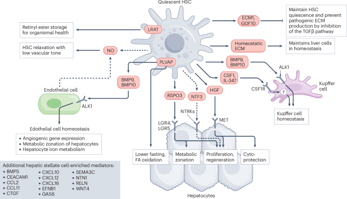
Sugimoto, A. et al. Hepatic stellate cells control liver zonation, size and functions via R-spondin 3. Nature 640, 752–761 (2025).
Article
CAS
PubMed
PubMed Central
Google Scholar
Bier, E. & De Robertis, E. M. EMBRYO DEVELOPMENT. BMP gradients: a paradigm for morphogen-mediated developmental patterning. Science 348, aaa5838 (2015).
Article
PubMed
Google Scholar
Gilmour, D., Rembold, M. & Leptin, M. From morphogen to morphogenesis and back. Nature 541, 311–320 (2017).
Article
CAS
PubMed
Google Scholar
Shilo, B. Z. & Barkai, N. Buffering global variability of morphogen gradients. Dev. Cell 40, 429–438 (2017).
Article
CAS
PubMed
Google Scholar
Simsek, M. F. & Ozbudak, E. M. Patterning principles of morphogen gradients. Open Biol. 12, 220224 (2022).
Article
CAS
PubMed
PubMed Central
Google Scholar
Brosch, M. et al. Epigenomic map of human liver reveals principles of zonated morphogenic and metabolic control. Nat. Commun. 9, 4150 (2018).
Article
PubMed
PubMed Central
Google Scholar
Gebhardt, R. & Hovhannisyan, A. Organ patterning in the adult stage: the role of Wnt/β-catenin signaling in liver zonation and beyond. Dev. Dyn. 239, 45–55 (2010).
Article
CAS
PubMed
Google Scholar
Martini, T., Naef, F. & Tchorz, J. S. Spatiotemporal metabolic liver zonation and consequences on pathophysiology. Annu. Rev. Pathol. 18, 439–466 (2023).
Article
CAS
PubMed
Google Scholar


Comments (0)