In this pilot study, we investigated the potential of CUDI for primary RCC diagnostics by implementing dedicated preprocessing steps including spatial resolution regularization, motion correction and SVD filtering, optimizing the CUDI algorithms and validating the results with pixel-based classification. The preliminary results demonstrate that the spatiotemporal similarity analysis outperforms the TIC fitting analysis in differentiating tumor and parenchyma regions, encouraging us to extend the dataset with a large number of reliable DCE-US acquisitions.
Compared to our previous experience on DCE-US acquisitions in the prostate, the DCE-US acquisitions in the kidney are more complex and heterogeneous, possibly because the image quality of the kidney can be affected by fat thickness, kidney size, tumor location and motion of surrounding organs. This increases the challenge for performing quantitative analysis. Limiting motion is beneficial for accurate CUDI analysis. The kidney was scanned transabdominally, which is more prone to motion artifacts as compared to the transrectal access employed for prostate imaging.
In clinical routine, clinicians prefer to use enhancement patterns of kidney DCE-US acquisitions to give a preliminary diagnosis, by describing the wash-in and wash-out of UCAs in the kidney [29]. These could be reflected by the parameters obtained from TIC fitting analysis, such as AT and FWHM, as well as time-to-peak and peak intensity as mentioned in [30]. However, the complexity and heterogeneity of kidney DCE-US acquisitions may hamper a robust and accurate analysis when only the temporal evolution of individual TICs is considered. This further emphasizes the importance of spatiotemporal analysis of neighboring TICs.
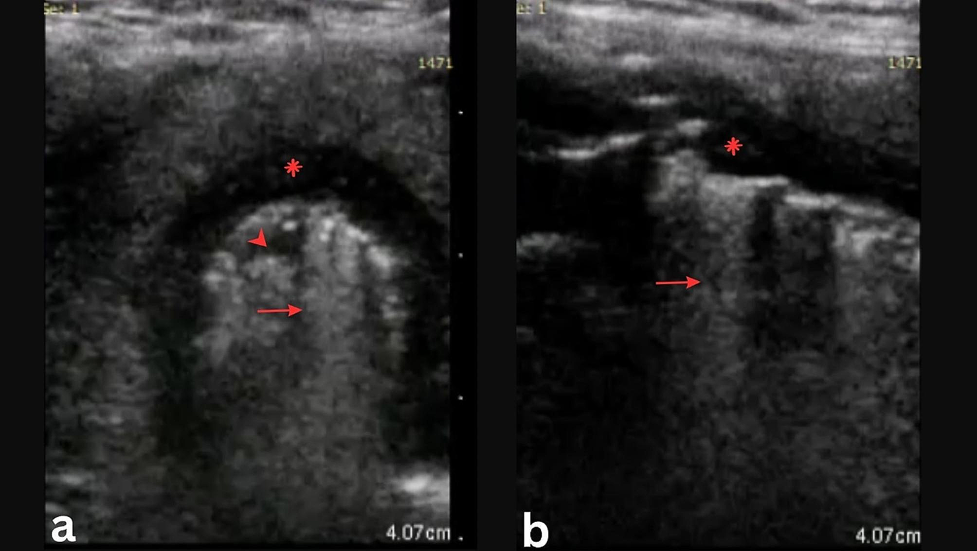
In our five acquisitions, the histological pRCC subtype can be easily differentiated from the ccRCC subtype due to a lack of enhancement in pRCC, hampering the feasibility of our CUDI analysis. The poor blood perfusion characteristics of the pRCC subtype are reflected into hypo-enhanced DCE-US imaging, as also reported in [16, 31]. For ccRCC, a heterogeneous hyperenhancement appears generally in the tumor regions, enabling the CUDI analysis. The obtained parametric maps demonstrate the difference between tumor and parenchyma regions, especially the higher spatiotemporal similarity values in the tumor regions, which is in line with our results on prostate cancer [19, 21, 22]. Tumor-driven angiogenesis is characterized by increased microvascular density and higher tortuosity [8, 32], which limits the dispersion of UCAs in the local measurement region. By modelling the UCA transport kinetics as a convective-dispersion process, lower dispersion is associated with higher similarity between neighbouring TICs [19, 21, 22]. Thus, spatiotemporal similarity analysis provides an indirect indicator of local dispersion, enabling tumor region detection. However, the (micro)vascular architecture in the kidney is complex, consisting of visible large vessels and dense microvessels. The TICs extracted from the large vessel regions show a high recirculation peak intensity and faster appearance time, which may influence the TIC fitting quality and the accuracy of extracted parameters such as AT and wash-in rate. The fitting quality directly influences the number of valid pixels for TIC analysis, which may further affect the classification performance. On the contrary, the spatiotemporal similarity analysis focuses on the shape similarity between neighboring TICs in a local region (kernel) without requiring TIC fitting, which can alleviate the impact of the presence of the high recirculation peak in individual TICs. Moreover, motion affects neighbouring TICs in a similar manner, especially in a local region; spatiotemporal similarity analysis is thus less affected by motion artefacts. This may explain why the spatiotemporal similarity analysis outperforms the TIC fitting analysis. In case 3, high similarity values also appear in the delineated parenchyma region, resulting in poor classification performance. It is hard to explain the reason based on the available ultrasound and CT images; therefore, histopathological results are necessary to shed some light into this peculiar case. Although the CUDI results obtained in this study can be interpreted by the underlying physiology of tumor-driven angiogenesis and the physics of the convective-dispersion process, the limited dataset constrains the generalizability of these findings in the context of RCC diagnosis. This limitation stems from three primary factors. First, the heterogeneity of RCC subtypes must be considered in CUDI analysis. Our dataset, being relatively small, only encompassed ccRCC and pRCC subtypes, leaving the applicability of CUDI to other subtypes unexplored. While ccRCC appears suitable for CUDI analysis, further validation of the diagnostic significance remains necessary based on histopathological reference and expanded ccRCC datasets, particularly given the unexplained poor classification performance observed in certain individual cases. Second, the UCA perfusion process in the kidney involves multiple phases, such as renal cortical enhancement and the final whole-kidney perfusion. This pilot study did not account for the potential impact of these multiphasic perfusion patterns on CUDI analysis, which may represent a significant limitation in diagnostic performance. Third, the complex microvascular architecture of the kidney, comprising both macroscopically visible vessels and dense microvascular networks, yields TICs with varying shapes. Future investigations with larger datasets should explore how optimization of the CUDI analysis for different vessel sizes could enhance both its accuracy and generalizability in the diagnosis of RCC.
In this pilot study, the patients were under anesthesia when scanning, which is challenging for clinical routine, especially for point-of-care ultrasound. Therefore, the potential of CUDI on routine kidney DCE-US acquisitions should also be investigated and proper measures should be taken to compensate for respiration motion. The recent development of 3D ultrasound imaging techniques can be beneficial for the mitigation of errors due to out-of-plane motion. Indeed, allowing for a more complete visualization of the kidney boundaries, 3D ultrasound imaging can lead to improved registration and compensation of motion artifacts due to respiration and free-hand scanning. Moreover, 3D imaging can provide comprehensive information on the hemodynamics of the whole kidney, describing more accurately the intrinsic behavior of blood flow and UCA perfusion in the kidney; therefore, we can directly model the 3D behavior of UCA transport through the kidney as a convective-dispersion process, which may allow us to extract more imaging markers, such as velocity vectors, dispersion, and vector-derived UCA transport tractography [33, 34]. In addition to hemodynamic parameters, ultrasound localization microscopy based on 2D and 3D DCE-US has recently been proposed to achieve resolutions beyond the diffraction limit in microvascular imaging, by detecting sparsely-distributed UCA microbubbles, tracking the centroids of their point spread functions over subsequent frames to reconstruct the microvascular networks where the microbubbles flow through. Several metrics related to the structure of the microvasculature can be extracted from ultrasound localization microscopy, such as vessel diameter, vessel density, vessel tortuosity quantified by distance metric, and fractal dimensions revealing the network complexity [35,36,37,38]. These may assess the tumor-associated angiogenesis in the kidney. However, the limited temporal resolution of most ultrasound scanners used in clinical routine as well as the motion artifacts during the scanning still hampers the implementation of ultrasound localization microscopy with regular clinical acquisitions in patients. Based on a set of quantitative parameters, our previous work also confirms that multiparametric ultrasound imaging achieved by training a machine-learning model to combine complementary parameters outperforms individual CUDI parameters for prostate cancer diagnosis whether using 2D or 3D imaging [39,40,41]. Hence, it is worth investigating in future studies the performance of multiparametric ultrasound imaging of the kidney, especially using histopathological results as the ground truth for tumor detection and subtype classification. While CE-CT is currently recommended for RCC diagnosis, the aforementioned advancements in ultrasound imaging and analysis techniques, combined with the inherent advantages of ultrasound imaging, including portability, high spatial resolution, real-time imaging capability, radiation-free operation in both static and dynamic imaging, along with its cost-effectiveness, establish a solid foundation for the clinical translation of CUDI as a point-of-care diagnostic tool for kidney cancer. Specifically, the portability of ultrasound systems, coupled with their cost-effectiveness, significantly reduces barriers to point-of-care diagnosis, making it accessible in diverse clinical settings. The high spatial resolution and real-time imaging capabilities enable visualization and hemodynamic analysis of tumor vascularity and perfusion patterns, providing both structural and functional information that complement tissue characterization. Furthermore, the radiation-free nature of ultrasound eliminates concerns about radiation exposure during dynamic acquisitions, a limitation inherent to CE-CT. In general, these advance the field of ultrasound-based cancer diagnostics.


Comments (0)