Experimental animals
M. nipponense prawns from a monophyletic sibling were used in this study, as described by Jiang et al. (Jiang et al. 2023).
Cloning of the full-length MniDmrt1B complementary DNA (cDNA)
Total RNA was extracted from the testis of adult M. nipponense by following the manufacturer’s instructions of TRIzol (Sangon Biotech, China). RNA quality was assessed by electrophoresis on a 1.0% agarose gel, and RNA concentration was measured using NanoDrop 2000 (Thermo Scientific, USA). The rapid amplification of cDNA ends (RACE) method was employed to clone the full-length cDNA of MniDmrt1B. The primers MniDmrt1B-F and MniDmrt1B-R designed to clone the intermediate fragment. These primers were based on a transcript annotated as Dmrt1B from the M. nipponense transcriptome. The transcript was obtained by analyzing the transcriptome libraries of the ovary of female prawns and compared with those of the testis of male prawns. The PrimeScript™ RT Reagent Kit with gDNA Eraser (Takara Bio Inc.), 3′-Full RACE Core Set with PrimeScript™ RTase, and SMARTer®RACE 5′/3′ Kit (Takara Bio Inc., Japan) were utilized for 3′- and 5′-RACE cloning. The specific primers were designed using Primer 5 software (Table 1).
Table 1 Sequences of primers usedThe PCR product was purified using the MiniBEST Agarose Gel DNA Extraction Kit Ver 4.0 (Takara Bio Inc., Japan). Subsequently, the extracted DNA was subcloned into a pMD19-T vector (Takara Bio Inc., Japan) and transformed into competent cells of Escherichia coli DH5 α for sequencing (Sangon Biotech, China).
Bioinformatics analysis
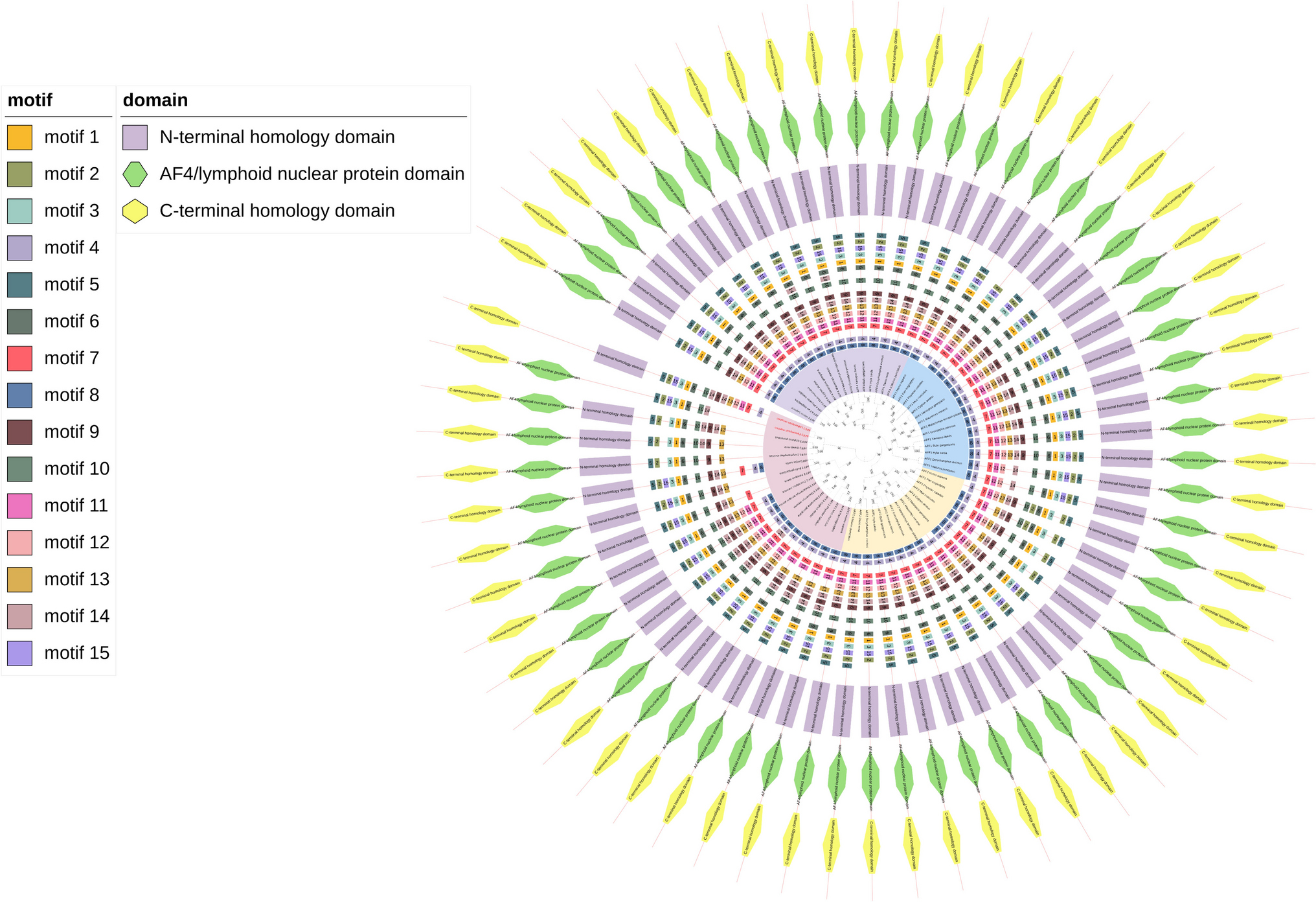
The ORF Finder program (https://www.ncbi.nlm.nih.gov/orffinder/) and BLASTX and BLASTN programs (http://www.ncbi.nlm.nih.gov/BLAST/) were used to deduce amino acid sequences (nonredundant protein sequence database was used for the BLAST database with E-value cutoffs of 0.05). The domains were predicted using InterProScan (http://www.ebi.ac.uk/Tools/pfa/iprscan/). Molecular weight and isoelectric point (pI) were predicted by using ProtParam (http://www.expasy.org/tools/protparam.html). The spatial structure was predicted by I-TASSER (https://zhanglab.ccmb.med.umich.edu/I-TASSER/). Amino acid sequences of different species were compared by performing multiple sequence alignment with DNAMAN 8.0. Multisequence alignment was conducted using ClustalW program with default parameters (Thompson et al. 1997). Phylogenetic trees were constructed using the neighbor-joining method (500 bootstraps) with MEGA7.0 software. All positions containing gaps and missing data were eliminated from the dataset (Tamura et al. 2007).
Tissue and temporal expression analysis by quantitative real-time PCR (qRT-PCR)
The mRNA expression levels of MniDmrt1B were measured in various tissues of adult nine males (body weight, BW 1.42 ± 0.05 g) and nine females (BW 0.53 ± 0.12 g) of M. nipponense by qPCR analysis. Prawns were collected and anesthetized on ice for 5 min before being sacrificed by dissection. The tissues tested, including the second pereiopod, hepatopancreas, eyestalk, muscle, ovary, and testis, were sampled by the sharp tweezers and scissors, respectively, and immediately immersed in liquid nitrogen. To ensure sufficient amounts of RNA samples, the same tissues from three prawns were pooled to form one biological replicate, and three replicates were sequenced for both males and females at each tissue.
The newly hatched zoeae from berried females were allocated into two 50-L polyethylene tanks for culturing. Subsequently, 10 healthy larvae were randomly collected at 20, 25, 30, and 35 days post-hatching (DPH), respectively. To avoid compromising larval survival, the second pereiopod of each prawn was dissected quickly and immediately frozen in liquid nitrogen and stored at − 80 ℃ until total RNA extraction. Each larva with a dissected second pereiopod was labeled and cultured separately in a 5-L tank until its gender could be identified. During the culturing period, enough Artemia was fed, and Ceratophyllum demersum L was provided as a shelter for M. nipponense. The water temperature was maintained at 30 ± 0.5 ℃, pH levels were between 7.5 and 8.0, and dissolved oxygen levels remained above 5 mg/L. The method for sex identification followed the description by New et al. (New et al. 2009).
The RNA samples were extracted using TRIzol reagent (Sangon Biotech, Shanghai, China), and the cDNA was synthesized using a PrimeScript™ RT Reagent Kit with gDNA Eraser (Takara Bio Inc.). The Agilent AriaMx Real-Time PCR System (Agilent) was used to carry out the SYBR Green RT-qPCR assay. The experimental procedures and analysis methods were well-described in our previous study (Jiang et al. 2023). The primers used for qPCR analysis are listed in Table 1. β-Actin was used as a reference gene, and the relative copy numbers of genes were calculated using the 2−∆∆CT comparative CT method (Livak and Schmittgen 2001).
Effects of MniDmrt1B on sex differentiation of M. nipponense
Larvae of different developmental time points (15, 20, 25, 30, and 35 DPH) were selected. Five larvae were pooled to create one biological replication. Three biological replicates were collected for each sample. The samples were treated in the same way as in the above description, and then, qRT-PCR was used to detect the expression levels.
Effects of temperature on the mRNA expression of MniDmrt1B
Jiang et al. have revealed that a lower temperature could induce masculinization in M. nipponense (Jiang et al. 2024). To investigate the impact of temperature on the mRNA expression of MniDmrt1B, the zoeae hatched from berried females were allocated into two 10-L polyethylene tanks and cultured at low (26 ± 0.5 ℃) and high (31 ± 0.5 ℃) temperatures, respectively. The size of the breeding tanks was gradually enlarged as the larvae grew, and the culture period lasted approximately 60 days. After the culturing period, the males (BW 0.45 ± 0.12 g) were sampled from the two temperature treatments respectively, and then, the testes were dissected. To ensure sufficient total RNA content, the testes from three male prawns were combined to form one biological replicate. Three biological replicates were collected for each sample. Following the same procedures as described above for sample preparation and cultivation, qPCR was used to measure the expression levels in the samples.
Statistical analysis
Data from qPCR were analyzed using the SPSS statistical software (version 25.0). Data were tested by one-way ANOVA and Duncan’s new multiple range test and shown as mean ± standard error.


Comments (0)