
Remember me


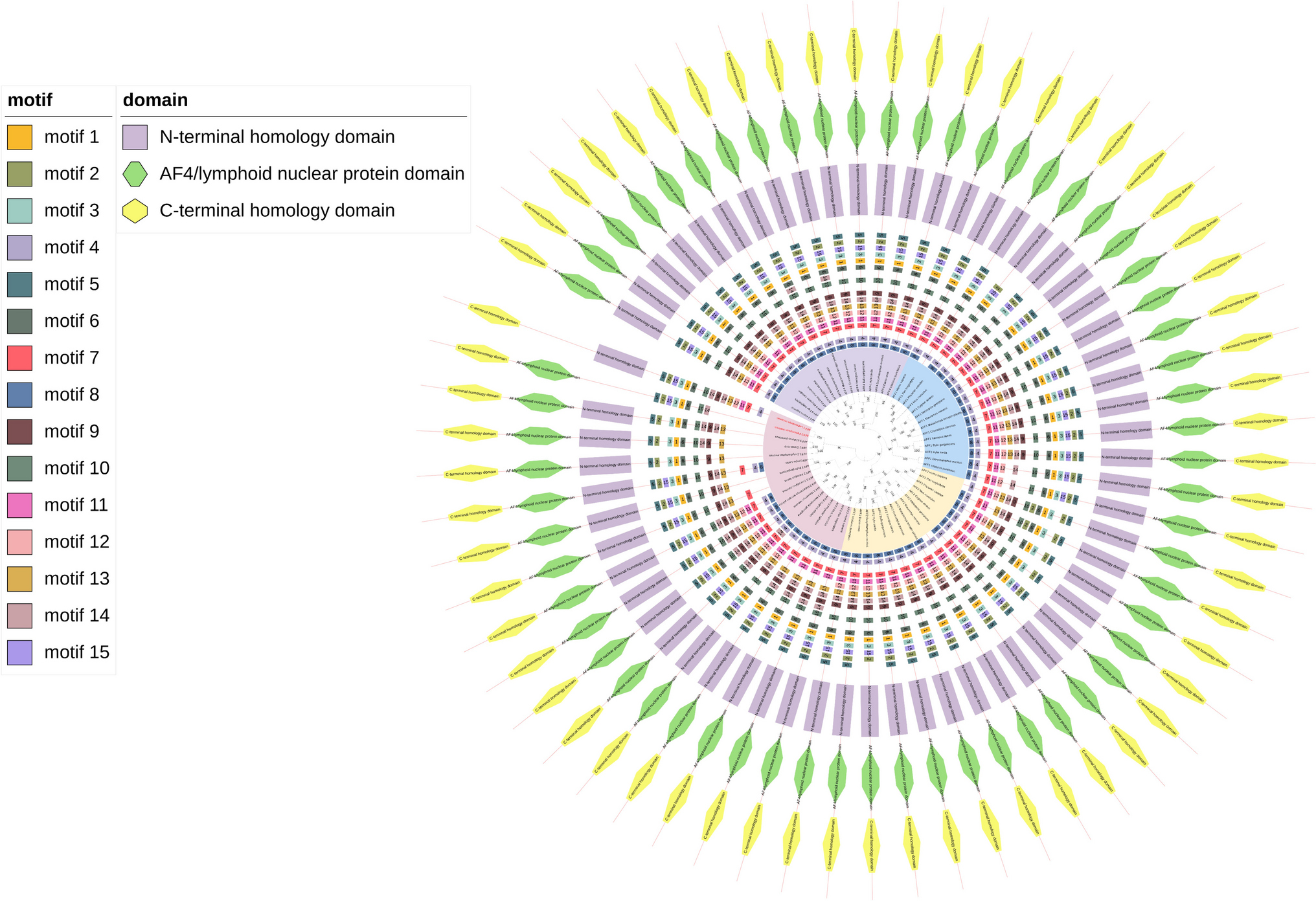
We have made a phylogenetic reconstruction of β-catenin sequences from a comprehensive sampling of higher-order flatworm taxa to clarify the interrelationships of these sequences, and to infer likely homology between sequences. Our search revealed a single homolog of β-catenin in catenulids and two homologs in rhabditophorans (Figs. 1, 2). Within Rhabditophora, Macrostomum lignano has three homologs and planarians four homologs of β-catenin. Rhabditophoran flatworm β-catenin sequences formed four distinct clades, sister to the single sequence found in the catenulids Stenostomum sthenum and Stenostomum leucops (Fig. 1). Macrostomum lignano homologs clustered with already described planarian β-catenin homologs. Using established nomenclature set in planarian flatworms [12,13,14], we named Macrostomum lignano homologs as Mlig-β-catenin1 (Mlig-βcat1), Mlig-β-catenin2a (Mlig-βcat2a), and Mlig-β-catenin2b (Mlig-βcat2b). Mlig-β-catenin1 clustered together with Smed-β-catenin1 and other planarian β-catenin1 sequences in a well-supported β-catenin1 clade. Mlig-β-catenin2a and Mlig-β-catenin2b were recovered as sister groups in a β-catenin2 clade, including all planarian β-catenin2 sequences. All represented planarians possess β-catenin3 and β-catenin4 sequences in highly supported clades, apart from Polycelis nigra, where both sequences appeared within the β-catenin3 clade.
Fig. 1
The consensus β-catenin maximum-likelihood phylogenetic tree illustrates the evolutionary relationships among 20 flatworm and 5 non-flatworm species, with Nematostella vectensis designated as the outgroup. Sequences are colour-coded based on the homolog they represent
Fig. 2
β-Catenin protein distribution across the flatworm species tree, drawn after Egger et al. [23] and Grosbusch et al. [52]
In summary, we have identified a single β-catenin homolog in catenulids, mostly two homologs in rhabditophorans, and two more homologs (β-catenin3 and β-catenin4) exlusive to triclads.
3.2 Macrostomum lignano has three distinct β-catenin homologsWe performed comparative analyses of the amino acid composition of the three M. lignano β-catenins against human and planarian β-catenin sequences to determine whether functional domains and residues for Wnt signal transduction and interaction with structural component are conserved. Full protein lengths of β-catenin homologs identified in M. lignano are 835 aa (Mlig-β-catenin1), 746 aa (Mlig-β-catenin2a), and 767 aa (Mlig-β-catenin2b). The three β-catenin proteins show evolutionary conservation of protein interaction domains compared to human β-catenin (Fig. 3a, b). More than 25% of the amino acid positions are identical and 40% are similar (Fig. 3c).
Fig. 3
Multiple sequence alignment of M. lignano homologs representing GSK3β, α-catenin and PDZ Domains. a Scheme of human β-catenin depicting functional domains and amino acid residues conserved in M. lignano homologs. b Domains from full alignment. b1 GSK3β-CK1α domain, arrow heads depict GSK3β-CK1α phosphorylation residues, note green arrowhead shows additional residue in Mlig-β-catenin1(Mlig_βcat1). b2 First and second α-catenin binding domains, identical amino acid positions in Mlig_βcat2a and Mlig_βcat2b when compared to human β-catenin are highlighted. b3 PDZ domain not conserved in M. lignano homologs. c Full length identity and similarity analysis of M. lignano homologs and S. mediterranea β-cat1 and β-cat2 against H. sapiens β-catenin. d Illustration of functional domains conserved in M. lignano β-catenin. Aligned species, prefix abbreviations: Hs: H. sapiens; Ce: C. elegans; Smed: S. mediterranea and Mlig: M. lignano
Only Mlig-β-catenin1 contains the GSK3β-ck1α domain at the N-terminal, which is not conserved in Mlig-β-catenin2a and Mlig-β-catenin2b (Fig. 3b1, d). Subsequent analysis of the GSK3β-CK1α domain revealed all residues necessary for concerted phosphorylation by GSK3 and CK1 kinases are conserved. But, unlike human β-catenin, Mlig-β-catenin1 has an insertion of an amino acid residue at the carboxyl end of the GSK3β recognition site (Fig. 3b1). From the alignment, we were able to identify the two α-catenin domains located downstream of the GSK3β-CK1α binding domain and corresponding to position 89–101 and 123–133 in human β-catenin (Fig. 3b2). The two α-catenin binding domains are conserved in Mlig-β-catenin2a and Mlig-β-catenin2b, but not in Mlig-β-catenin1. When compared with human β-catenin, Mlig-β-catenin2a and Mlig-β-catenin2b conserved more than 60% identical amino acid residues in the first and second α-catenin binding domain (Fig. 3b2). Crystal structure and functional studies have shown that proteins involved in signalling and cell adhesion competitively bind to Armadillo repeat (ARM) of β-catenin. In our analysis, we identified the 12 ARM repeats found in human β-catenin [42] being conserved in M. lignano β-catenins (Fig. 3d, Fig. S1). Within ARM repeats Axin, APC, E-cadherin, and Tcf compete for the four residues Phe-253, Phe-293, Lys-312, and Lys-435 of β-catenin [8, 43]. The three β-catenin homologs in M. lignano conserved Lys-312 and Lys-435. Mlig-β-catenin1 and Mlig-β-catenin2a, but not Mlig-β-catenin2b conserved Phe-253 and Phe-293 (Fig. S1). We also found that Leu-156 and Leu-159 residues, known to be the binding site for the co-activator BCL9 [44], were conserved in the three Macrostomum homologs. Furthermore, we assessed residues known to have distinct functions; for instance, phosphorylation of Tyr-142 has been associated with reducing the binding affinity of α-catenin to β-catenin [8]. This residue is also conserved in both, Mlig-β-catenin2a and Mlig-β-catenin2b, but not in Mlig-β-catenin1. Similarly, phosphorylation of Try-654 by Src kinase dislodges E-cadherin from binding to β-catenin [45]. This amino acid position is also conserved in Mlig-β-catenin1 and Mlig-β-catenin2a but substituted with Threonine in Mlig-β-catenin2b (Fig. S1).
Although Mlig-β-catenin2a and Mlig-β-catenin2b are more similar to each other than to Mlig-β-catenin1, there is a difference in the fourth ARM repeat, where Mlig-β-catenin2b has insertions of 34 amino acids at position (252–286) and 23 amino acids in the ninth ARM repeat at position (504–526), which are missing in Mlig-β-catenin2a (Fig. S1). Whether these insertions influence the interaction with the binding partners remains unknown.
We could not identify the PDZ domain at the carboxyl end in any of the three M. lignano β-catenin homologs (Fig. 3b3). In summary, M. lignano β-catenin1 domains indicate that it is potentially regulated by Wnt signalling, while Mlig-β-catenin2a and Mlig-β-catenin2b may provide structural support.
3.3 Knockdown analysis at the transcript levelWe performed RT-qPCR analysis to determine the knockdown efficiency of Mlig-β-catenin homologs. In the first approach, gene expression was absolutely quantified, revealing a downregulation of Mlig-β-catenin1 by 75% in RNAi-treated groups in comparison to the control (Fig. S7g). We then repeated qPCR analysis for all three Mlig-β-catenin genes using relative quantification. All worms were amputated through the gnoads and treated for total of 9 days (Fig. 4a). Here, the knockdown efficiency was 86% for Mlig-β-catenin1, 83% for Mlig-β-catenin2a and 65% for Mlig-β-catenin2b (Fig. 4b). These results indicate that all three genes were strongly downregulated.
Fig. 4
a Scheme of knockdown approaches used in most RNAi treatments. b Mlig-β-catenin1, Mlig-β-catenin2a and Mlig-β-catenin2b RNAi knockdown efficiency quantified with qPCR employing the amputation level through the gonads. Bars represent the mean (± SD) (n = 3)
3.4 Knockdown of Mlig-β-catenin cannot rescue a head in anterior regenerates of M. lignanoWe set to investigate whether knockdown of any M. lignano β-catenin homolog is able to rescue head regeneration in anterior regenerates, as seen in some planarians with β-catenin1 knockdowns [17, 18]. Knocking down Mlig-β-catenin1, we tested 9 and 14 days of RNAi treatment, and all worms were amputated through the gonads (Fig. 5b, c). No phenotype differences in worms treated for a total of 9 or 14 days were observed. All the anterior regenerates closed the wound several hours after amputation, but could not form a blastema, completely failed to regenerate any anterior structure including the head and were not different from controls (Fig. 5d, e, Table 1—2.1–2.3).
Fig. 5
Single, double and triple knockdowns of Mlig-β-catenin1 (Mlig-βcat1), Mlig-β-catenin2a (Mlig-βcat2a) and Mlig-β-catenin2b (Mlig-βcat2b) were unable to rescue a head in anterior regenerates. a Experimental setup. b, c Amputation through the gonads. d Luciferase used as control. e–g Single knockdowns. h–j Double knockdowns and k triple knockdowns. Anterior end with the wound is at the top. The scale bars correspond to 100 µm
We then examined whether Mlig-β-catenin1 RNAi-treated worms may regenerate anterior structures if more tissue was left after amputation. We therefore tested two additional amputation levels: amputation just behind the pharynx, and amputation just behind the eyes (Fig. S2b, c). In the two amputation levels, all the anterior regenerates closed the wound, but no anterior organs were regenerated (Table 1—3.0–4.1).
Additionally, we evaluated single knockdowns of Mlig-β-catenin2a and Mlig-β-catenin2b, double knockdowns (Mlig-βcat1 + 2a, Mlig-βcat1 + 2b, Mlig-βcat2a + 2b) and triple knockdowns (Mlig-βcat1 + 2a + 2b) in worms amputated through the gonads and treated for a total 9 days (Fig. 5a–c). In these knockdowns, all the worms closed the wound and were not different from Mlig-β-catenin1 RNAi or controls (Fig. 5f–k).
Altogether, these results demonstrate that knockdown of Mlig-β-catenin1, Mlig-β-catenin2a and Mlig-β-catenin2b individually or in combination cannot induce a head at any level along the A-P axis in M. lignano.
3.5 Effects of β-catenin knockdowns on posterior regenerates3.5.1 Single knockdown of Mlig-β-catenin1 blocked tail regenerationTo study the regeneration response of posterior regenerates following knockdown of Mlig-β-catenin1, we implemented multiple knockdown approaches in worms amputated through the gonads (Fig. 6b, c). Initially, worms were treated with 15 ng/µl dsRNA (Table 1—1.1). In this group, no visible phenotypic changes were observed during treatment. Next, we increased the dsRNA concentration to 18 ng/µl dsRNA (Table 1—1.2). At 1 day post-amputation (dpa), all Mlig-β-catenin1 RNAi-treated worms (n = 24) closed the wound. At 4 dpa, 14 worms had regenerated a tail, while 10 worms failed to form a blastema and did not regenerate a tail (Fig. S3c, d). In contrast, at 4 dpa, all controls (n = 16) had regenerated a tail with posterior structures, including adhesive organs becoming visible (Fig. S3a, b). By 7 dpa, the worms treated with 18 ng/µl Mlig-β-catenin1 dsRNA that had regenerated a tail at 4 dpa were now fully regenerated, while the remaining 10 worms did not form a blastema and did not regenerate a tail (Fig. S3g, h). At this time point, untreated controls were fully regenerated and attaching to the surface (Fig. S3e, f).
Fig. 6
Knockdown of Mlig-βcatenin1, Mlig-β-catenin2a and Mlig-β-catenin2b. During treatment, Mlig-β-catenin1 blocked tail regeneration in posterior regenerates. a–c Experimental set up and amputation level through gonads. d–f Control (Luciferase). g–i Mlig-β-catenin1 at 2 dpa, 4 dpa and 8 dpa no tail regeneration was observed whereas in Mlig-β-catenin2a (j–l) and Mlig-β-catenin2b (m–o) a newly regenerated tail was visible 4 dpa and at 8 dpa like control animals. The scale bars correspond to 200 µm
In the next set of experiments, we further increased the concentration to 120 ng/µl dsRNA (Table 1—1.3). In this group, all (n = 46) Mlig-β-catenin1 RNAi-treated worms did not form a blastema and did not regenerate any posterior structure as shown in (Fig. S3c, d, g, h). Worms were also observed post-treatment in the two treatment conditions (Table 1—1.2–1.3). 7 days after stopping the RNAi treatment, all the worms which had failed to regenerate during treatment with Mlig-β-catenin1 dsRNA (18 ng/µl and 120 ng/µl) began to regenerate a tail and 7 days later (14 days without RNAi), the tails were fully restored, but with instances of heteromorphoses (see below).
Next, we reduced the RNAi incubation time from a total of 14 days to a total of 9 days (Fig. 6a, Fig. S6a) and changed the RNAi measurement. We observed that all Mlig-β-catenin1 treated worms inhibited tail regeneration (Table 1—1.4); however, when the experiment was repeated (Table 1—1.5), 9 of the Mlig-β-catenin1 treated worms regenerated a tail while the knockdown was still ongoing. In the same treatment, 6 individuals did not regenerate a tail during knockdown.
In all four Mlig-β-catenin1 treatment conditions (Table 1—1.2–1.5), a total of 86 individuals failed completely to regenerate any posterior structures during treatment, while 27 worms regenerated the tail. However, after treatment, all 86 worms regenerated a tail, with 50 (59%) worms regenerating misplaced tails, 11 (13%) individuals with multiple tails, while 25 (28%) had normal tails. The individuals with multiple tails survived up to 94 dpa with no change in phenotype (Fig. 8a–c).
Three additional RNAi experiments (Table 1—1.6–1.8) were performed to test whether the misplaced tail regeneration observed above resulted from amputation. We implemented a 9 days RNAi treatment approach (Fig. 6a), and only worms with unruffled wounds after amputation were selected. Posterior regenerates in all three biological repeats closed their amputation wound within several hours; since the wound was still fragile, we did not take pictures. At 1 dpa, we observed that 4 out of 20 worms (Table 1—1.7) had formed unexpected phenotypes not observed in controls (Fig. S6b), which included the emergence of finger-like appendages and loss of eyes (Fig. S6c). These worms were separated, and at 2 dpa (Fig. S6d), the heteromorphic phenotype was still present, but all the worms died by 6 dpa.
We continued treating the remaining worms (n = 16), and the tail inhibition phenotype was consistent across all three treatment conditions (Table 1—1.6–1.8), similar to the previous experiments. At 2 dpa, no blastema was visible in Mlig-β-catenin1 treated worms (Figs. 6g, 7d), and by 4 dpa, no posterior organs such as adhesive glands or the stylet (male copulatory organ) had formed (Fig. 7e). At 8 dpa (4 days with and 4 days without RNAi), the Mlig-β-catenin1 treated worms were still unable to regenerate a tail or any of the posterior structures (Figs. 6i, 7f). The first stylet, an indicator of complete tail regeneration, was observed 23 dpa (19 days without RNAi) in treatment in (Table 1—1.8). 90% of all the posterior regenerates required up to 35 dpa to regenerate a tail plate. As before, we observed 5 individuals (8%) out of the 61 remaining worms regenerated misplaced tails, while 56 individuals regenerated normal tails; none regenerated multiple tails (Fig. 9).
Fig. 7
Detail of the tail plate of after 2, 4 and 8 dpa of untreated controls and knockdown animals. At 2 dpa, a blastema is visible in controls a and knockdowns of Mlig-β-catenin2a+b g, j, while no blastema has formed in Mlig-β-catenin1 knockdowns d. At 4 dpa, controls b and knockdowns of Mlig-β-catenin2a+b h, k have regenerated a tail plate, while in Mlig-β-catenin1 knockdowns, still no blastema has formed e. At 8 dpa, controls c and knockdowns of Mlig-β-catenin2a+b i, I show a stylet (arrowheads), while in Mlig-β-catenin1 knockdowns, still no blastema has formed f. Anterior is to the left. The scale bars correspond to 50 μm
In summary, single knockdown of Mlig-β-catenin1 consistently blocked tail regeneration during and some time after knockdown. In several cases, heteromorphoses such as multiple or skewed tails were observed after knockdown.
3.5.2 Single knockdown of Mlig-β-catenin2a and Mlig-β-catenin2b showed no phenotypeNext, we examined whether posterior regeneration was affected by Mlig-β-catenin2a and Mlig-β-catenin2b RNAi. We analysed worms amputated through the gonads and treated for a total of 9 days (Fig. 6a–c). Posterior regenerates in controls, as well as in Mlig-β-catenin2a and Mlig-β-catenin2b RNAi-treated worms closed the wound several hours after amputation, and at 2 dpa, a regenerative blastema was already visible (Figs. 6d, j, m, 7a, g, j). At 4 dpa all worms had regenerated a tail, but with an incomplete stylet (Figs. 6e, k, n, 7b, h, k). At 8 dpa (4 dpa with and 4 dpa without dsRNA), the tail was fully restored (Fig. 6f, l, o), and a fully formed stylet could be observed in all treatment conditions (Fig. 7c, i, l). These findings indicate that knockdown of Mlig-β-catenin2a and Mlig-β-catenin2b does not affect posterior regeneration.
3.5.3 Double knockdownAs Mlig-β-catenin2a and Mlig-β-catenin2b conserved similar amino acid residues, we performed double knockdown experiments to examine whether they may compensate for each other. The worms were treated for a total of 10 days. In our knockdown, we did not observe any effect on posterior regenerates (Fig. S4a–f).
To test whether other pairs of β-catenin are able to compensate for or to inhibit each other, we performed double knockdowns of Mlig-β-catenin1 together with Mlig-β-catenin2a, and of Mlig-β-catenin1 together with Mlig-β-catenin2b. Posterior regenerates in all knockdown conditions closed their wounds at 2 dpa (Fig. S5a, d, g). At 4 dpa, luciferase treated controls had regenerated a tail (with incomplete stylet) and were fully regenerated by 8 dpa (4 days with and 4 days without treatment) (Fig. S5b-c). Compared to controls at 4 dpa, the double knockdown of Mlig-β-catenin1 together with Mlig-β-catenin2a or Mlig-β-catenin2b (Fig. S5e, h) resulted in a slight delay in tail regeneration. A newly formed tail was visible 8 dpa (Fig. S5f, i) and only fully regenerated by 10 dpa.
3.5.4 Triple knockdownWe then performed triple knockdowns (Mlig-β-catenin1 + Mlig-β-catenin2a + Mlig-β-catenin2b) to test for interactions between the three homologs in the context of posterior regeneration. The worms were treated for a total of 10 days and amputated through the gonads. The wound was closed by 2 dpa (Fig. S5j) and at 4 dpa, a newly regenerated tail was visible in the triple knockdown regenerates (Fig. S5k) and at 8 dpa (4 days without treatment), all worms were fully regenerated and posterior organs such as stylet was visible (Fig. S5l).
3.6 Cell proliferation in Mlig-β-catenin1 knockdownsTo determine whether Mlig-β-catenin1 knockdown influences cell proliferation at the wound site, we examined the incorporation of the S-phase marker BrdU in knockdown and control posterior regenerates of worms amputated at the level of the female genital opening (Fig. 10b). Two days after amputation, much fewer BrdU-labelled nuclei were observed in the entire body of Mlig-β-catenin1 knockdowns from eye to testes level (Fig. 10d) compared to controls (Fig. 10c): 136 vs 231 BrdU-positive nuclei, respectively. When comparing the posterior-most parts of the regenerates, we found that Mlig-β-catenin1 knockdowns had no blastema and only moderate proliferation in the posterior-most body part, while controls showed high proliferation within the blastema (Fig. 10e, f): 49 ± 15 vs 155 ± 26 BrdU-positive nuclei, respectively (n = 3).
3.7 Expression of Mlig-β-catenin1 in controls and regenerating M. lignanoThe expression pattern of Mlig-β-catenin1 was examined in homeostatic worms (intact) and regenerating worms using whole mount in situ hybridisation (WISH). Mlig-β-catenin1 expression in homeostatic worms was ubiquitous, with the exception of the epidermis and tail plate. The signal was strong in the testes, ovaries, and the developing eggs (Fig. S7a). In posterior regenerates, amputated through the gonads, the overall expression remained similar to the intact worms but with a slight increase at the posterior end at 1 dpa (Fig. S7b) compared to the tail plate of intact worms (Fig. S7a). Conversely, the anterior regenerates closed the wound and the expression remained unchanged 1 dpa (Fig. S7c). Probe specificity was confirmed using sense probes, which weakly labelled the testes in homeostatic and regenerating controls (Fig. S7d-f).
3.8 Mlig-β-catenin1 knockdown in intact animalsWe then examined whether silencing Mlig-β-catenin1 can induce changes in the anterior/posterior (A/P) identity of intact Macrostomum lignano, as demonstrated in planarians [12, 14]. Knockdown of Mlig-β-catenin1 in intact worms was performed continuously for 19 days. In our knockdowns, we did not observe any morphological defects compared to controls.
Comments (0)