2.1. Cells culture
The breast cancer human cell line Hs578t (American Type Culture Collection, ATCC, Virginia, United States) and the breast cancer-associated fibroblast (CAF) were cultivated in Dulbecco’s modified Eagle’s medium (DMEM, 41,966,029, Gibco, Thermo Fisher, Massachusetts, United States) containing 10% fetal bovine serum (FBS) and 1% penicillin (100 U/mL) and streptomycin (100 µg/mL) at 37 °C in 5% CO2 atmosphere. CAF cells were kindly donated by Prof. Charlotte Kuperwasser (Tufts University School of Medicine, Boston, US). Cell lines were tested routinely for mycoplasma contamination using MycoAlertTM (LT07-118, Lonza).
2.2. 2D and 3D cell culture models
Spheroids of Hs578t and CAF were generated using 10,000 cells/well, which were seeded out in ultra-low attachment round bottom 96-wells plates (Corning, Merck, Germany) and cultured for 3 to 4 days. Parallel sets of cell monolayers were prepared alongside using an equal number of cells seeded in adherent flat bottom 96-wells plates.
2.3. Immunofluorescence
Spheroids or monolayers were washed with phosphate-buffered saline (PBS), fixed with 4% paraformaldehyde for 15 min, permeabilized with 0.3% Triton X-100 (Art11869, Merck, Germany) for 15 min. Afterward, cultures were stained with 50 µg/mL Phalloidin fluorescein isothiocyanate labeled (P5282, Merck) for 45 min and Hoechst 33,258 diluted 1:1000. All solutions were diluted in PBS. Imaging was done using an ImageXpress Micro confocal microscope (Molecular Devices, USA) and processing by FIJI [12] or Imaris for 3D reconstruction (Oxford Instruments, UK).
2.4. qPCR
RNA samples from monolayers and spheroids were made using TRIsure (BIO-38033, GC Biotech, UK) following the manufacturer’s specifications. cDNA was synthesized using 1 µg RNA and Invitrogen SuperScript II Reverse Transcriptase (Thermo Fisher Scientific, USA). And qPCR was performed using primers (Table 1) and SYBR Select Master Mix (Thermo Fisher Scientific, USA) on a CFX384 Touch Real-Time PCR detection system (Bio-Rad, USA).
Table 1 Primer sequences used for qPCR2.5. EVs quantification
Cells were washed two times with PBS and pre-adapted to serum-free DMEM for 1 or 2 h. The density of EVs released into the media was measured by evaluating the phosphatidylserine (PS) concentration using the Zymuphen MP-activity kit (#521,096, BioMed, Neuville-sur-Oise, France) according to the manufacturer’s instructions.
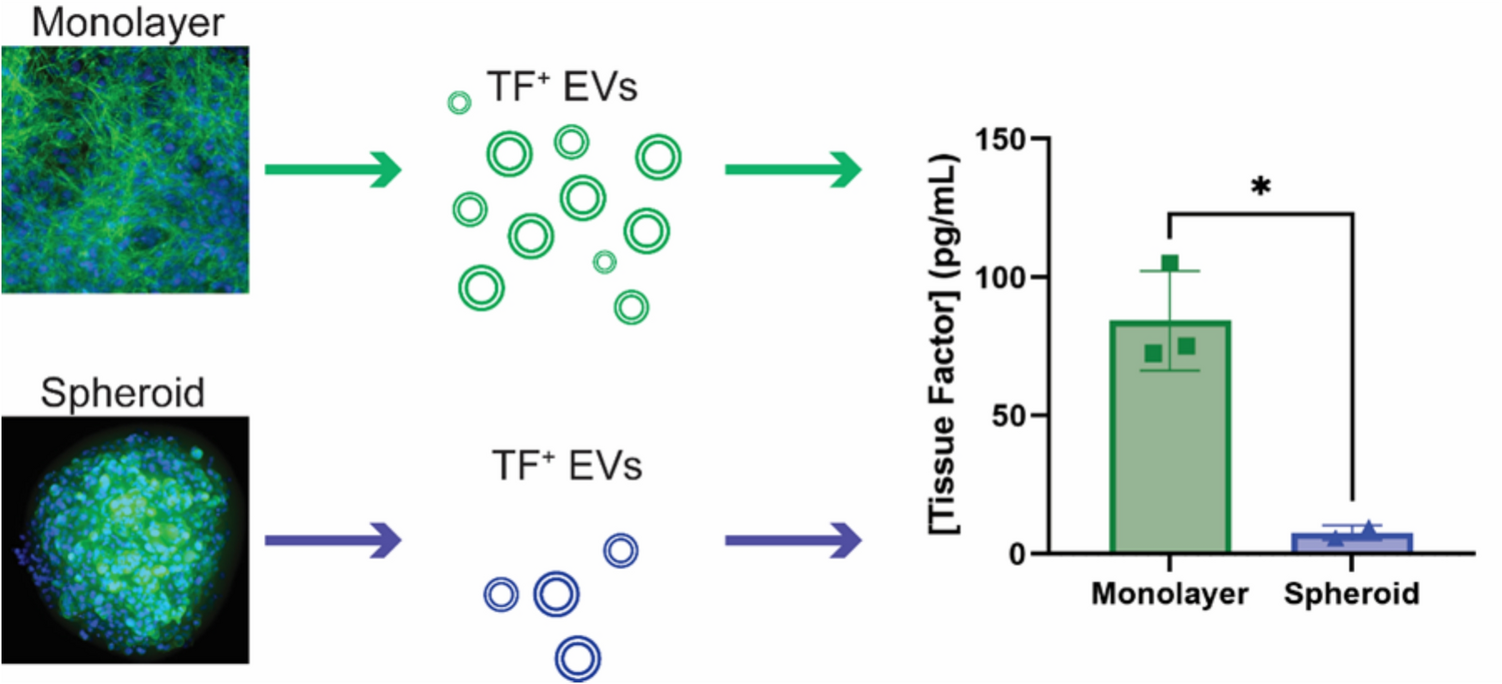
2.6. TF-ELISA (enzyme-linked immunosorbent assay)
Cells were washed with PBS and serum-free DMEM was added. After 2 h of incubation, cell culture supernatants were collected and centrifugated to remove cell debris at 1,200 g for 5 min. The concentration of the released TF antigen was analyzed using the Quantikine human TF-ELISA (DCF300, R&D systems, Minneapolis, United States) kit according to the manufacturer’s instructions.
2.7. Western blot
Cells were seeded in monolayers or spheroids and after 4 days in culture, cells were washed with PBS and 100 µL of 2X Sample Buffer Tris–Glycine SDS (Novex, Thermo Fisher) was added to the wells. Samples were sonicated 10 s at 10 A, heated at 95 °C for 5 min and quantified using nanodrop (Protein A280). 20 µg of samples were loaded in Bolt 4–12% Bis–Tris Plus Gels (Invitrogen) and SeeBlue Plus2 Pre-stained Protein Standard (Thermo Fisher) was used for size evaluation. The proteins were transferred to a PVDF membrane and blocked for one hour at room temperature with 5% milk in tris-buffered saline tween (TBS-T). The membranes were probed with primary antibody: tissue factor (RabMab95, kindly donated by Dr. Vladimir Y. Bogdanov, University of Cincinnati), PAR2 (D61D5, Cell Signaling Technology) and anti-GAPDH rabbit polyclonal (D16H11, Cell Signaling Technology), and incubated overnight at 4 °C. After four washes with TBS-T, HRP-conjugated secondary antibodies (Abcam) were incubated for 1 h at room temperature. The membranes were developed using Western Lightning Plus-ECL (PerkinElmer) and visualized with the ChemiDoc Touch Imaging System (Bio-Rad).
2.8. EVs purification and nanoparticle tracking analysis
Cells were washed two times with PBS and serum-free DMEM was added for 2 h. Cell culture medium was collected and centrifuged at 1,000 g for 10 min to remove cell debris. Then, the supernatant was centrifuged 2 times at 20,000 g for 1 h each using 26.3 mL polycarbonate bottles (Beckman Coulter, Woerden, The Netherlands) on a 50.2 Ti rotor using an Optima XE-90 ultracentrifuge (Beckman Coulter). For the second centrifugation, PBS was added to the pellet. The EVs samples were diluted in filtered PBS and measured using a Nanosight NS300 microscope (Malvern, UK). The concentration and size of particles were determined by the analysis of three movies per sample. The temperature was between 19 and 22 °C; the camera was sCMOS. Viscosity was between 0.988 and 0.990 cP, and camera levels were between 13 and 15. The syringe pump speed was 50 AU. EVs that were identified in the PBS were found from the other EVs samples.
2.9. Procoagulant activity using clotting assay
A clotting assay was performed to assess the procoagulant activity of EVs using platelet-poor plasma, as previously described [13, 14]. 50 µL of plasma was incubated for 1 min at 37 °C in a coagulometer equipment (STart, Stago, FR). Then, 0.1 ug of EVs from monolayers and spheroids were added and the assay was initiated by adding 100 µL of 6.25 mM CaCl2.
2.10. Statistical analysis
All experiments were repeated at least three separate experiments with three replicates per group. Results were analyzed by ANOVA and unpaired Student’s t-test using GraphPad Prism 10.2.3 (GraphPad Software, CA, USA). Asterisks indicate a significant difference compared to the control: p < 0.05 (*), p < 0.01 (**), p < 0.001 (***) and p < 0.0001 (****).


Comments (0)