Plant material
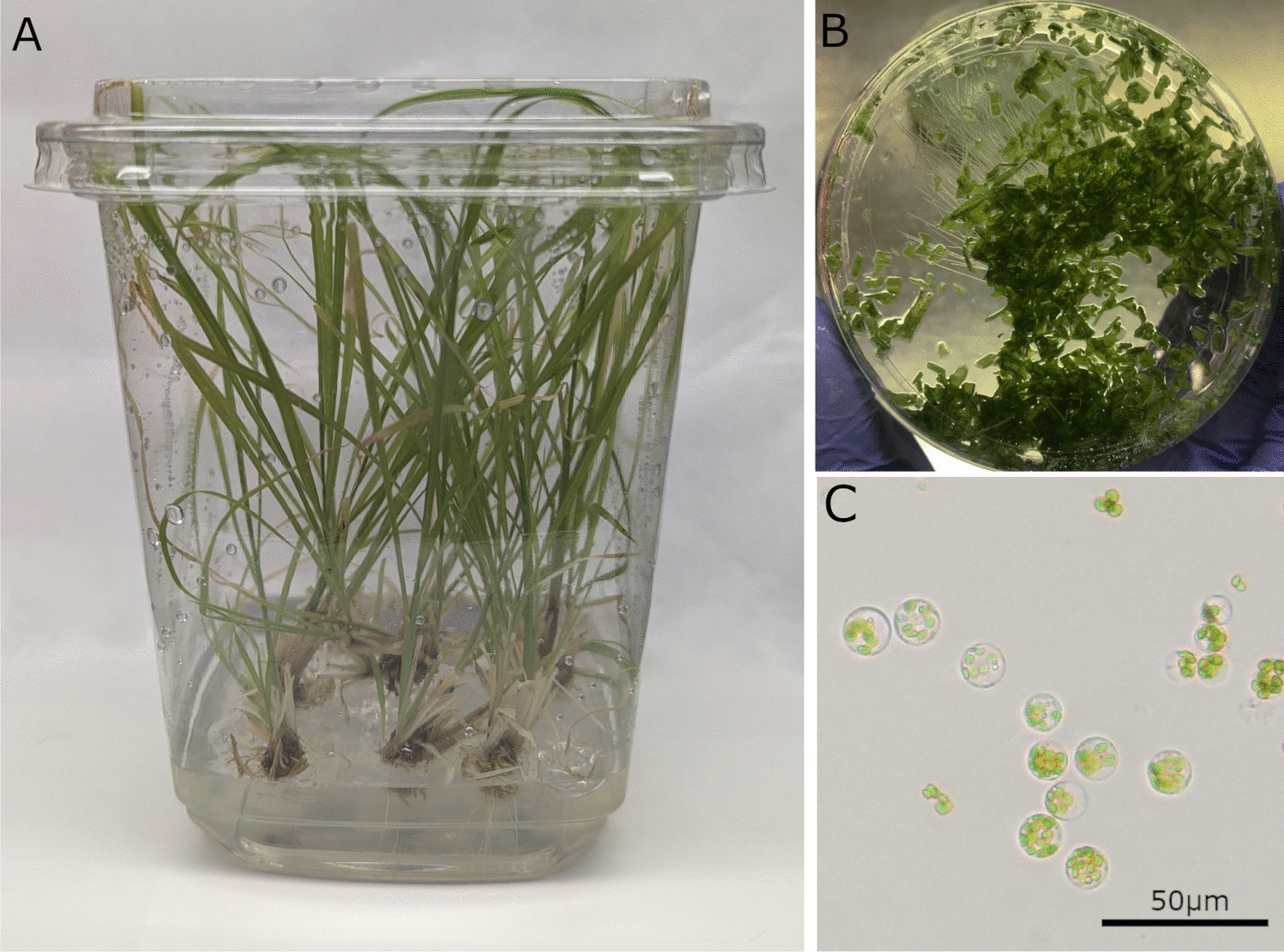
Tissue culture leaf material was derived from the Rhodes grass cv. Tolgar clonal line Tolgar 108 (T108), which was established in a previous work (Maybery-Reupert et al. 2024). Shoots were maintained and multiplied in 947-mL tubs containing approx. 75 mL Murashige-Skoog (MS) medium in light growth conditions (16-h light photoperiod, photon flux density of 80 µmol.m−2 s−1 at 24°C), with 1–2-wk-old leaves from these plants used as tissue sources for protoplast isolation.
Protoplast isolation and quantification
All protoplast isolation and transformation steps were conducted under aseptic conditions. Six combinations of conditions were initially assessed for overnight digestion (Table I). For each set of conditions, 0.5 g fresh weight leaf tissue from 1–2-wk-old tissue culture plants was collected and cut into 0.5–1-mm strips in a 90-mm-diameter Petri dish.
Table I. Rhodes grass (Chloris gayana) protoplast isolation conditions and resulting cell yield and viability.
The digestion mixes consisted of 50 mL of a base solution (0.6 M mannitol, 10 mM mannitol, adjusted to pH 5.7 and 0.22-µm filter sterilized) with Cellulase R10 and Macerozyme R10 (both enzymes manufactured by Yakult Pharmaceutical Industry Co., Ltd, Tokyo, Japan) in varying quantities. Enzyme digestion mix 1 (E1) contained 1.5% (w/v) Cellulase R10 and 0.3% (w/v) Macerozyme, and enzyme digest mix 2 (E2) contained 4% (w/v) Cellulase R10 and 0.8% (w/v) Macerozyme. E2 was based on enzyme quantities from the established bermudagrass protoplast isolation protocol (Chen et al. 2023). To dissolve the enzymes, the digestion mix was heated at 50°C for 10 min in a water bath, then cooled to room temperature. The following components were added: 1 mM calcium chloride dihydrate (CaCl2·2H2O), 1% (w/v) bovine serum albumin, 0.025% (v/v) pluronic acid (10%), and 5 mM β-mercaptoethanol; with pH adjusted to 5.7 and sterilized with a 0.22-µm filter. Petri dishes were placed in a vacuum chamber (250–450 psi) for 0 or 30 min (Table I), then sealed and placed in an incubator overnight (approx. 16 h, 24°C, no agitation).
Protoplasts were collected by gentle swirling of the Petri dish, with the digestion mix collected in a sterile 50-mL tube after passing through 100-µm and 40-µm sieves (pluriStrainer, pluriSelect Life Science, Leipzig, Germany). Leaf wash buffer (WB: 0.6 M mannitol, 20 mM KCl, 4 mM MES hydrate; Sigma-Aldrich (St. Louis, Missouri), pH 5.6 and filter sterilized) was added, and the digested leaf material was washed by gentle swirling and agitation of the tissue by gentle mixing through aspiration into a 25-mL pipette (Greiner Bio-One, Kremsmünster, Austria). Protoplasts suspended in wash buffer were then passed through 100-µm and 40-µm sieves and collected in a 50-mL tube. The suspended protoplasts were then transferred to round-bottom 12-mL tubes and centrifuged (70 g for 10 min at 15°C), with subsequent removal of supernatant. Protoplasts were resuspended in 7 mL WB, then gently agitated and centrifuged (70 g for 10 min at 15°C). The supernatant was removed, and the wash was repeated. Following removal of the supernatant, protoplasts were resuspended in 1 mL Qiao’s medium (liquid MS, 3 mM MES hydrate, 0.6 M glucose, 4.5 µM 2,4-D; pH 5.7).
Cells were counted using a Countess II FL automated Cell Counter (Thermo Fisher, Waltham, MA) as per the manufacturer’s instructions. To accurately set the Countess II cell counting protocol for the Rhodes grass cell size, protoplasts were initially, manually counted with a Reichert Bright-Line hemacytometer (0.1-mm depth, Hausser Scientific, Horsham, PA) with an Olympus BX41 microscope (Olympus Life Science, Tokyo, Japan) after staining with Evans blue (Huang et al. 1986). The same sample was then used to set parameters for total cell count and viability assessment in the Countess II, with viability measured as a proportion of fluorescent cells post staining of a 10 µL protoplast sample, with 2.5 µM Sytox Red Dead Cell stain (Thermo Fisher). For cell counting, 10 µL protoplast samples were similarly stained, with two samples measured per isolation. Total cells and dead cells were measured as the average of the two measurements, with cell viability calculated as the total number of cells minus dead cells.
PEG-mediated trasformation
The green fluorescent protein (GFP) reporter vector (pDPI-94) which contains the turbo GFP expression cassette (ZmUbi-p:turboGFP:nos-t) was used for transformation (Supplementary Figure 1).
For transformation, leaf tissue was digested with 10–15 mL of E2 for 16 h in dark conditions, with isolation and cell counting completed as earlier described. The transformation factors PEG concentration, DNA quantity, and transformation time were set at three levels each, with combinations of the factors determined according to Taguchi’s orthogonal array (Table II). According to cell counting results, 1×10⁶ cells were aliquoted into a 12-mL round-bottom tube, along with 10, 20, or 30 µg of plasmid DNA (Table II) and made up to 230 µL with WB. Subsequently, 1 mL of PEG (0.6 M mannitol, 135 mM CaCl2, 30, 40, or 50% (w/v) polyethylene glycol, Table II) was added dropwise with gentle agitation of the tube until components were homogenous. The transformation mix was left at room temperature for 10, 15, or 20 min (Table II), and then 1 mL wash buffer was added with simultaneous gentle agitation to stop the reaction. Following this, 2 mL WB was added and gently mixed as before, and then a further 6 mL WB was similarly added. The nine distinct tubes containing the orthogonal array treatment combinations were centrifuged (70 g for 10 min at 15°C), the supernatant was removed, and the protoplasts were resuspended in 10 mL of WB then centrifuged and washed as described previously. The supernatant was removed, and the cells were resuspended in 1 mL of Qiao’s medium and incubated for 48 h (dark conditions, 24°C). The same nine transformation condition combinations were repeated on three separate days, with each repetition termed a biological replicate.
Table II. Orthogonal (L9) Rhodes grass (Chloris gayana) protoplast transformation factor optimization and transformation efficiency determined by replicate and overall mean values.
Following incubation, total and dead cell counts were determined with the Countess II as previously described. To determine transformation efficiency, the protoplasts were centrifuged (70 g for 10 min at 15°C) and 10 mL concentrated protoplasts were microscopically counted with a hemacytometer as previously described. The number of transformed cells was determined by counting the total number of GFP-expressing cells, and the transformation efficiency was determined as the proportion of GFP-expressing cells of the total viewable in the hemocytometer. Lysed and egg-shaped or otherwise misshapen cells were excluded from the cell count.
Statistical analysis
The data used for statistical analyses were the cell viability percentages (calculated as the total number of cells minus the number of dead cells) and the transformed cell percentages (calculated as the proportion of GFP-expressing cells). Cell viability values were analyzed via a single-factor ANOVA in Excel (Microsoft, Redmond, WA) of both raw and z-transformed data.
Initial analysis of the transformation data calculated the S/N ratio for each treatment. Relevant terms for this analysis are as follows:
Treatment: combination of the three factors evaluated here (e.g., treatment 1 is 10-min transformation time, 30% PEG and 10 µg DNA)
Factor: one experimental variable in a given treatment (e.g., PEG concentration)
Level: amount of a given factor (e.g., transformation time has levels of 10, 15, and 20 min)
The S/N formula used was for experiments where larger values are preferred (here, a higher/larger value for transformation efficiency is preferred), with S/N ratio for each treatment (T1-9) calculated as per (Rashid 2023):
$$\frac=-10\;}_\left[\frac\times _^\frac_^}\right]$$
where n is the number of biological replicants (here, n = 3), i is the experiment number, and x is the response variable (here, transformation efficiency).
Following this, the average S/N for each level of each factor was determined as the sum of response variables for a level of a factor (e.g., the S/N for 30% PEG is the average S/N for treatments 1, 2, and 3). Subsequently, the S/N values for each level of each factor were tabulated, with the range (R) determined as the maximum S/N minus the minimum S/N for a factor (Supplementary Table I).
Transformation efficiency data were also analyzed using an LMM, which was fitted using restricted maximum likelihood (REML), with the individual test being unit of analysis. The LMM used to analyze transformation efficiency data can be written in the following form:
$$y = \mu + DNA + PEG + }}}} + rep + \varepsilon$$
where y is the response variate (transformation efficiency), μ is the overall mean, and DNA, PEG, and Time are the fixed main effects. The rep and ε were random effects, with rep being the effect of biological replicate and ε the random error term. The random effects were assumed to follow normal distribution with zero mean and constant variance. Only the main effects were fitted and assessed based on Wald’s F-tests, because the Taguchi’s orthogonal array design does not allow for testing of the interactions due to interactions being confounded. The P-values less than 0.05 were considered statistically significant. Histograms of residuals and plots of residuals versus fitted values were examined for normality of distribution with constant variance. All LMM statistical analyses were undertaken using Genstat (Genstat release 23, VSN International Ltd., Hemel Hempstead, UK).


Comments (0)