
Remember me
Carbon-based nanostructures (CNs) such as graphene and its derivatives, carbon nanotubes (CNTs), fullerenes, carbon quantum dots, carbon nanohorns and nanodiamonds (NDs), and their hybrids are becoming a matter of increasing importance in the field of neurology, neuro-oncology, and neuropharmacology. This is because of their specific intrinsic physicochemical, biopharmaceutical, and pharmacological properties such as good bioavailability, relatively low toxicity, low weight and large surface area for active agent loading and functionalization with (intra)cellular component targeting ligands, and extremely small size for crossing the blood–brain barrier (BBB) and targeted delivery to the brain. The hydrophobic nature of the CNs offers good membrane permeability. Through chemical modifications (i.e., oxidation) of their surface, CNs with optimal hydrophobic/hydrophilic properties and increased dispersibility can be obtained as preconditions for biocompatibility and low immunogenicity. Also, improved electronic, mechanical, and thermal properties as preconditions for (photo)thermal and photodynamic therapy can be obtained . It has been shown that CNs have an anti-amyloid aggregation activity, and some of them (i.e., carbon nanotubes (CNTs) and graphene) are able to interface with neurons and neuronal circuits and play an important role in the modulation of neurobiological processes, including neuroregeneration, neuronal differentiation, and stimulation of neuronal electrical signalization and brain activity. Thus, they are promising materials for new products regarding tissue engineering and prosthetic neuronal devices . There is also an evidence that CNs manifest antiproliferative, antimigratory, and antiangiogenic activities in gliomas due to their direct killing effect through inducing apoptosis and the capability to inhibit genes involved in oxidative phosphorylation and to downregulate genes essential for cytoskeleton formation . In addition, an effect on the ectopic (acentrosomal) microtubule nucleation was observed, with disassembly of the centrosome and a cytoskeletal reorganization that trigger the generation of ineffective biomechanical forces, which leads to migration defects, and ultimately to spindle-assembly checkpoint blockage and apoptosis . It was also shown that the CNs impair extracellular adhesion and regulate adhesion-dependent pathways (such as EGFR/AKT/mTOR and β-catenin) , inhibit angiogenesis via the NF-κB pathway , inhibit several signal transduction pathways (WNT/β-catenin, Notch, and JAK-STAT) , promote apoptosis via activation of reactive oxygen species (ROS)-, caspase-, and mitochondrion-dependent pathways, such as p53-mPTP , and reduce the expression of voltage-dependent ion channel genes and extracellular receptors in glioma cells, damaging the cell membrane and changing its potential . These findings are supported by several publications in which CNs were evaluated as potential drug carriers or inherent drugs for brain targeting and (synergistic) treatment of Alzheimer disease , Parkinson disease , and brain tumors , and as agents for the detection of brain ischemic stroke and photodynamic and photothermal therapy of brain tumors .
Brain tumors are characterized by very high morbidity and mortality rates; among them, the most frequent primary tumors are malignant gliomas, representing more than 80% of all brain tumors, of which more than 40% are glioblastomas. Glioblastoma multiforme (GBM), grade 4 astrocytoma, is the most aggressive and deadly brain tumor, representing 16% of primary brain cancers and up to 54% of all gliomas. The mean survival time estimate for patients with GBM is 14.6 months, the two-year and five-year survival rates are less than 30% and 5%, respectively, while for the patients with unresectable GBM, the prognosis is worse . The fast proliferation and aggressive invasion of GBM in the surrounding brain tissue complicate the total resection of the tumor. In addition to the heterogeneity of GBM, the immunosuppressive surrounding, high levels and different mechanisms of drug resistance and radiation tolerance, as well as a blocking effect of the blood–brain–tumor barrier (BBTB) for drug permeation into the brain extracellular matrix lead to high recurrence rates (in up to 90% of patients) even when complete treatment is applied. The standard of care includes surgical resection followed by combined chemo- and radiotherapy (RT), together with non-conventional adjuvant treatment with inhibitors targeting one or a family of biomarkers included in signalization or activation of oncogenic pathways, immunotherapy, gene therapy, and photodynamic therapy.
Cytotoxic drugs acting by preventing DNA replication and apoptosis via methylation of guanine-rich areas in DNA and by cross-linking guanine and cytosine in DNA strains are a standard in chemotherapy. Among them, the prodrug temozolomide (TMZ) with its active metabolite 5-(3-methyl-1-triazene-1-yl)imidazole-4-carboxamide) (MITC) is a first-line therapy for primary and recurrent GBM, especially O6-methylguanine-DNA methyltransferase (MGMT)-promotor-methylated tumors. MITC, acting via its metabolite methyl diazonium cation generated at physiological pH (>7), develops O6-methylguanine lesions and/or mutations that ultimatively avoid mismatch repair system and lead to apoptosis, autophagia, and cell senescence. Aside from the methylation in the O6 position of guanine, TMZ has shown an ability to alkylate other macromolecules with an important role in many cell processes related to changes in the structure of RNA, protein chromatin structure, gene expression and replication, and synthesis and repair of DNA . It has also been shown that TMZ has radiosensitizing effects , increasing the degradation of DNA strains and cell death when combined with RT, which is a reason for the National Comprehensive Cancer Network to recommend standard RT and adjuvant TMZ plus alternating electric field therapy for patients with GBM at ages up to 70 years with good performance status and having either a “methylated” or “indeterminate” MGMT promoter status . However, the treatment with TMZ is associated with several disadvantages such as excessive toxicity for the surrounding tissues, short half-life of 1.8 h (which requires frequent administrations for achieving therapeutic concentrations at the site of action and leads to unfavorable safety profile and tolerability), and limited efficacy (due to the high level of DNA repair by MGMT and other mechanisms of resistance) . In addition, the high hydrolytic instability of TMZ at pH ≥ 7 limits its efficacy, because the formed MTIC exhibits high systemic toxicity and cannot pass the BBTB and accumulate in the GBM, unlike TMZ. Therefore, it is of paramount importance to prolong the half-life of TMZ at physiological pH and to increase its accumulation in the tumor cells prior to degradation in the central circulation.
Several strategies have been exploited to stabilize TMZ and overcome the disadvantages. Among them is the incorporation of TMZ in organic and inorganic nanomaterials and their hybrids, designed in a wide variety of shapes such as nanoparticles (NPs), conjugates, dendrimers, and liposomes . With various bioengineering techniques, the nanomaterials’ size, shape, and surface properties were modified to improve formulation, biopharmaceutics/release kinetics, and pharmacokinetics of TMZ. Also, surface functionalization attempts with multiple targeting ligands were made to deliver TMZ to the site of interest, exploiting the site-specific expression or overexpression of specific molecules on BBTB and GBM cells to enable transport and delivery to brain tumors. Inorganic nanostructures as TMZ carriers have shown several advantages compared to organic ones with respect to physicochemical stability and potency/cytotoxic activity, overcoming their main disadvantages, that is, hydrophobicity/fluidity and toxicity by chemical modifications and/or coupling with hydrophilic polymers. TMZ was successfully incorporated in magnetic NPs , mesoporous silica NPs , and NPs made of silver , zinc oxide , and gold , all of them showing high accumulation in tumor cells and cytotoxic activity in vitro and in vivo.
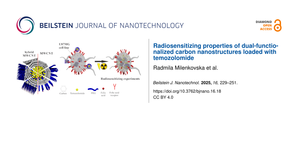
Despite the beneficial properties, CNs have not been extensively researched as inherent drugs or drug/TMZ carriers for the treatment of GBM. In two papers, a hybrid made of carbon quantum dots functionalized with chitosan, polyethylene oxide, and carboxymethyl cellulose–polyvinyl alcohol provided controlled release of TMZ . In another publication , the suitability of graphene oxide (GO) functionalized with folic acid (FA) for controlled release of TMZ and the inhibition of glioma growth was confirmed in vivo. To our knowledge (and stated also in the paper of Petrenko et al. ), our group was the first one that incorporated TMZ in multiwalled CNTs (MWCNTs), and their hybrid with graphene (MWCNT-G), non-covalently functionalized with polyethylene glycol (PEG), and pointed to their suitability for targeted and controlled brain delivery, based on their physicochemical and biopharmaceutical properties . In this paper, TMZ was incorporated in the same carriers, this time covalently dual-functionalized with PEG (average Mw 6000 g/mol) and FA, with an overall aim to prolong the TMZ circulation time (through the stealth effect of PEG) and to increase permeability through the BBTB and accumulation in the brain tumor cells (through the active targeting of the formulations towards membrane folate receptors of GBM). Detailed physicochemical and biopharmaceutical characterizations of the prepared TMZ-loaded dual-functionalized CNs was performed, and the cytotoxicity/radiosensitizing properties of the functionalized CNs with and without TMZ were investigated and compared in vitro, using human glioblastoma cell line exposed to irradiation with a dose rate used in clinical settings for most of the patients with GBM. Simultaneously, the formulations exposed to irradiation under the same conditions were characterized in terms of their physicochemical and biopharmaceutical properties.
Results and Discussion Biopharmaceutical characterization of temozolomide-loaded carbon nanostructures Loading efficacy, drug content, surface charge, and particle size distributionIn the study, relatively high values for TMZ loading efficacy and content were achieved, ranging from 42% to 67% and from 11% to 18%, respectively (Table 1), which are in the scope of values reported for nanocarriers of TMZ (27% to 89% and 4% to 11%, respectively) . The higher values achieved for plain (non-functionalized) formulations relative to the functionalized ones (Table 1) could be explained by the formation of strong covalent bonds between CNs and PEG6000, the competition between PEG6000 and TMZ regarding interactions with MWCNTs-COOH and MWCNTs-G-COOH, and the physical entrapment of TMZ in the tubes. One can assume that TMZ is both physically entrapped in the tubes and wrapped around the CNs because of electrostatic and hydrogen bond interactions with CNs and PEG6000. This assumption is supported by the similar values for loading efficacy and drug content of covalently PEGylated TMZ-loaded MWCNTs and hybrid MWCNTs-G (Table 1), which differ in the fraction of tubes. When analyzing the data for dual-functionalized formulations with TMZ, one can see that FA acts like an additional competitor for drug loading (although interactions between TMZ and FA cannot be excluded), indicated by the lower values for loading efficacy and content in these formulations (around 42% and 11%, respectively, for MWCNTs-PEG6000-FA-TMZ and 46% and 13%, respectively, for MWCNTs-G-PEG6000-FA-TMZ; Table 1).
Table 1: Loading efficacy, drug content, size distribution, and surface charge of TMZ-loaded carbon nanostructures.
Series Parameters loading efficacyaMWCNTs-COOH (oxidized MWCNTs); bMWCNTs-PEG6000 (MWCNTs covalently functionalized with PEG6000); cMWCNTs-PEG6000-FA (MWCNTs dual-functionalized with PEG6000 and FA); dI-MWCNTs-PEG6000-FA (irradiated MWCNTs dual-functionalized with PEG6000 and FA); eMWCNTs-TMZ (temozolomide-loaded MWCNTs); fMWCNTs-PEG6000-TMZ (temozolomide-loaded MWCNTs covalently functionalized with PEG6000); gMWCNTs-PEG6000-FA-TMZ (temozolomide-loaded MWCNTs dual-functionalized with PEG6000 and FA); hI-MWCNTs-PEG6000-FA-TMZ (irradiated temozolomide-loaded MWCNTs dual-functionalized with PEG6000 and FA); iMWCNTs-G-COOH (oxidized MWCNTs/graphene hybrid); jMWCNTs-G-PEG6000 (MWCNTs/graphene hybrid covalently functionalized with PEG6000); kMWCNTs-G-PEG6000-FA (MWCNTs/graphene hybrid dual-functionalized with PEG6000 and FA); lI-MWCNTs-G-PEG6000-FA (irradiated MWCNTs/graphene hybrid dual-functionalized with PEG6000 and FA); mMWCNTs-G-TMZ (temozolomide-loaded MWCNTs/graphene hybrid); nMWCNTs-G-PEG6000-TMZ (temozolomide-loaded MWCNTs/graphene hybrid covalently functionalized with PEG6000); oMWCNTs-G-PEG6000-FA-TMZ (temozolomide-loaded MWCNTs/graphene hybrid dual-functionalized with PEG6000 and FA); pI-MWCNTs-G-PEG6000-FA-TMZ (irradiated temozolomide-loaded MWCNTs/graphene hybrid dual-functionalized with PEG6000 and FA).
When the CNs were PEGylated, an increase in zeta potential was observed (−38.38 and −46.05 mV vs −21.40 and −22.30 mV for MWCNTs-PEG6000 and MWCNTs-G-PEG6000, respectively) (Table 1) due to modifications of the carboxylic groups on the surface of CNs. The additional functionalization with FA decreased the zeta potential (−33.10 and −38.10 mV for MWCNTs-PEG6000-FA and MWCNTs-G-PEG6000-FA, respectively), which can be ascribed to the ionized carboxyl groups of FA in the corresponding medium. Incorporation of TMZ in the CNs did not change their zeta potential significantly, confirming the assumptions regarding the TMZ loading (Table 1).
The mean particle size for the parent CNs (MWCNTs-COOH and MWCNTs-G-COOH) was 136 and 222 nm, respectively (Table 1). The most significant changes in the particle size was observed after covalent PEGylation (increase to 231 and 322 nm, respectively). The larger mean particle size observed for covalently modified CNs vs non-covalently modified ones supports the assumption of a higher content of polymer on the surface of the covalently modified CNs (observed also with thermogravimetric analysis (TGA)). After coupling with FA, an additional increase in the mean particle size was observed, with values of 270 and 305 nm for MWCNTs-PEG6000-FA and MWCNTs-G-PEG6000-FA, respectively. This points to surface attachment of FA, although its physical entrapment into the tubes cannot be excluded. After incorporation of TMZ, the mean particle size was increased by 30–40 nm, suggesting again the embedment of TMZ not only into the tubes, but also on the surface of the CNs (as shown in the SEM images in this study and the SEM and TEM images in ). In all series, a relatively unimodal particle size distribution was observed, with PDI values not higher than 0.541.
In the irradiated series, the mean particle size ranged from 222 nm (I-MWCNTs-PEG6000-FA) to 347 nm (I-MWCNTs-G-PEG6000-FA-TMZ) (Table 1). Generally, the particle size was smaller compared to the size of the corresponding non-irradiated formulations, which can be attributed to the penetration power and destructive effect of the gamma rays and the dissociation of certain molecules from the nanocarriers into the surrounding medium. Such a trend was observed in a study of Jun et al. in which MWCNTs conjugated with chitosan oligomers and with incorporated tea polyphenols for cancer treatment were irradiated by gamma rays from 60Co for 30 min with a dose of 1.5 Gy. The irradiation also led to changes in zeta potential to lower values (i.e., more positive than those of non-irradiated series), ranging from −21.05 mV (I-MWCNTs-PEG6000-FA) to −16.50 mV (I-MWCNTs-G-PEG6000-FA-TMZ) (Table 1).
In general, in the present study, carbon-based TMZ carriers with suitable surface charge for the established aim were prepared, considering that particles with negative (and neutral) zeta potential have a higher capability to escape opsonization and accumulation in liver and spleen (which is a precondition for prolonged circulation time of the particles/TMZ). In addition, particles with suitable size (between 50 and 400 nm) for permeation through the BBTB and uptake into the tumor cells were obtained, as a precondition for higher extent and rate of their internalization and optimal risk/benefit ratio of the treatment with TMZ. This knowledge is based on a lot of publications in which nanoparticulated formulations of different materials designed for brain delivery, including carbon-based, with moderately negative (between −1 and −15 mV) or highly negative zeta potentials (between −15 and −45 mV) and particle sizes between 100 and 400 nm, showed the capability to cross the BBTB and accumulate in the brain tumor cells in vitro and in vivo .
MorphologyIn the SEM images of the MWCNTs-COOH (presented in our previous study ), a dense structure of randomly aggregated, convoluted, and highly tangled tubes was observed. The image of MWCNTs-G shows a hybrid structure of multiwalled nanotubes dispersed within the graphene sheets, formed by interactions between the hydrophobic regions of graphene and the side walls of MWCNTs. Also, in the SEM and TEM images of TMZ-loaded non-modified CNs, entrapment of TMZ into the tubes and wrapping around the CNs was visible . In the present study, the covalent PEGylation and dual functionalization of MWCNTs (Figure 1a–d) was visible by enlarged tubes/thicker walls and non-uniform surfaces of the tubes. The images of PEGylated and dual-functionalized hybrid MWCNTs-G (Figure 1e–h) showed, in addition to the thicker side walls and rounded ends of the tubes, spherical structures attached to the graphene sheets, attributed dominantly to PEG6000. No clear distinction regarding the morphology of single- and dual-functionalized CNs and structures with and without TMZ could be made with the used imaging technique. Also, no morphological differences were observed between the irradiated and non-irradiated CNs.
![[2190-4286-16-18-1]](https://www.beilstein-journals.org/bjnano/content/figures/2190-4286-16-18-1.jpg?scale=2.0&max-width=1024&background=FFFFFF)
Figure 1: SEM images of (a) MWCNTs-PEG6000, (b) MWCNTs-PEG6000-FA, (c) MWCNTs-PEG6000-TMZ, (d) MWCNTs-PEG6000-FA-TMZ, (e) MWCNTs-G-PEG6000, (f) MWCNTs-G-PEG6000-FA, (g) MWCNTs-G-PEG6000-TMZ, and (h) MWCNTs-G-PEG6000-FA-TMZ.
In vitro releaseAll functionalized CNs loaded with TMZ manifested a biphasic release profile, with an initial burst release and a phase of continuous release (observed also in non-covalently PEGylated formulations in our previous study ). In the initial phase of 2 h, 16% and 27% of the loaded TMZ were released from MWCNTs-PEG6000-TMZ and MWCNTs-G-PEG6000-TMZ formulations, respectively (Figure 2a,b), which can be attributed to the release of TMZ located on the surface of the CNs. A lower percentage of TMZ was initially released from the covalently PEGylated CNs compared to the non-covalently PEGylated ones prepared in our previous study (35% and 41%, respectively). This can be explained by the higher content of PEG6000 in the covalently functionalized formulations (confirmed by TGA also) and its barrier role in the TMZ release from the CNs. However, when considering the whole process of drug release, faster release was observed from covalently PEGylated formulations compared to non-covalently PEGylated formulations. Namely, the total content of TMZ loaded in MWCNTs-PEG6000-TMZ and MWCNTs-G-PEG6000-TMZ was released after 72 h (Figure 2a) and 48 h (Figure 2b), respectively, in contrast to non-covalently PEGylated formulations from which the complete release of TMZ occurred over a period of 192 h . The reason is the lower content of TMZ entrapped into the tubes, that is, the higher content of surface-bound TMZ in covalently PEGylated formulations. This can also explain the difference in drug release between the covalently PEGylated and non-PEGylated MWCNTs; over a period of 72 h, around 71% of the drug was released from MWCNTs-TMZ and 99% from MWCNTs-PEG6000-TMZ (Figure 2a). The same trend was observed for the dual-functionalized formulations, MWCNTs-PEG6000-FA-TMZ and MWCNTs-G-PEG6000-FA-TMZ, from which initially similar or slightly lower quantities of TMZ were released (17% and 18%, respectively) compared to the covalently PEGylated (and non-covalently PEGylated) formulations, but from which the total TMZ content was released faster, that is, after 48 h. One can suppose that this release profile can be also attributed to the additional barrier (consisting of PEG6000 and FA) for the entrapment of TMZ into the tubes. When comparing the drug release profiles of the covalently PEGylated MWCNTs and MWCNTs-G formulations, whether modified with FA or not, one can see a faster release from the hybrid structure, which can be again explained by lower TMZ content in the tubes and higher content of surface-bound TMZ in the hybrid structure and the resulting faster diffusion in the dissolution medium. The higher content of surface-bound TMZ in the non-modified hybrid structure could also explain the faster TMZ release from this formulation in comparison with the functionalized ones (Figure 2b).
![[2190-4286-16-18-2]](https://www.beilstein-journals.org/bjnano/content/figures/2190-4286-16-18-2.png?scale=2.0&max-width=1024&background=FFFFFF)
Figure 2: Cumulative release of TMZ from (а) plain, covalently PEGylated, and additionally FA modified MWCNTs, (b) plain, covalently PEGylated, and additionally FA modified MWCNTs-G, and (c) non-irradiated and irradiated MWCNTs and MWCNTs-G covalently PEGylated and modified with FA.
After irradiation, the TMZ release from the dual-functionalized CNs was slightly faster, which can be attributed to the changes in the size of the CNs, faster dissociation of TMZ into the surrounding medium, and structural changes of the CNs caused by irradiation. In the initial 2 h, 30% and 40% of TMZ from I-MWCNTs-PEG6000-FA-TMZ and I-MWCNTs-G-PEG6000-FA-TMZ, respectively, were released, followed by complete release after 24 and 48 h, respectively (Figure 2c).
Carbon-based TMZ carriers with a favorable release profile have been prepared considering the importance of controlled and sustained drug release to prevent fast TMZ degradation, postsurgical sensitization on radiotherapy, less frequent drug administration, and improved risk/benefit ratio in patients with malignant (recurrent) glioma. These findings are supported by several studies in which the controlled release of TMZ was provided by loading in nanoparticulated carriers, with subsequent improved brain uptake, increased potency, and lower systemic toxicity . Controlled release was also provided through loading TMZ in a hybrid compound of carbon quantum dots, chitosan, polyethylene oxide, and carboxymethyl cellulose–polyvinyl alcohol (CS-PEO-CQDs/CMC-PVA) via coaxial spinning, and a transport system of CMC-PVA coating and CS-PEO-CQDs core was formed . In a study of Wang et al. , formulations made of TMZ-loaded and FA-functionalized GO provided pH-dependent and controlled TMZ release, and the favorable release profile was further confirmed in vivo by successful inhibition of glioma growth. In our previous study , in which non-covalent PEGylation of the same TMZ-loaded CNs was performed using PEG1500, PEG4000, and PEG6000, sustained release over extended periods of time was also observed, with no significant difference in the drug release profile between the different PEGylated formulations. Therefore, in the current study, PEG6000 was used for covalent functionalization, based on the assumption that the polymer with the higher molecular weight would provide longer circulation time and, thus, higher uptake ratio in the brain tumor cells.
Physicochemical characterization of temozolomide-loaded carbon nanostructuresFor physicochemical characterization of all formulations regarding interactions of the components and their stability during the preparation procedures, different techniques were used including infrared (IR), ultraviolet–visible (UV–vis), and Raman spectroscopy as well as TGA. For analyzing potential structural changes in the CNs after exposure to irradiation, X-ray powder diffraction (XRPD) was used. The stability of TMZ under these conditions was determined unsing attenuated total reflectance Fourier-transform infrared (ATR-FTIR) and UV–vis spectroscopy. Most of the procedures and techniques were used in our previous study in which physicochemical properties of non-covalently PEGylated CNs loaded with TMZ were characterized.
Ultraviolet-visible absorption spectroscopyUV–vis absorption spectroscopy turned out to be a useful tool for characterizing functionalization with FA and for confirming the TMZ loading through the characteristic absorption peaks of FA (at 280 nm) and TMZ (at 255 and 328 nm, corresponding to the active hydrolytic metabolite MTIC and the prodrug TMZ, respectively) in water solution and the concurrent absence of peaks in water solution of free PEG6000 and unloaded non-PEGylated and PEGylated CNs (Figure 3).
![[2190-4286-16-18-3]](https://www.beilstein-journals.org/bjnano/content/figures/2190-4286-16-18-3.png?scale=2.0&max-width=1024&background=FFFFFF)
Figure 3: UV–vis spectra of (а) TMZ, PEG6000, FA, and blank and TMZ-loaded single- and dual-functionalized MWCNTs; (b) TMZ, PEG6000, FA, and blank and TMZ-loaded single- and dual-functionalized MWCNTs-G; and (c) irradiated blank and TMZ-loaded dual-functionalized CNs.
In the UV–vis spectra of TMZ-loaded covalently PEGylated CNs, the two characteristic peaks of TMZ were present, as well as in the spectra of MWCNTs-PEG6000-FA-TMZ (Figure 3a) and MWCNT-G-PEG6000-FA-TMZ (Figure 3b), in which the characteristic peak of FA was also visible. No difference between the UV–vis spectra of irradiated and non-irradiated formulations was observed, and no changes in the spectra of TMZ released from I-MWCNTs-PEG6000-FA-TMZ and I-MWCNTs-G-PEG6000-FA-TMZ were observed when comparing with the spectra of TMZ released from the corresponding non-irradiated formulations.
Infrared spectroscopyIR spectroscopy was used to analyze the presence of different functional groups in the CNs and interactions formed during single and dual functionalization and TMZ loading. The IR spectra of PEG6000, MWCNTs-COOH, MWCNTs-G-COOH, and TMZ were already characterized in our previous study , and they are also presented in Figure 4.
![[2190-4286-16-18-4]](https://www.beilstein-journals.org/bjnano/content/figures/2190-4286-16-18-4.png?scale=2.0&max-width=1024&background=FFFFFF)
Figure 4: Comparison of IR spectra of individual components and blank and TMZ-loaded (a) PEGylated MWCNTs, (b) dual-functionalized MWCNTs, (c) PEGylated МWCNTs-G, and (d) dual-functionalized MWCNTs-G. (е) Comparison of ATR-FTIR spectra of TMZ and irradiated blank and TMZ-loaded MWCNTs-COOH and dual-functionalized MWCNTs-COOH.
When analyzing the IR spectra of PEG6000, oxidized MWCNTs-COOH, and covalently PEGylated MWCNTs-PEG6000 (Figure 4a), one can see that upon covalent functionalization, the spectrum of MWCNTs-COOH significantly changed. Namely, the bands at 3423 and 3390 cm−1 in the spectrum of MCWNTs-COOH, assigned to O–H stretching vibrations, were significantly more intense in the spectrum of MWCNTs-PEG6000. Bands at 2917 and 2852 cm−1 emerged in the spectrum of MCWNTs-PEG6000, which are attributed to asymmetric and symmetric C–H stretching vibrations of the CH2 groups in PEG6000. The band at 1730 cm−1 (originating from C=O stretching vibrations) in the spectrum of MWCNTs-COOH has a lower intensity in the MWCNTs-PEG6000 spectrum as a result of interaction between MWCNTs-COOH and PEG6000. In addition, a new band evolved in the spectrum of MWCNTs-PEG6000 at 1048 cm−1 (attributed to stretching vibrations of C–O bonds in PEG6000), which disappeared in the spectrum of MWCNTs-COOH. In summary, the changes in the spectrum of MWCNTs-PEG6000 relative to the spectrum of MWCNTs-COOH indicate the successful covalent PEGylation of MWCNTs. The covalent PEGylation was also evident in the spectrum of MWCNTs-G-PEG6000 (Figure 4c) by the bands between 2950 and 2850 cm−1, originating from C–H stretching vibrations of PEG6000, which are absent in the spectrum of MWCNTs-G-COOH. Furthermore, the characteristic band at 1467 cm−1 found in the spectra of both PEG6000 and MWCNTs-G-PEG6000 is assigned to CH2 deformation vibrations. A characteristic band also occurred at 1078 cm−1, which can be assigned to asymmetric C–O–C stretching vibrations in PEG6000. The new band in the spectra of MWCNTs-PEG6000 and MWCNTs-G-PEG6000 at 1730 cm−1, which is shifted from 1703 cm−1 in the spectra of MWCNTs-COOH and MWCNTs-G-COOH (assigned to C=O vibrations from the carboxyl groups of PEG6000) probably implies on ester bond and confirms the covalent PEGylation of the CNs.
Upon FA functionalization, the spectra of MWCNTs-PEG6000-FA (Figure 4b) and MWCNTs-G-PEG6000-FA (Figure 4d) showed a new band at 1679 cm−1, probably from C=O stretching vibrations within the FA molecular structure. The spectrum of FA exhibits a band at 1639 cm−1 from the amide C=O group, which is shifted to 1604 and 1637 cm−1 in the spectra of MWCNTs-PEG6000-FA and MWCNTs-G-PEG6000-FA, respectively. The spectra of MWCNTs-PEG6000-FA and MWCNTs-G-PEG6000-FA exhibit another novel band at 1728 and 1732 cm−1, respectively, resulting probably from C=O stretching vibrations of the ester RCOOR′ group formed between the carboxyl groups of FA and hydroxy groups of PEG6000. The bands at 1423 cm−1 in the spectrum of MWCNTs-PEG6000-FA and at 1428 cm−1 in the spectrum of MWCNTs-G-PEG6000-FA correspond to deformation vibrations of C–H bonds in the aromatic ring of FA, pointing to a successful functionalization of the PEGylated CNs with FA.
The IR spectrum of TMZ (presented also in our previous work ) depicts three wide bands at 3339, 3381, and 3426 cm−1, attributed to the NH2 and OH stretching vibrations, as well as two bands at 2927 and 2855 cm−1, related to asymmetric and symmetric stretching vibrations of aliphatic methylene groups. All these bands are stronger in the spectra of MWCNTs-PEG6000-FA-TMZ (Figure 4b) and MWCNTs-G-PEG6000-FA-TMZ (Figure 4d), but are slightly shifted to 3346, 3389, 3423, 2921, and 2853 cm−1, serving as a spectroscopic evidence for the existence of non-covalent interactions (electrostatic, hydrogen bond, and/or van der Waals forces) between CNs and TMZ.
From comparison of the ATR-FTIR spectrum of TMZ with the spectra of the irradiated formulations I-MWCNTs-COOH, I-MWCNTs-TMZ, I-MWCNTs-PEG6000-FA, and I-MWCNTs-PEG6000-FA-TMZ (Figure 4e,f), it becomes obvious that the specific bands of TMZ appear in the spectra of the drug-loaded formulations. The most evident features are the bands at 1106 and 1046 cm−1, resulting from C–H deformation vibrations in the aromatic ring of TMZ, as well as the band at 699 cm−1, originating either from rocking vibrations of PEG6000 or C–H out-of-plane deformation vibrations in TMZ. The appearance of bands in the spectrum of TMZ and their absence in the spectra of blank CNs indicate that the structure of TMZ was preserved during irradiation. Upon its incorporation in the CNs, in vitro dissolution studies, in which UV–vis spectra of TMZ after its release from the irradiated CNs were recorded, were carried out.
Raman spectroscopyThe Raman spectra of single- and dual-functionalized MWCNTs and MWCNTs-G, blank and TMZ-loaded, are presented in Figure 5. When analyzing these spectra, the following features were taken into consideration: the D (“disorder”) band, usually positioned around 1350 cm−1 and related to the degree of structural defects, deteriorations, and sp3-hybridization of the carbon atoms in the CNTs; the position and intensity of the G band related to the E2g phonon mode of sp2-bonded carbon atoms in the 1600–1500 cm−1 region; the bands from the so-called low-frequency radial breathing mode (below 300 cm−1), sensitive to the diameter and chirality of the CNTs; the bands from the Raman modes that result from the vibration of all carbon atoms in the CNTs; and the 2D band occurring between 2600 and 2800 cm−1, which is sensitive to the number of graphene layers and their arrangement. The intensity ratio between the D and the G band was analyzed because of their significance for identifying the structural defects .
![[2190-4286-16-18-5]](https://www.beilstein-journals.org/bjnano/content/figures/2190-4286-16-18-5.png?scale=2.0&max-width=1024&background=FFFFFF)
Figure 5: Raman spectra of individual components and blank and TMZ-loaded (a) MWCNTs functionalized with PEG, (b) MWCNTs functionalized with PEG and FA, (c) МWCNTs-G functionalized with PEG, and (d) MWCNTs-G functionalized with PEG and FA.
In the Raman spectra of MWCNT–COOH and MWCNTs-G-COOH (Figure 5a and Figure 5c, respectively), the intensity ratios of D and G band (IG/ID) were calculated as 0.59 and 0.65, respectively. The analogous calculation for modified MWCNTs-PEG6000 and MWCNTs-G-PEG6000 gave D/G intensity ratios of 0.64 and 0.75, respectively. The increase in ID/IG ratios of the PEGylated CNs compared to the pristine ones indicates that the functionalization with PEG6000 induced a slight destructive effects on the surface of CNTs and confirmed the successful introduction of functional groups on their surfaces, that is, chemical modification of their outer layers . The spectra of covalently functionalized MWCNTs-G with PEG6000 follow this behavior and are also accompanied by a broadening of the G band. The broadening and its intensity increase is indicative for a breakage of the graphene sheet symmetry, which is associated with the introduction of functionalities onto/into the structure of the MWCNTs-G hybrid.
A decrease in the intensity of all Raman bands was observed upon FA functionalization (Figure 5b,d). In addition, after dual functionalization and TMZ loading, further changes of the ID/IG band ratio were observed. Namely, in the MWCNTs-based formulations, the values of 0.64 and 0.96 for MWCNTs-PEG6000 and MWCNTs-PEG6000-FA, respectively, increased to 1.17 and 0.98 for MWCNTs-PEG6000-TMZ and MWCNTs-PEG6000-FA-TMZ, respectively. These results point to increase and expansion of surface defects and successful functionalization. Functionalization by FA of MWCNTs-G-PEG6000 yielded an increase of the ID/IG ratio from 0.75 to 0.85, while the corresponding lower values determined for MWCNTs-G-PEG-6000-TMZ and MWCNTs-G-PEG6000-FA-TMZ (0.60 and 0.79, respectively) point to a reverse trend during TMZ loading in the hybrid carrier, suggesting the formation of stable complexes between the drug and the carrier.
Thermogravimetric analysisThe TGA findings are presented in Figure 6. As mentioned in our previous study and also seen in the actual study (Figure 6a,b), the weight of pure PEG6000 decreased sharply with increasing temperature, showing almost 100% weight loss at ca. 400 °C. The same trend can be seen in the covalently functionalized MWCNTs-PEG6000 (Figure 6a,c), that is, 37% weight loss was observed at 400 °C (corresponding to the elimination of the functional groups with oxygen, i.e., H2O, CO2, and CO) and an additional 5% weight loss at 800 °C (42% weight loss in total). In the TGA diagram of covalently PEGylated MWCNTs-G (Figure 6b,d), a weight loss of 23% at 400 °C was observed, while at 800 °C, 39% of the weight remained (61% weight loss in total). For comparison, the TGA diagrams of non-covalently PEGylated MWCNTs and MWCNTs-G showed lower weight losses in total (29% and 52%, respectively) , pointing to a higher content of PEG6000 in the covalently PEGylated CNs.
![[2190-4286-16-18-6]](https://www.beilstein-journals.org/bjnano/content/figures/2190-4286-16-18-6.png?scale=2.0&max-width=1024&background=FFFFFF)
Figure 6: TGA diagrams of individual components and blank and TMZ-loaded (a) MWCNTs functionalized with PEG, (b) MWCNTs-G functionalized with PEG, (c) MWCNTs functionalized with PEG and FA, and (d) MWCNTs-G functionalized with PEG and FA.
In the thermogram of FA (Figure 6c,d), one can see a weight loss of 8% at 200 °C and up to 85% at 800 °C. After functionalization with FA, in the thermograms of both dual-functionalized CNs (Figure 6c,d), weight losses at two levels were observed, as in the thermogram of FA. Namely, in the thermograms of MWCNTs-PEG6000-FA (Figure 6c) and MWCNTs-G-PEG6000-FA (Figure 6d), 95% and 97% of the weight, respectively, remained at 200 °C, while at 800 °C, weight losses of 75% and 32% were observed, respectively, due to the decomposition of PEG and FA. After TMZ loading, in the thermograms of all formulations, non-significant or small differences in the weights were observed at 800 °C (Figure 6a–d) compared to the thermograms of corresponding formulations without TMZ (18%, 53%, 13%, and 59% for MWCNTs-PEG6000-TMZ, MWCNTs-PEG6000-FA-TMZ, MWCNTs-G-PEG6000-TMZ, and MWCNTs-G-PEG6000-FA-TMZ, respectively). This points to the formation of stable complexes and the absence of additional defects on the surface of CNs after TMZ loading.
X-ray powder diffractionSeveral literature data are focused on the changes in structure and size of CNs after their exposure to gamma radiation. In one study , upon exposure of MWCNTs to gamma radiation and subsequent oxidation, the content of functional groups increased because of the increase in the effective surface for functionalization (or decrease in size) and the increase in number of defective sites on the MWCNTs created by the gamma photons. In addition, an exposure of SWCNTs to gamma radiation led to a significantly higher extent of functionalization with single-stranded DNA . It was also shown that gamma radiation with low doses, 25 and 50 kGy, improved the graphite arrangement of MWCNTs; higher doses, 100 and 150 kGy, led to major structural deteriorations, while very high doses above 200 kGy distorted the structure . There is also a great possibility that the changes in size and structure of CNs impact their physicochemical and biopharmaceutical properties (including solubility and permeability) .
The changes in the structure of the CNs after exposure to radiation were analyzed using XRPD. In the diffractograms of non-irradiated and gamma ray-irradiated CNs (Figure 7), two peaks important for the graphite arrangement of MWCNTs were recorded at 2θ angles of 26° and 43°. For the irradiated MWCNTs-COOH, a rise of the intensity of the 26° peak was observed pointing to an increase of its crystallinity, as previously reported by Nie and coauthors . Furthermore, an increase of the intensity of the peak at 43° was also observed in addition to an occurrence of two new peaks at 2θ angles of 31° and 45°. The peak at 31° can be assigned to the (002) interlayer spacing in nanotubes, while the other at 45° is related to changes in the crystallinity or arrangements in the nanotubes after irradiation. In the X-ray pattern of MWCNTs-G-COOH, the two characteristic peaks at 2θ angles of 26° and 43° were also present; however, the intensity of the peak at 26° decreased in the irradiated sample (pointing to structural imperfections), while the peak at 43° remained unchanged. No shifts of the peaks were observed in the X-ray patterns of both CNs pointing to non-significant changes in the structure of the CNs.
![[2190-4286-16-18-7]](https://www.beilstein-journals.org/bjnano/content/figures/2190-4286-16-18-7.png?scale=2.0&max-width=1024&background=FFFFFF)
Figure 7: Comparison of XRPD diffractograms of non-irradiated and irradiated (a) MWCNTs-COOH and (b) MWCNTs-G-COOH.
Cytotoxic activity of temozolomide-loaded carbon nanostructuresA number of publications contain information on the potential mechanisms of CNs’ cytotoxicity, pointing to physical destruction, oxidative stress/ROS generation, DNA damage, cell autophagia and lysosomal membrane damage followed by mitochondrial dysfunction, pyroptosis followed by activation of inflammasomes, apoptosis via mitochondrial pathway and scavenger receptors, and necrosis . For example, it has been shown that when single-wall CNTs (SWCNTs) enter into cells, they interact with the actin filaments, rearranging the actin, disrupt the mitochondrial function, and cause abnormalities in cell division; MWCNTs interact with tubulin and actin, causing cell apoptosis, which was confirmed in vitro and in vivo . In addition, genotoxic
Comments (0)