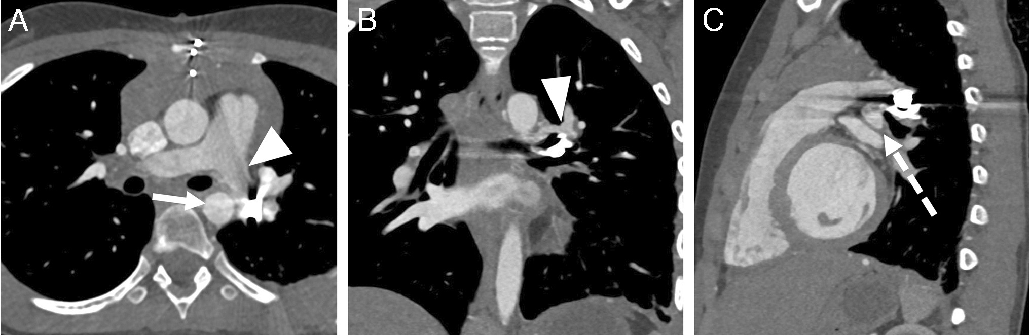
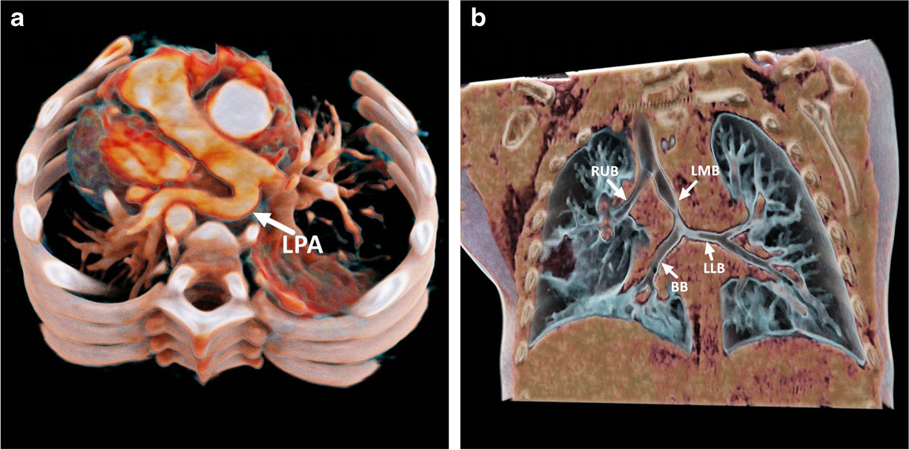
Type IIA pulmonary artery sling with tracheobronchial anomalies
A 9-year-old girl presented with wheezing and fever (temperature, 38.5 °C) after catching a cold 1 month prior. After receiving supportive treatment at a local hospital, her symptoms improved. After 10 days, she was hospitalized again because of severe stridor and tachypnea. Chest radiography revealed bilateral pneumonia. She was subsequently referred to our hospital for better management. Superior-view chest computed tomography (CT) angiography confirmed a pulmonary sling, with the left pulmonary artery crossing leftward between the trachea and esophagus to the left pulmonary hilum to form a vascular sling around the trachea (a). Thus, a tracheobronchial tree anomaly was suspected. Coronal CT revealed that the bronchus of the right upper lobe originated separately from the distal trachea, with mild stenosis in its middle segment. The bridging bronchus and bronchus of the left lower lobe were connected via a narrowed left main bronchus (b). Bilateral pneumonia was also present. BB bridging bronchus, LLB left lower lobe bronchus, LMB left main bronchus, LPA left pulmonary artery, RUB right upper lobe bronchus.


Comments (0)