
Remember me


All reagents were purchased from commercial vendors. All solvents were of American Chemical Society grade or higher. 4-phenylbutanoic anhydride [28] and cis, cis, trans-[Pt(NH3)2Cl2(OH)2] [29] were synthesized as previously described.
Physical measurementsNuclear magnetic resonance (NMR) samples were prepared in DMSO-d6 and analyzed on a 400 MHz Bruker AV 3HD spectrometer with BBFO broadband probe. 1H NMR chemical shifts were referenced internally to tetramethylsilane (δ = 0 ppm). High-resolution electrospray mass spectrometry (HR-ESI-MS) measurements were obtained on an Exactive Orbitrap mass spectrometer in negative ion mode (ThermoFisher Scientific, Waltham, MA). Elemental analysis (CHN) was performed by Atlantic Microlab Inc. (Norcross, GA, USA).
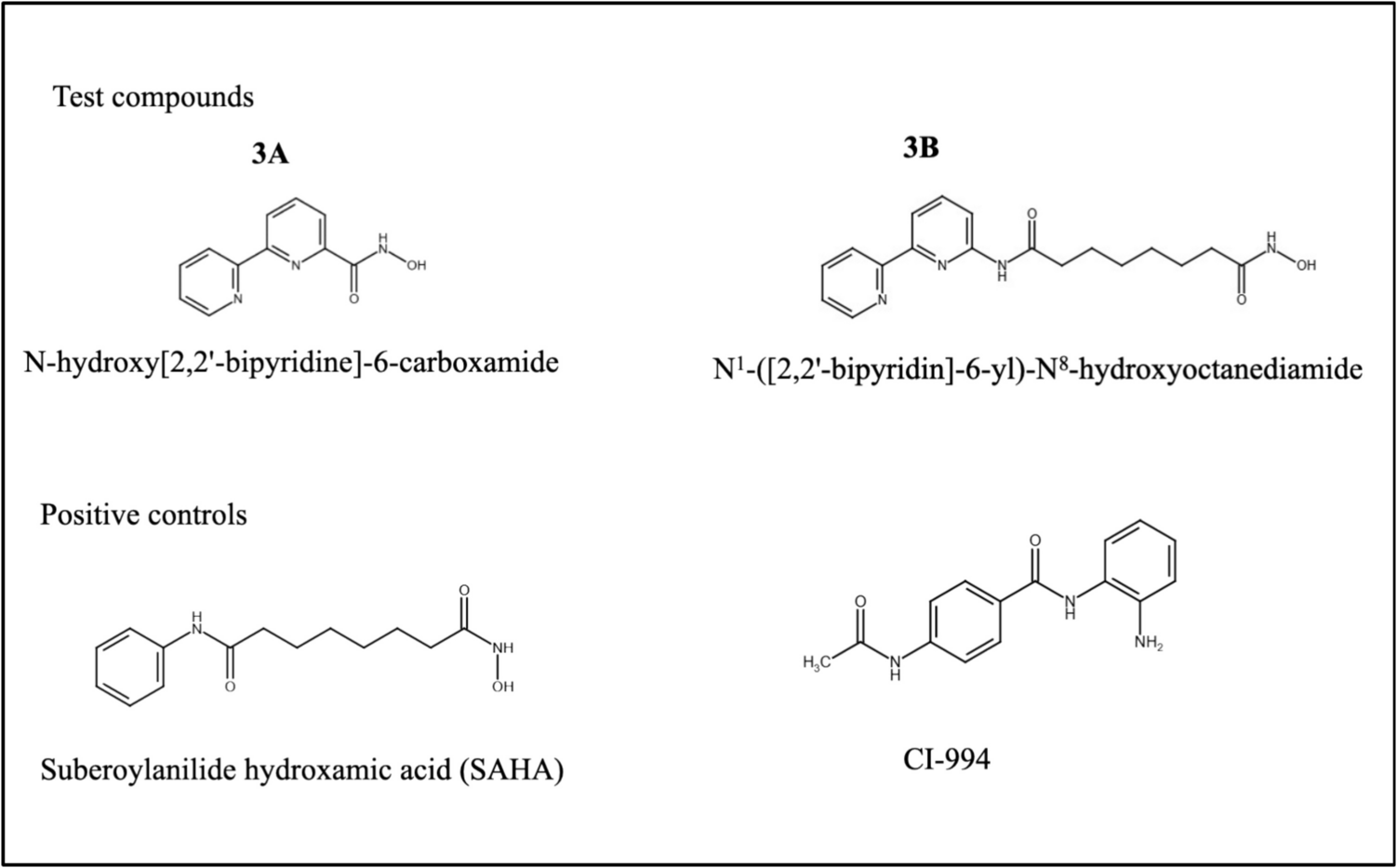
Synthesis of cis, cis, trans-[Pt(NH3)2CI2(PBA)2]Compound B was prepared using modifications to a previously reported procedure (Fig. 1) [30]. To a stirring solution of 4-phenylbutanoic anhydride (244 mg, 0.748 mmol) in dimethyl formamide (DMF) (2 mL) was added cis, cis, trans-[Pt(NH3)2Cl2(OH)2] (100 mg, 0.298 mmol). The mixture was first heated at 40 °C for 2 h and then stirred at room temperature for 22 h. At the end of the stirring, all solids were fully dissolved. The DMF was removed under reduced pressure, and the remaining residue was resuspended in 3 mL of acetone and filtered after sonication. The filtrate was added dropwise to a stirring solution of diethyl ether (30 mL), precipitating the desired product as a white solid, which was isolated by vacuum filtration, washed with diethyl ether (3 × 10 mL), and dried in vacuo. Yield: 100 mg (0.160 mmol, 54%). 1H NMR (400 MHz, DMSO-d6): δ 7.29–7.15 (m, 10 H), 6.53 (br s, 6 H), 2.59 (t, J = 7.6 Hz, 4 H), 2.21 (t, J = 7.4 Hz, 4 H), 1.74 (quint, J = 7.5 Hz, 4 H). ESI-MS (negative ion mode): m/z 624.0998 ([M–H]–, calculated 624.0996). Analytical calculated for C20H28Cl2N2O4Pt: C, 38.35; H, 4.51; N, 4.47. Found: C, 38.62; H, 4.59; N, 4.48.
Fig. 1
Synthesis schema of cis, cis, trans-[Pt(NH3)2Cl2(PBA)2] [Compound B] Addition of 4-phenylbutanoic anhydride reagent to cis, cis, trans-[Pt(NH3)2Cl2(OH)2] results in the substitution of PBA HDAC inhibitor ligands for the hydroxides on cis, cis, trans-[Pt(NH3)2Cl2(OH)2]. See Supplementary Fig. S1 and S2 for NMR and HR-ESI-MS validation
Zebrafish MaintenanceWild-type AB zebrafish (Cat. #: ZL1) were obtained from the Zebrafish International Resource Center (Eugene, OR). wt1b:eGFP zebrafish were obtained from Dr. Iain Drummond (MDI Biological Laboratory, Bar Harbor, ME) and brn3c (pouf4f3):eGFP fish were obtained from the University of Oregon, Eugene, OR. Zebrafish embryos were maintained in petri dishes with E3 water media (5 mM NaCl, 0.17 mM KCl, 0.33 mM CaCl2, 0.33 mM MgSO4, 1 ppm methylene blue (all Sigma Aldrich, St. Louis, MO)) at 28 °C with a 14-hour light and 10-hour dark cycle according to established protocols [31]. All zebrafish experiments were conducted in accordance with the guidelines of the Animal Care and Use Committee of the University of Mississippi Medical Center (UMMC), Jackson, MS, and approved by the UMMC Institutional Biosafety and IACUC Review Committees (IACUC protocol number: 2021 − 1161).
Cell CultureA2780 (cisplatin-sensitive; Cat.#: 93112519) and A2780cis (cisplatin-resistant; Cat.#: 93112517) ovarian cell lines were obtained from Sigma-Aldrich (St. Louis, MO) were cultured in RPMI-1640 media (Sigma) with 2 mM glutamine (Sigma), 10% fetal bovine serum (FBS, Gibco, Gaithersburg, MD) and 1:100 penicillin/streptomycin (Invitrogen, Carlsbad, CA) and incubated at 37 °C. To maintain cisplatin resistance, the A2780cis cell line was also treated with 1 µM cisplatin every 2–3 passages.
Cell viability assayThe 3-(4,5-dimethylthiazol-2-yl)-2,5-diphenyltetrazolium bromide (MTT) assay was performed according to the procedure in [32]. Cells were seeded at 50,000 cells per well in 24-well plates and incubated for 24 h at 37 °C in 5% CO2. Then, wells were treated for 48 h in replicates of three in three separate experiments with a concentration series (500, 50, 5, 0.5, 0.05 µM) of cisplatin in media vehicle or compound B in 0.3% dimethyl sulfoxide (DMSO) vehicle. A negative control (media only), 0.3% DMSO vehicle control, positive control (Triton X-100), and set of blanks (media only with no cells) were also simultaneously performed in replicates of three in three separate experiments. The MTT assay was conducted for 2 h, with plates read using a spectrophotometer (BioTek Gen5, Winooski, VT) at 570 nm and 690 nm absorbance wavelengths. IC50 values were calculated in PRISM (GraphPad version 10, La Jolla, CA) using a sigmoidal, four parameter logistic equation. Standard deviation values were calculated using ED50 plus v1.0 online software (Mexico City, Mexico).
Xenograft migration assay2-day postfertilization (dpf) AB zebrafish embryos were dechorionized, anesthetized with 0.2 mg/mL tricaine methanesulfonate (MS-222) (Sigma), and placed into petri dishes with E3 water media. Ovarian cancer cell suspensions were collected from cell culture dishes using 0.025% trypsin (Gibco). Samples of 1 million cells/ml in cell culture media with 5 µl of Vybrant Dil, (Thermo Fisher Scientific, Waltham, MA) per mL of cell suspension were prepared and set aside on ice. Next, 100 K per µl of DiI labelled ovarian cancer cells were prepared in phosphate buffer saline (PBS; Invitrogen, Carlsbad, CA), and loaded into a borosilicate glass capillary needle (World Precision Instruments, Sarasota, FL) pulled by a micropipette puller (Narishige, PN-30, Amityville, NY). Loaded needles were then placed into a Narishige stereotaxic apparatus and connected to a Tritech Research microinjector (Los Angeles, CA) with the following settings: injection pressure, 10 kPa; holding pressure, 0 kPa; clear pressure, 10 kPa; and injection time, 0.1 s. Embryos were positioned on an agarose stage covered with E3 water placed under a Labomed Luxeo 6z stereo microscope (Fremont, CA) and were then injected with ~ 100–200 cells into their yolk sac. Embryos were then placed back into the incubator, and after 1-day post-injection (dpi), zebrafish xenografts with the same tumor sizes were randomly assigned to treatment and control groups. At 1 dpi, embryos were treated with either E3 media control, 0.3% DMSO/E3 solvent control, 0.9% NaCl/E3 solvent control, cisplatin in 0.9% NaCl/E3 solvent or Compound B in 0.3% DMSO/E3 solvent and were then placed back into the incubator. After 3 dpi, embryos were fixed in paraformaldehyde (Sigma) and kept in a 4 °C refrigerator overnight for imaging using an Axio Imager 2 (10X) and a Nikon Eclipse E600 (Tokyo, Japan). ImageJ (National Institutes of Health, MD) was used to calculate the corrected total cell fluorescence (CTCF) of samples using the formula: integrated density – (area of selected cell × mean fluorescence of background) [24].
General toxicity assayGeneral toxicity was analyzed by quantifying apoptosis as previously described in [19, 20]. Therefore, 72 h post fertilization (hpf) AB embryos were treated for 48 h with an IC50 value of one of the platinum compounds or a control (0.9% sodium chloride (NaCl) [cisplatin control], 0.3% DMSO [compound B control], or 10 nM carbonyl cyanide m-chlorophenyl hydrazine [CCCP], a positive control). Then, embryos were anaesthetized using MS-222, fixed in 4% paraformaldehyde overnight at 4 °C, and transferred and stored in 100% methanol. Whole mount in situ hybridization followed by caspase-3 antibody (primary: ABCAM (Cambridge, UK); secondary: ThermoFisher Scientific) staining was performed on stored embryos. Images were taken using an Axio Imager 2 and processed with Adobe Photoshop software (Adobe Incorporated, San Jose, CA). For each experiment, 15 embryo sample sets were evaluated in three independent experiments, and two-way ANOVAs were performed with a Dunnett’s multiple comparison test with data presented as means ± SEM with p < 0.05 being significant.
Ototoxicity assayTo quantify ototoxicity, the brn3c (pouf4f3):eGFP zebrafish transgenic, which expresses GFP in neuromast hair cells, was used [21]. Embryos were treated as described in the migration assay and were then placed into a 28ºC incubator for 48 h before analysis. Embryos were then anaesthetized with MS-222 and microscopically examined using an Axio Imager 2 (10X) and a Nikon Eclipse E600 (60X), and the number of hair cells in 10 randomly selected cranial and caudal neuromasts were counted and recorded. For each experiment, 15 embryo sample sets were evaluated in three independent experiments, and two-way ANOVAs were performed followed by a Fisher’s multiple comparison test with data presented as means ± SEM with p < 0.05 being significant.
Nephrotoxicity assaysAnalysis of treatment effect on renal area was performed using (wt1b):eGFP transgenic zebrafish that express GFP in the fused glomerulus and the pronephric tubules [15]. Embryos were treated with an IC50 value of either cisplatin, compound B, solvent vehicle negative control (0.9% NaCl [cisplatin control], or 0.3% DMSO [compound B control]) and were then placed into a 28ºC incubator for 48 h before analysis. Treated embryos were then anaesthetized with MS-222 and microscopically imaged using an Axio Imager 2. Renal area was measured using the Zeiss region-of-interest tool. Experiments were repeated in triplicate with 15 embryos per treatment group, and statistical analysis used two-way ANOVAs followed by a Fisher’s multiple comparison test with data presented as means ± SEM with p < 0.05 being significant.
Analysis of treatment effect on renal function was performed using AB wild-type zebrafish according to a procedure adapted from [14] that allows us to measure the clearance of rhodamine labelled dextran from a defined region of the zebrafish embryo circulatory system. Therefore, at 48 hpf, dechorionized zebrafish were anesthetized with 0.2 mg/mL tricaine methanesulfonate (MS-222) and embryos were positioned on an agarose stage covered with E3 water placed under an Olympus S2 microscope (Tokyo, Japan). Embryos were microinjected with 50 ng of rhodamine-labeled dextran (10 kDa, Invitrogen, Carlsbad, CA) into their sinus venosus using the same procedure, equipment and settings as in the xenograft migration assay. Drug and control treatment, microscopy and statistical analysis was the same as for the renal area experiments except that fluorescent measurements were made using ImageJ (National Institutes of Health, Bethesda, MD). CTCF) was calculated as in the xenograft migration assay.
Comments (0)