
Remember me
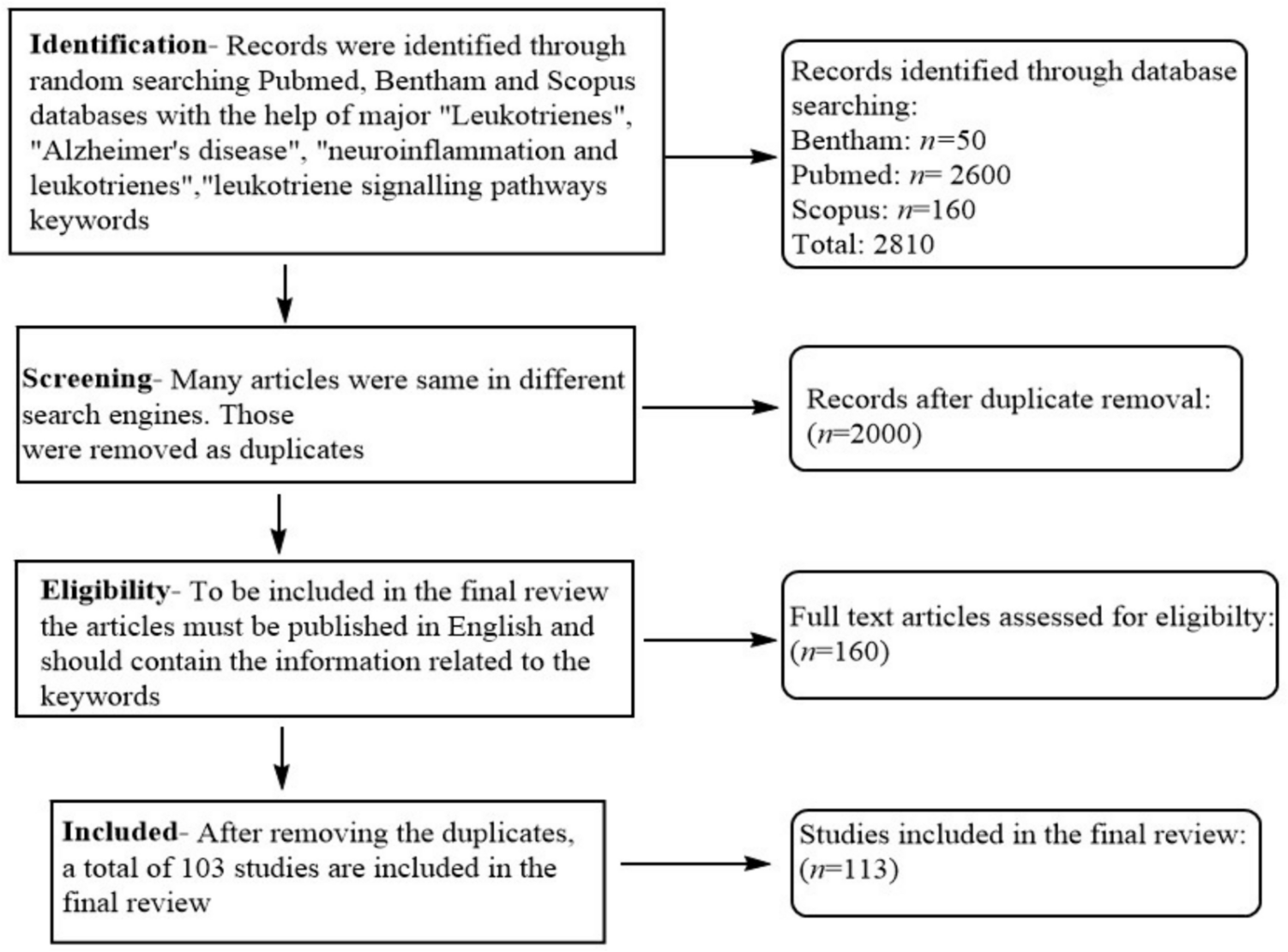


To mimic LPS-induced lung injury, the effect of LPS at various concentrations (ranging from 0.001 ng/mL to 1 µg/mL) on the viability of THP1-Blue™ (vendor and location) positive cells was determined by MTS assay. After 48 h, the viability was significantly decreased by LPS stimulation (0.1–1000 ng/mL), whereas stimulation for 6, 12, or 24 h did not affect cell viability (p > 0.05) except at concentrations of 100 and 1000 ng/mL (Figs. 1A–D). These results suggested that LPS suppressed the growth of THP1-Blue™ cells in a dose- and time-dependent manner.
Fig. 1
LPS and progestogen as modulators of TLR-4/NF-κB/AP-1 signaling. A–D The viability of THP1-Blue™ cells stimulated by LPS at different concentrations (0.001–1000 ng/mL) for 6, 12, 24, or 48 h. E–H NF-κB/AP-1 activation in response to different doses of LPS. Effects of a concentration gradient of DEX, P4 and NES on I the viability of THP1-Blue™ cells, J the viability of THP1-Blue™ cells after LPS (10 ng/mL) stimulation, and K the NF-kB/AP-1 activation after 24 h. The results are presented as the mean ± SD from three separate experiments with triplet repeat represented by each data point. #p < 0.05, ##p < 0.01, ###p < 0.001 vs. vehicle, and *p < 0.05, **p < 0.01, ***p < 0.001 vs. LPS
To confirm that LPS had an inhibitory effect on TLR-4/NF-κB signaling, we employed a reporter cell system to monitor NF-κB/AP-1 activation as described previously (Yang et al. 2015). As shown in Fig. 1B, LPS (10 and 100 ng/mL) exhibited the greatest effect on increasing NF-κB activation after 24 h compared to the other times and concentrations tested. Based on these results, we chose 10 ng/mL as the optimal LPS concentration for stimulation and 24 h as the optimal time for further experiments.
NES (0.001–10 µmol/L) had no significant effect on cell viability at 24 h when used independently (p < 0.05, Fig. 1I). In addition, treatment with DEX, serving as a positive control in this study, caused significant inhibition of the proliferation of THP-1 cell-derived macrophages in a dose-dependent manner (10, 100, and 1000 µmol/L). Simultaneously, the MTS cell viability assay demonstrated that the reduction in viability following a 24-h stimulation with LPS (10 ng/mL) was ameliorated by pre-treatment with NES or P4 at concentrations of 1, 10, and 100 µmol/L, with statistical significance (p < 0.05, Fig. 1J). These data suggested that NES had a protective effect on THP1-Blue™ cells against injury induced by LPS (10 ng/mL) at 24 h.
TLR-4 initiates signaling through both MyD88-dependent and MyD88-independent (TRIF-dependent) pathways. Initially, we assessed the impact of NES on the MyD88-dependent pathways of NF-κB activation induced by LPS at a concentration of 10 ng/mL (Fig. 1K). Two hours before LPS stimulation, the reporter cells were treated with DEX, P4, or NES (0.01–100 µmol/L). We found that NES (at concentrations of 1–100 µmol/L) significantly reduced the LPS-induced NF-κB/AP-1 activation. This inhibitory effect was not attributable to cytotoxicity (Figs. 1I and J).
NES protects the viability of THP1-Dual™ positive cells against damage induced by LPS and enhances anti-inflammatory activity in vitroThe MyD88-independent pathway signals through the TIR domain containing adapter inducing interferon-beta (TRIF) leading to activation of interferon regulatory factor 3 (IRF-3) together with late-phase activation of NF-κB driving secretion of IFN-β and other cytokines.
We evaluated the effect of NES on the MyD88-independent pathways of IRF triggered by LPS, by measuring cell viability and IRF activation in THP1-Dual™ cells at different time-points. MTS assay showed that when stimulated for 6, 12, or 24 h, cell death caused by LPS was similar at different concentrations (0.001 to 1000 ng/mL, viability (% of vehicle) > 95%, p > 0.05). However, after stimulating for 48 h, LPS (10, 100, and 1000 ng/mL) caused a significant increase in cell death compared to vehicle (Figs. 2A–D). LPS did not cause cellular toxicity at the experimental concentrations of 0.001–1000 ng/mL after 6, 12, or 24 h of treatment (Figs. 2A–D).
Fig. 2
NES as a modulator of TLR-4/IRF signaling. A–D The viability of THP-1 cell-derived macrophages after stimulation with 0.001–1000 ng/mL LPS for 6, 12, 24, or 48 h. E–H The effect of different doses of LPS on IRF activation at 6, 12, 24, and 48 h. I–K The effect of DEX, P4, and NES on THP1-Dual™ cells with or without LPS (10 ng/mL) stimulation for 24 h: I viability of THP-1 cell-derived macrophages was assessed at 24 h after treatment with different concentrations (0.0001–1000 µmol/L) of DEX, P4, or NES. J Viability of THP-1 cell-derived macrophages treated with the indicated concentrations (0.01–100 µmol/L) of DEX, P4, or NES for 24 h after 2 h of LPS (10 ng/mL) stimulation. K Effects of DEX, P4, and NES (0.01–100 µmol/L) on LPS (10 ng/mL)-induced IRF activation. Values shown are from triplicate wells per data point obtained in three separate cultures. #p < 0.05, ##p < 0.01, ###p < 0.001 vs. vehicle, and *p < 0.05, **p < 0.01, ***p < 0.001 vs. LPS
Next, we employed a reporter cell system to monitor activation of IRF in the TLR-4 pathway at all analyzed time-points. As shown in Fig. 2G, LPS (10 ng/mL) stimulation for 24 h induced the greatest IRF activation compared to other concentrations or time-points. Taking these results together with the cell viability results, we chose LPS (10 ng/mL) stimulation for 24 h as the optimal treatment for further experiments.
The MTS cell viability assay showed that NES (0.0001–100 µmol/L) had no significant effect on cell viability at 24 h when used alone (Fig. 2C), while at a concentration of 1000 µmol/L, cell death was significantly enhanced (p < 0.001 compared to vehicle). These data suggested that a high concentration of NES (1000 µmol/L) was severely toxic to cells. Next, we evaluated whether NES had a protective effect against LPS-induced death of THP1-Dual™ positive cells. As expected pretreatment with NES (0.1–100 µmol/L) increased the viability of THP1-Dual™ cells after stimulation with LPS (10 ng/mL) at 24 h, while 0.01 µmol/L NES had no effect (p > 0.05, Fig. 2G).
To evaluate the inhibitory effect of NES on LPS-induced THP1-Dual™ positive cells, we determined IRF activation, which is activated by the MyD88-independent pathway. Figures 2E–H show the LPS dose responsiveness of IRF activation. Two hours before LPS stimulation, the THP1-Dual™ reporter cells were treated with NES (0.01–100 µmol/L). We found that NES at these concentrations significantly reduced LPS-induced IRF activation. The inhibitory effect was not due to cellular toxicity (Fig. 1A). These observations substantiated that NES exhibited significant inhibitory effects on both signaling pathways downstream of TLR-4. Among them, NES strongly inhibited NF-κB activation, while it had a weaker inhibitory effect on IRF activation.
Effect of NES on the inhibition of TLR-4 signaling and associated inflammatory responseThe anti-inflammatory activity of NES was next verified in A549 type II alveolar epithelial cells, and THP1-Dual™ cell-derived macrophages. The levels of pro-inflammatory cytokines were analyzed after treatment with LPS alone or together with DEX, P4, or NES for 24 h (Fig. 3). Through assessment of the pro-inflammatory cytokine levels, we found that all three drugs were able to reduce LPS-induced KC (keratinocyte-derived chemokine, or chemokine (C-X-C motif) ligand 1, CXCL-1), IL-6, tumor necrosis factor-α (TNF-α), and monocyte chemoattractant protein-1 (MCP-1) production, where NES exhibited a much stronger inhibitory effect on IL-6, KC, TNF-α, and MCP-1 secretion than DEX or P4 (Fig. 3). There was no statistically significant difference observed in the inhibitory activity between DEX and P4. Although these results suggested that concentration was important for the inhibitory activity of the drugs, increasing the dose (100 µmol/L) did not help further enhance this inhibitory effect. As a result, the in vitro evaluation revealed that the inhibitory activity of NES on LPS-induced NF-κB/AP-1 and IRF activation, as well as IL-6, KC, TNF-α, and MCP-1 secretion, was significantly higher than that of DEX or P4 at the same concentration (1, 10, or 100 µmol/L).
Fig. 3
The anti-inflammatory effect of NES in type II alveolar epithelial A549 cells and THP1-Dual™ cell-derived macrophages. The levels of pro-inflammatory cytokines IL-6, KC, TNF-α, and MCP-1 in A549 cells (A–D) or THP-1 cell-derived macrophages (E–H) stimulated with LPS (10 ng/mL) for 24 h in the presence and absence of DEX, P4 or NES treatment. These results were typical of three independent experiments. *p < 0.05, **p < 0.01, ***p < 0.001 vs. LPS
NES shows potent therapeutic activity in an LPS-induced ALI mouse modelSince P4 exhibited strong ability to ameliorate inflammation and eliminate excessive oxidative stress(Kolatorova et al. 2022; Taraborrelli 2015), we anticipated that NES as one of the progestogens, which has a high affinity for progesterone receptors, might effectively reduce LPS-triggered lung injury in vivo. Herein, we further validated this hypothesis in an LPS-induced ALI mouse model. To exclude the influence of progesterone, we chose male mice as the animal model for this study. We adopted a classical mouse model of ALI caused by a 10 mg/kg dose of LPS administered intranasally (i.n.) to induce acute injury within lung tissue. NES at doses of 0.1, 1, and 10 mg/kg, DEX at a dose of 5 mg/kg, or PBS as the control were administered via intratracheal (i.t.) instillation 2 h prior to LPS stimulation. Subsequent analyses were performed 24 h after the LPS challenge (Fig. 4A).
Fig. 4
The inhibitory activity of NES in an ALI mouse model induced by LPS. A Schematic diagram of the animal experimental protocol. Mice were pretreated with DEX (5 mg/kg) or NES (0.1, 1, and 10 mg/kg) 2 h before LPS challenge, then sacrificed 24 h after stimulation with LPS (10 mg/kg), and biological measurements including differential BALF cell counting and epithelial permeability were performed for further analysis. C–F BALF in each group was collected for evaluation of C the total number of cells, as well as numbers of D neutrophils, E macrophages, F lymphocytes, G total protein content, and H lung W/D ratio. n = 8/group, *p < 0.05, **p < 0.01, ***p < 0.001 relative to the LPS group
First, we quantified inflammatory cell infiltration and secretion of certain cytokines within BALF to evaluate the airway inflammatory response. After 24 h of LPS infusion, we found that NES (0.1, 1, and 10 mg/kg) and DEX (5 mg/kg) significantly reduced the total numbers of inflammatory cells, neutrophils and macrophages, but slightly reduced the lymphocytes in BALF of ALI mice compared to the control group (Figs. 4B–D). These results suggested that NES pretreatment promoted macrophage infiltration while suppressing neutrophil recruitment.
As a key element in the pathogenesis of ALI, we then assessed the permeability of the alveolocapillary membrane by measuring total proteins in BALF and the wet-to-dry (W/D) weight ratio of the right lung. Treatment with progestogen effectively inhibited the LPS-induced increase in the content of total proteins in BALF (Fig. 4F), especially at a high concentration of NES (10 mg/kg). As shown in Figs. 4H and I, NES pretreatment significantly decreased the W/D ratio within lungs of ALI mice. These results demonstrated that progestogen was potent in ameliorating the injury caused to the alveolocapillary membrane in the early stage of ALI. NES was more potent than DEX in the management of lung inflammation. Taken together, these data suggest that progestogen may regulate inflammatory responses and mitigate diffuse alveolar damage in the context of ALI.
Anti-inflammatory effects of NES in lung tissue of LPS-treated ALI miceWe further evaluated the histopathological characteristics of lung tissue following an LPS challenge, both with and without pretreatment using NES and DEX. Progestogen was able to reduce total cell counts and neutrophil counts in the BALF, and markedly ameliorated pathological changes of lung tissues. The severity of lung injury and inflammation was manifested by five independent histological indexes: neutrophils in the alveolar space, neutrophils in the interstitial space, formation of hyaline membranes, proteinaceous debris filling the airspaces, and alveolar septal thickening (Figs. 5A–E), and the lung injury score was obtained based on these features. It was found that the average injury score of the NES-treated group (1 mg/kg) was significantly lower than that of the groups treated with DEX (0.1 mg/kg) or NES (10 mg/kg) (Fig. 5F). Specifically, NES at 1 mg/kg resulted in fewer interstitial neutrophils and less alveolar septal thickening than NES at 10 mg/kg (Figs. 5G and H). These data suggested that NES (1 mg/kg) was superior to either DEX (5 mg/kg) or NES (0.1 and 10 mg/kg) in inhibiting inflammatory responses and reducing diffuse alveolar damage in lungs with ALI.
Fig. 5
The protective effect of NES on lung damage in LPS-induced ALI mice. A–F Representative histological images of lung sections from mice, stained with H&E for histological evaluation, are presented at magnifications of ×10 and ×40: A sham control group (administered PBS only), B LPS + PBS group, C LPS + DEX group (5 mg/kg), D LPS + NES group (0.1 mg/kg), E LPS + NES group (1 mg/kg), and F LPS + NES group (10 mg/kg). Arrows (red) denote representative regions, highlighting features such as alveolar septal thickening, the presence of neutrophils within the alveolar and interstitial spaces, hyaline membranes, and proteinaceous debris. The scale bar in the left panel = 200 μm while that in the right panel = 50 μm. Lung damage was assessed by examination of five pathophysiological features to obtain the total injury score (G–K). G NES markedly decreased the neutrophil count within the alveolar space. H Higher doses of NES (1 and 10 mg/kg) markedly decreased the neutrophil count within the interstitial spaces, I diminished the formation of hyaline membranes and J proteinaceous debris, and K attenuated alveolar septal thickening. n = 8 per group, ns not significant
Anti-inflammatory effects of NES in blood and BALF of mice with LPS-induced ALINext, an analysis to evaluate the global impacts of NES on cytokine production was performed in both BALF and serum of mice with LPS-induced ALI using a multiplex Luminex assay (vendor and address) (Figs. 6 and 7), to investigate the underlying mechanisms of the protective effects of NES. As shown in Figs. 6 and 7, we observed significant alterations in the expression of various pro-inflammatory cytokines and chemokines across the control group, the LPS group, the DEX-treated group (5 mg/kg), and the NES-treated group (0.1, 1, and 10 mg/kg). In comparison to the LPS challenge group, pre-treatment with NES, particularly at a dose of 1 mg/kg, or DEX, resulted in a significant reduction in the levels of pro-inflammatory cytokines and chemokines, including TNF-α, IL-6, IFN-γ, MCP-1, IL-1α, IL-1β, and KC. Additionally, there was a tendency for increased levels of IL-10, IL-17α, and granulocyte-colony stimulating factor (G-CSF) in both BALF and serum. Of note, the levels of G-CSF, IL-1β, and IFN-γ have been documented to have associations with neutrophils. Collectively, these findings indicate that, beyond its TLR-4 inhibitory properties, the anti-inflammatory effects of NES pretreatment may also play a role in neutrophil-mediated inflammatory processes in ALI/ARDS.
Fig. 6
The effect of NES on cytokine production in the BALF of mice with LPS-induced ALI. A Heat map showing cytokine profiles of the BALF under LPS challenge (10 mg/kg, 24 h) with/without NES pre-treatment (0.1, 1, and 10 mg/kg, 2 h before LPS stimulation) using a multiplexed cytokine assay. Different color codes represent the mean fold changes normalized to the PBS group, n = 6 (six were randomly selected from the BALF of eight mice in each group). B–G The levels of selected cytokines in the BALF: B IL-6, C G-CSF, D IFN-γ, E KC, F MCP-1, and G TNF-α. #p < 0.05, ##p < 0.01, ###p < 0.001 vs. control group; *p < 0.05, **p < 0.01, ***p < 0.001 vs. LPS group. ns not significant
Fig. 7
The effect of NES on cytokine production in the serum of mice with LPS-induced ALI. Multiplexed cytokine array profile of serum (A) from mice stimulated with LPS with or without NES (0.1, 1, and 10 mg/kg) pre-treatment, different color codes represent the mean fold changes normalized to the PBS group. B–G The levels of selected cytokines in the serum: B IL-6, C G-CSF, D IFN-γ, E KC, F MCP-1, and G TNF-α; n = 6. #p < 0.05, ##p < 0.01, ###p < 0.001 vs. control group; *p < 0.05, **p < 0.01, ***p < 0.001 vs. LPS group. ns not significant
Survival rateDEX is one of the corticosteroid drugs that have shown a reduction in mortality of COVID-19 (Horby et al. 2021). In hospitalized patients with COVID-19 pneumonia without additional oxygen requirements and at risk of progressing to severe disease, DEX treatments might lead to a decrease in the development of ARDS and thereby reduce death. Considering that NES shows significant binding to glucocorticoid receptors (GR) in vivo (Lee et al. 2022), we investigated whether NES administration could affect the short-term and long-term survival rates in mice subjected to lethal doses of LPS (20 mg/kg) (Fig. 6A). The administration of the drugs, DEX and NES, occurred 2 h prior to the initial and subsequent LPS challenges, followed by administration on alternate days. Daily observations and recordings were made regarding changes in body weight and the survival status of the mice. As shown in Fig. 8C, the survival rates for the groups pretreated with NES at doses of 0.1, 1, and 10 mg/kg were 70.83% (17/24), 91.67% (22/24), and 87.50% (21/24), respectively, while in the DEX (5 mg/kg) group it was 83.34% (20/24), all of which were significantly higher survival rates than that in the LPS group at 66.67% (16/24, p < 0.001). Interestingly, it was observed that NES-treated mice that survived the initial LPS shock subsequently developed a sustained tolerance to a second lethal LPS challenge (25 mg/kg). This phenomenon occurred with the higher dose of NES (1 and 10 mg/kg) group, but not with the lower dose of NES (0.1 mg/kg, 50.00%, 12/24) or DEX (5 mg/kg, 62.50%, 15/24); the survival rate in the LPS group was only 37.50% (9/24, p < 0.001 vs. the other groups). The body weight and mental state of the PBS control mice were always good.
Fig. 8
Effect of NES or DEX on the short-term and long-term survival of LPS-treated ALI mice (A–D): A A scheme of the prophylactic treatments of NES (0.1, 1, and 10 mg/kg) or DEX (5 mg/kg) in an LPS-induced ALI model. The mice received an intratracheal pretreatment with NES, DEX or PBS 2 h prior to the initial LPS challenge (20 mg/kg) on Day 0. Subsequently, the surviving mice were administered a second LPS dose (25 mg/kg) on Day 10. The remainder of the time (days 2–8 and days 12–20) treatments were administered at a fixed time (9:00 am) every other day; B the mouse body weights before and during the experiments; C the effects of NES and DEX on the short-term survival of mice with severe ALI; D the long-term survival of mice with severe ALI after secondary LPS (25 mg/kg) challenge with prophylactic treatments with NES or DEX. n = 8 per group, repeated three times. E and F The augmented tolerance to LPS, elicited by DEX and NES in reporter THP-1 cell-derived macrophages, is characterized by the suppression of E NF-κB activation and F IRF activation following a second LPS exposure 24 h subsequent to the initial LPS stimulation. n = 3, repeated three times. Values shown were obtained in three separate experiments. *p < 0.05, **p < 0.01, ***p < 0.001 vs. double LPS stimulus
In addition, the weight loss observed in mice treated with LPS (20 mg/kg) appeared to be exacerbated (Fig. 6D). These findings suggest that NES (1 mg/kg) not only enhances short-term survival rates but also induces a long-term protective effect against LPS challenge, while mitigating the side effect of weight loss. This indicates the potential clinical applicability of NES in the treatment of ALI/ARDS.
Such unexpected finding is consistent with previous studies (Dorneles et al. 2023) on LPS tolerance, suggesting that NES also has the ability to promote LPS tolerance. Subsequently, we assessed the effect of LPS tolerance by utilizing THP-1-derived macrophages equipped with reporting systems for NF-κB/AP-1 or IRF activation. In both the presence and absence of DEX or NES, the cells were initially stimulated with LPS at a concentration of 10 ng/mL. After a 24-h incubation period, the cells were subjected to a subsequent exposure to LPS at the same concentration. Twenty-four hours later, the reporter enzyme reaction was measured by colorimetry to quantify the activation of NF-κB/AP-1 or IRF (Figs. 8E and F). A previous study (Mauvais-Jarvis et al. 2020) found that cells showed tolerance to a second LPS stimulation after LPS-pretreatment, as evidenced by a significantly reduced activation of NF-κB/AP-1 compared to a one-time LPS exposure (Fig. 8E). Without intervention, the tolerance effect of LPS 24 h after the second stimulation was more pronounced with regard to NF than IRF. For IRF activation, DEX and NES had a slight effect on LPS tolerance, but the results were not significantly different. These results indicated that the LPS tolerance effect on IRF activation basically disappeared after 24 h of rest. Interestingly, only larger dose of NES (10 μmol/L and 100 μmol/L) showed an enhanced LPS tolerance effect in both arms of the signaling pathways, while DEX could not no matter which arm of the two signaling pathways was evaluated. This observation suggested that the NES concentration was important to induce stronger LPS tolerance in vitro, supporting our in vivo findings (Figs. 8C and D).
Comments (0)