Bacterial strains
The bacterial strains used in this study are listed in Table S1. Tryptic soy broth (TSB) and tryptic soy agar (TSA) were used for growing P. aeruginosa, while 15 µg/ml gentamicin was added to select P. aeruginosa transformants. C. jejuni strains were routinely grown on blood agar base II (Oxoid) supplemented with 5% calf blood (BA) at 42 °C under microaerobic conditions (MA: 6% CO2, 6% O2, 84.5% N2, and 3.5% H2). Luria–Bertani broth (LB) and LB agar (LA) (Difco) were used for growing E. coli, while E. coli transformants were selected in 100 μg/ml kanamycin and 50 μg/ml chloramphenicol.
Construction of campycins
Plasmids responsible for campycin 2 expression were constructed similarly to previous work (Zampara et al. 2021) (Table S2). Plasmid pM50, previously engineered to carry the coding sequence of the N-terminal domain of R2-pyocin Prf15 (amino acids 1 to 164), was used as a backbone for inserting the RM1221 CJIE1 prophage H-fiber (CJE0231) along with or without the predicted chaperone (CJE0230). Specifically, pM50 was linearized via inverse PCR using R2N primers (Table S3). The fragment comprising the coding sequence of the C-terminus of the H-fiber (amino acids 151 to 359) and the downstream predicted chaperone gene was amplified from RM1221 (CP000025) by specific primers (Table S3), adding overhangs identical to the distal ends of the linearized pM50. In-Fusion® cloning kit (Takara Bio) was used for the recombination, creating plasmids pM179 and pM182, respectively (Table S2). P. aeruginosa PAO1 Δprf15 was transformed with either of the plasmids, and transformants were selected on TSA plates supplemented with 15 µg/ml gentamicin as described previously (Choi et al. 2006). As a result, a tail fiber–deficient R2-pyocin derivative (∆prf15) was substituted in trans with the RM1221 C-terminal H-fiber along with or without the predicted chaperone. All PCRs were performed using the CloneAMP HiFi PCR premix (Takara Bio) using recommended cycling conditions, and all PCR products and constructs were verified by sequencing.
Campycin expression and purification
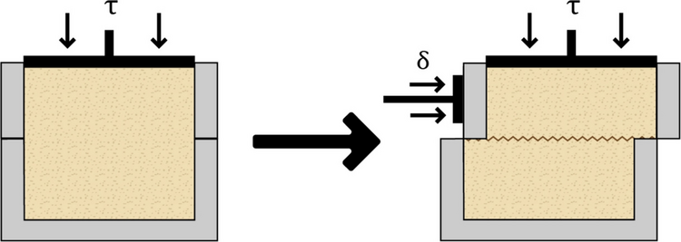
Expression and purification of R2-pyocin and campycins were conducted as previously described (Williams et al. 2008; Zampara et al. 2021). Briefly, P. aeruginosa PAO1 expressing the native R2-pyocin or campycins was grown overnight in TSB at 37 °C and 100-fold diluted in G medium (Ikeda and Egami 1969), which was supplemented with 15 µg/ml gentamicin in case of campycins. Cultures were incubated at 37 °C until reaching an OD600 of 0.25 and treated with mitomycin (3 µg/ml) and isopropyl-β-D-thiogalactopyranoside (0.25 mM) for the induction of expression. After 2.5 h of incubation at the same conditions, DNase I (Invitrogen) was added at a final concentration of 5 U/ml, and the culture was further incubated for 30 min at the same conditions. Centrifugation of cultures (at 18,000 g at 4 °C for 1 h) was followed to remove cell debris, and supernatants were treated with saturated ammonium sulfate solution (final concentration of 1.6 M) while stirring on ice. After overnight incubation at 4 °C, suspensions were centrifuged (18,000 g for 1 at 4 °C), and pellets were resuspended in 1/10th of the start volume with ice-cold TN50 buffer (50 mM NaCl and 10 mM Tris–HCl adjusted to pH 7.5). Ultracentrifuge was followed for 1 h at 60,000 g, and pellets containing the precipitated campycins/pyocins were harvested and resuspended in 1/20th of the start volume with ice-cold TN50 buffer.
Campycin quantification
Quantitative campycin assays were performed by counting surviving bacteria using a method slightly modified from that described by Kageyama and Egami (1962). Accordingly, CAMSA2147 or RM1221 was used to identify the concentration of campycin 1 (co-expressed with downstream chaperone) or campycin 2, respectively. Cells were harvested using 3 ml calcium brain–heart infusion (CBHI) broth and adjusted the optical density OD600 to 0.2, which equals approximately 109 colony forming units per milliliter (cfu/ml). Campycins were fivefold diluted in TN50 buffer, and 100 μl of each diluted campycin was mixed with 100 μl of adjusted C. jejuni in a deep-well plate and incubated for 3 h at 42 °C under MA conditions. TN50 buffer was used as a negative control. Proper dilutions were made in TN50, and samples were spotted on BA plates and incubated under MA conditions at 42 ºC. It was previously estimated for pyocins that a microtiter well that has an average of 1 pyocin particle per bacterium will yield at or near equilibrium 37% survivors, and a well with an average of 2.3 pyocin particles per bacterium will yield 10% survivors (Scholl et al. 2009). Since campycins are engineered pyocins, we also used this calculation to quantify campycins. A typical density of purified, concentrated campycins was 1 × 10⁹ per ml.
Antibacterial spectrum of campycins
To determine the spectrum of campycins, a lysis spot assay was performed on Campylobacter bacterial lawns as previously described (Gencay et al. 2017). Briefly, cells were streaked on BA plates and harvested with CBHI broth. Suspensions were further adjusted to an OD600 of 0.35 and incubated for 4 h at 42 °C under MA conditions. Cell cultures (500 μl) were mixed with molten NZCYM overlay agar (5 ml of NZCYM broth [Sigma] with 0.6% agar [Sigma]) and poured on NZCYM basal agar (with 1.2% agar [Sigma]) plates, supplemented with 10 μg/ml vancomycin (Sigma). After 20 min, plates were dried for 45 min in the flow hood, and campycins (5 μl) were spotted on top, followed by incubation for 24 h at 42 °C under MA conditions. As negative controls, the native R2-pyocin or fiber mutant derivative (∆prf15) were also spotted. Bacterial lawns were inspected for the formation of a distinct, clear zone due to cell killing.
Antibacterial activity of campycins in vitro
To assess the killing efficiency of campycins under different conditions, C. jejuni strains were grown on BA plates, harvested with CBHI broth, and adjusted to the final concentration of 106 cfu/ml. For each cell culture, 100 μl (106 cfu/ml) was mixed with 100 μl of each campycin (109 campycins/ml) for 3 or 24 h under MA conditions or 24 h under anaerobic conditions using an anaerobic gas generator (AnaeroGen), in plastic containers at 5 °C. Cells were treated with TN50 instead of campycins as a negative control. After incubation, samples were tenfold diluted in TN50 buffer and spotted on BA plates that were incubated for 48 h under MA conditions. Colonies were enumerated, and colony-forming units per milliliter was calculated. Data represent the mean from biological triplicates.
Construction of plasmids for H-fiber expression
To express CJIE1 H-fibers of CAMSA2147 or RM1221, the responsible genes were cloned into vector pET28-a ( +) using In-Fusion® cloning (Takara Bio). Specifically, CAMSA2147 (GCA_003095855) was used as a template for the amplification of the fragment comprising the coding sequence of the C-terminus of the H-fiber (amino acids 151 to 343) by specific primers (Table S3). The pET28-a ( +) vector was digested with NdeI and XhoI, and recombination resulted in the pCRYS1 plasmid. Similarly, RM1221 (CP000025) was used as a template for amplification of the fragment comprising the coding sequence of the C-terminus of the H-fiber (amino acids 151 to 359) with or without the downstream predicted chaperone gene. Specific primers were used for the amplification (Table S3), and recombination of the fragments with the linearized pET28-a ( +) resulted in the pCRYS4 and pCRYS5 plasmids, respectively (Table S2). E. coli BL21-CodonPlus-(DE3)-RIL were used for transformation, and transformants were selected on LB agar plates in the presence of kanamycin (100 μg/ml) and chloramphenicol (50 μg/ml). All PCRs were performed using the CloneAMP HiFi PCR premix (Takara Bio) using recommended cycling conditions, and all PCR products and constructs were verified by sequencing.
H-fibers were expressed with or without the predicted chaperones by growing cells in 1 L of LB at 37 °C until reaching optical density OD600 = 0.6. Protein expression was induced by adding isopropyl-β-D-thiogalactopyranoside (0.5 mM), and cultures were incubated for 18 h at 16 °C at 120 rpm. Pellets of cultures were harvested by centrifugation (8000 × g, 10 min, 4 °C) and resuspended in 10 ml of lysis buffer (20 mM NaH2PO4-NaOH, 0.5 M NaCl, 50 mM imidazole, pH 7.4). Sonication (Bandelin Sonopul HD 2070 homogenizer) with 10 bursts of 30 s (amplitude of 50%) and 30 s intervals allowed cell lysis, and samples were further filtered twice with 0.22-μm pore size filters. His GraviTrap™ gravity flow columns (GE Healthcare) were used for protein purification. The lysis buffer (10 ml) was used for the wash step during purification, and proteins were eluted with 6 ml of elution buffer (20 mM NaH2PO4-NaOH, 0.5 M NaCl, 500 mM imidazole, pH 7.4). The elution buffer was further exchanged with Thermo Scientific BupH Tris-buffered saline (TBS) by using Amicon Ultra-15 Centrifugal Filter Units with Ultracel-10 membrane cutoff (Merck Millipore), and protein concentration was measured with a Qubit 2.0 fluorometer using the kit Qubit™ Protein Assay Kit (Invitrogen).
Determination of the H-fiber receptor
For identifying the receptor, we used the Pierce™ His Protein Interaction Pull-Down Kit that purifies protein interactors of any His-tagged fusion proteins. To do so, we used 700 μl of 200 μg purified H-fibers fused with a His-tag as bait. First, the H-fibers were immobilized onto cobalt chelate resin and could thereby further trap their prey, which was present in the cell lysates. CAMSA2147 or RM1221 provided the prey protein, i.e., the receptor. Specifically, the strains were grown on BA plates, and cells were harvested using a loop and dissolved in 1 ml of TBS. Cell lysates were further prepared by following the manufacturer’s instructions. The H-fibers or the cell lysates alone were run in the columns as negative controls. Finally, 30 μl of each eluted sample was analyzed by SDS-PAGE analysis, and the bands of interest were cut from the gel for further analysis. Alphalyse A/S and Proteome Factory further identified the proteins using LCMS-based high-resolution mass spectrometric techniques.
Bioinformatic analysis
To investigate the diversity of CJIE1 H-fibers, CAMSA2147 H-fiber (WP_002878910.1) was used for searching CJIE1 H-fiber homologs in our CAMSA bacterial collection, and sequences were further aligned using CLC Main Workbench 22 (QIAGEN). To analyze the MOMP diversity, CAMSA2147 MOMP (WP_002875862.1) was used as a query sequence to search C. jejuni homologous proteins in the National Center for Biotechnology Information (NCBI) genome database through BLASTP (Sayers et al. 2022). In total, 5000 MOMP sequences were retrieved that were further aligned, including default settings in mafft (Katoh et al. 2019), and a neighbor-joining phylogenetic tree was created using 100 bootstraps. Interactive Tree Of Life (iTOLv6) was used for the display, manipulation, and annotation of the phylogenetic tree (Letunic and Bork 2021). Structural prediction of proteins and protein complexes was performed using Colab AlphaFold and Colab AlphaFold Multimer v3 with default settings (Mirdita et al. 2022) using the webpage https://colab.research.google.com/github/sokrypton/ColabFold/blob/main/AlphaFold2.ipynb#scrollTo=ADDuaolKmjGW and visualized using PyMOL (Schrodinger 2015).
Statistical analysis
GraphPad Prism 7 software (Version 7.0d) was used for statistical analysis. To test the significance of the logarithmic bacterial reduction of cells treated with campycin compared to the cells treated with TN50 buffer, we used the Paired-Samples t-test with 95% confidence interval percentage based on biological triplicates.


Comments (0)