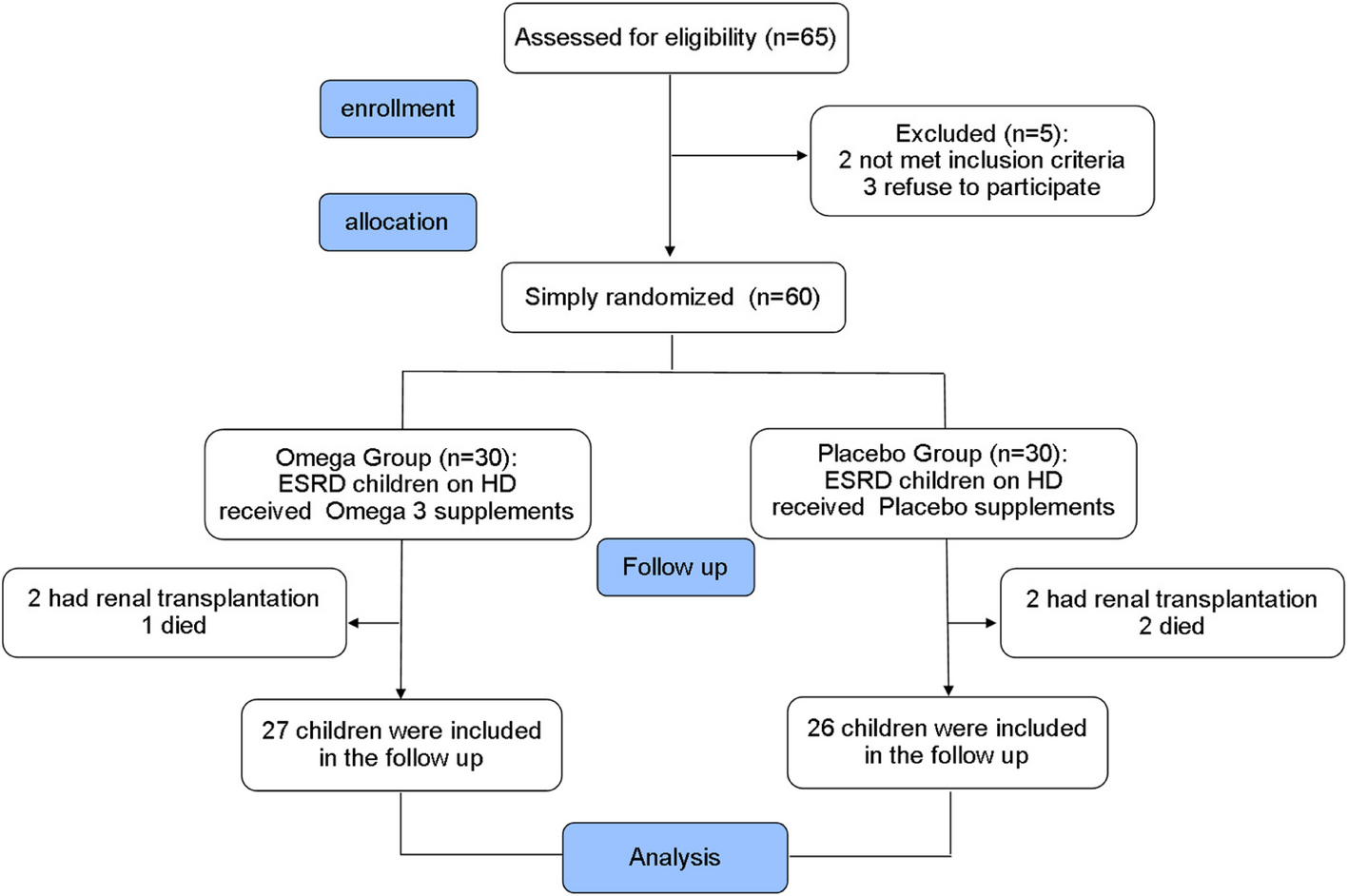
Our hybrid study design comprised two phases: the cross-sectional analysis for detecting asthma prevalence, and the case–control study for investigating the genetic association reveals that 12% of school-aged children in El Manzala City, Dakahlia, Egypt, have asthma. Through the analysis of serum biomarkers, asthmatic children have shown a significantly higher eosinophil count and total serum IgE level compared to controls. Moreover, no significant differences existed in CCR3-T51C genotypes or alleles between asthmatic children and controls.
The study findings reported that most of the studied children were aged between 7 and 16 years. Additionally, most of the subjects evaluated were male, comprising over half of the total population. Asthma prevalence differs by sex across the lifespan, where boys have higher rates than girls before puberty, but this reverses in adolescence [29].
According to a study using an ISAAC questionnaire, the Sohag Governorate found that 12.5% of children aged 6 to 12 years old in public primary schools had asthma, which is consistent with our result [30]. On the other hand, this percentage was greater than in the Nile Delta area of Egypt in 2009; overall asthma prevalence was 7.7%, with a slightly higher prevalence in males (50.7%) [6]. Another survey in Cairo demonstrated that the asthma prevalence rate among school children aged 3–15 years was 8.2% [31]. Meanwhile, a study conducted in 1996 by Khallaf et al. revealed an asthma prevalence rate of 4.8% in Egypt [32]. It is worth noting that the disparities in prevalence rates may be attributed to differences in populations and environmental factors, leading to varying degrees of allergen exposure.
Also, this study revealed that 12% of the children exhibited previous wheezes, and 66.7% of them had experienced wheezing during the last year. A previous nationwide survey of school-aged children indicated that 26.5% of them had persistent wheezing, and 14.7% experienced wheezing in the last year of the disease [33]. Similarly, another national study focused on primary school children aged 8 to 12 years, which found that the prevalence of wheezing was 16.83%, which increased to 26.74% over the past 12 months [34]. The findings of these studies are crucial and provide insight into the prevalence of this disease, highlighting the need for continued research to better understand and manage the disease.
Additionally, this study shows that only 8% of children suffered from night cough last year and wheezed during exercise. Also, Kajbaf et al. reported that 7.2% and 3.4% of Iranian primary school students had night coughs and exercise-induced wheezing, respectively [35]. Exercise-induced wheezing may be caused by factors such as dryness of the mucosal lining, increased osmolality that triggers mast cell degranulation, and rapid airway rewarming after exercise, which can lead to vascular congestion, enhanced permeability, and edema [36].
This study revealed no notable differences in age, sex, residence, and consanguinity between asthmatic and control groups. However, a positive family history of asthma was significantly more common in asthmatic patients as compared to the control group. This is consistent with a previous study that documented a higher positive family history of atopy among asthmatic cases compared to controls. Also, it was found that 88.6% of responsive and 80% of resistant asthma patients had positive family histories, respectively, while controls had 0% [37]. Children with a family history of asthma have a 4.2 times higher risk of developing asthma (95% CI 3.91–4.50) than those without [30].
Furthermore, it was observed that asthmatic patients were less likely to have been breastfed and more artificially fed than the control group. This is consistent with Zedan et al., who revealed that only 51.7% of asthmatic cases received exclusive breastfeeding versus 86% of controls [38]. It is worth noting that breastfeeding exclusively for 6 months may prevent asthma or atopic diseases [39].
In the current study, asthmatic children had a higher rate of paternal smoking, and a crowding index of more than two children per room than controls. Prior research has established that students who are exposed to smoking and reside in houses with high crowding indices have a higher prevalence of asthma than the control group [38, 40]. This could be due to the decrease in Forkhead/winged helix transcription factor (FoxP3) levels and tumor growth factor-β caused by passive smoking, which is linked with T-reg cells, and the increase in interleukin-17A and interleukin-23, which are connected with Th17 cell [41]. Besides, the study by Islam et al. suggests that overcrowding enhances respiratory infection transmission and elevates the risk of asthma [42].
This study demonstrated that asthmatic children exhibit a lower absolute eosinophil count but a higher relative eosinophil count in comparison to controls. Likewise, patients with asthma have higher eosinophil counts in their blood [43], suggesting a pivotal role for eosinophils in asthma development and that an increased eosinophil count can lead to irreversible changes in lung function and airway remodeling [44]. Basophil populations in the blood of individuals with allergic asthma respond to IgE-mediated activation [45].
Eosinophil accumulation in the bronchial wall is the primary mechanism of action. Inflammatory cells’ secretory granules stain dark pink in eosin and hematoxylin preparations. These inflammatory cells have secretory granules that stain dark pink in eosin and hematoxylin preparations. The content of these granules can be released locally through different mechanisms. When activated, human eosinophils undergo cytolysis in the tissues, leading to the extracellular release of membrane-bound granules [15].
These granulocytes release the eosinophil cationic protein and eosinophil peroxidase, which can damage surrounding tissues. Around two-thirds of patients with asthma have an allergic component, and 50% of those with severe asthma have an allergic component. The most significant known contributing factor to the development of asthma is an increase in IgE production due to exposure to environmental allergens (atopy), especially when sensitization occurs in early life [46]. On the other hand, the expression of the high-affinity IgE receptor, Fc ƐRI, on mast cells and basophils has been shown to be sensitive to the presence of IgE or cytokines previously [47].
In this study, patients with asthma showed significantly increased total serum IgE levels compared to those without asthma, which aligns with previous findings by Zedan et al. [38]. This suggests a potential role for the IgE-related mechanism in the up-regulation of neurokinin-1 receptor (NK1R) expression [48], where IgE at 1 μg/mL increases mean fluorescent intensity (MFI) of NK1R expression and NK1R mRNA expression in KU812 cells at 2 h following incubation, which was blocked by coincubation with anti-IgE antibody for 30 min.
Furthermore, it was observed that the cough, wheezy, and cough with wheezy phenotypes exhibited significantly lower absolute eosinophil counts compared to the control group, with no significant variance among the three. Conversely, the relative eosinophil count was notably higher in the wheezy and cough with wheezy phenotypic groups compared to the control group. Similarly, groups with cough-predominant asthma phenotype and wheezy phenotype showed a greater serum eosinophil count than the control group, with no significant difference between the cough and wheezy phenotypes [27]. These findings suggest that different phenotypes of asthma have varying impacts on eosinophil counts, with cough and wheezy phenotypes presenting higher serum eosinophil counts than the control group.
CCR3 is the primary receptor for eotaxins, stimulating eosinophil migration, degranulation, and activation via G protein-dependent mechanisms [49]. This study revealed no significant differences in the CCR3-T51C genotypes between asthmatic and control children; however, the TT genotype was more common among asthmatics. A previous study found no significant difference in CCR3-T51C genotype and allelic frequency between asthmatic cases and controls. However, asthmatic patients had a higher prevalence of the TT genotype [38], consistent with our study. According to a study conducted in Taiwan, the CCR3-T51C polymorphism was not found to be associated with asthma [50]. However, it is likely that CCR3 + and CD123 + HLA-DR − cells play a role in AA and AR through SP and NK1R. The cells that express up-regulated SP in CCR3 + and CD123 + HLA-DR − granulocytes may be a subtype of basophils that is different from those found in PBMC. It is possible that this subtype may also include other types of cells. Previous research has shown that AA patients have unique basophil populations in their peripheral blood, and CCR3 + cells in blood granulocytes can include both eosinophils and basophils [45]. Although there is no information available about CD123 + HLA-DR − cells in blood granulocytes, CD123 + granulocytes are composed of eosinophils, immature neutrophils, and basophils [48].
In Saudi Arabia, CCR3-T51C polymorphism was significantly correlated with increased asthma risk [51]. However, the association was observed in the British population (odds ratio = 2.35, P < 0.01) and not in the Japanese [28]. Conversely, a Japanese study found that the 64Ile and 51C variations in CCR2 and CCR3 genes are linked with cedar pollinosis. The haplotype 64Ile/780C/51C frequency was higher in individuals with pollinosis, suggesting that these genes are candidates for the condition [52]. Differences in genetic variation may contribute to complex traits like asthma [12].
The effect of allergens on CCR3-T51C genotype expression in cough, wheezy, and cough-wheezy phenotypes has yet to be investigated. This study showed no statistically significant difference in laboratory biomarkers among the CCR3-T51C genotypes in cough, wheezy, and cough-wheezy groups, except for relative eosinophil count, which was significantly lower in CT cough phenotypes than the wheezy phenotype. CCR3 mRNA and protein expressions are increased in the bronchial mucosa of asthmatic patients and correlated with airway hyperresponsiveness [53].
The allergen extract affects eosinophil behavior in allergic asthma patients during late-phase airway inflammation [54]. The bronchial HDME challenge increases sputum eosinophils and ECP in allergic asthma patients [55]. Eosinophils express CCR3, which suggests that SP + CCR3 + granulocytes may be eosinophils [48, 56]. However, further studies are needed to confirm or disprove this association with other allergic sensitizations. This study is noteworthy for being the first to investigate the relationship between the CCR3-T51C gene polymorphism and asthma phenotype as well as various inflammatory biomarkers, such as absolute eosinophil count, relative eosinophil percentage, and IgE levels. Adding new genetic data about polymorphisms, from different ethnic groups, could be of great value in personalized medicine.
In conclusion, our study reveals that asthma affects 12% of the school-aged children. The CCR3-T51C genotype or allelic polymorphism frequency did not differ between asthmatics and controls; however, the TT genotype was more frequent in asthmatic children. Eosinophil count, serum IgE and gene polymorphism of CCR3-T51C appeared similar among different asthmatic phenotypes.


Comments (0)