Brancatelli G, Federle M, Grazioli L, Carr B. Hepatocellular carcinoma in noncirrhotic liver: CT, clinical, and pathologic findings in 39 U.S. residents1. Radiology. 2002;222:89–94. https://doi.org/10.1148/radiol.2221010767.
Article
Google Scholar
Mamone G, Marrone G, Caruso S, Carollo V, Gentile G, Crino F, et al. Intrahepatic mass-forming cholangiocarcinoma: enhancement pattern on Gd-BOPTA-MRI with emphasis of hepatobiliary phase. Abdom Imaging. 2015;40(7):2313–22. https://doi.org/10.1007/s00261-015-0445-5.
Article
Google Scholar
Lim CH, Moon SH, Cho YS, Choi JY, Lee KH, Hyun SH. Prognostic value of (18)F-fluorodeoxyglucose positron emission tomography/computed tomography in patients with combined hepatocellular-cholangiocarcinoma. Eur J Nucl Med Mol Imaging. 2019;46(8):1705–12. https://doi.org/10.1007/s00259-019-04327-2.
Article
Google Scholar
Shen YT, Yue WW, Xu HX. Non-invasive imaging in the diagnosis of combined hepatocellular carcinoma and cholangiocarcinoma. Abdom Radiol. 2023;48(6):2019–37. https://doi.org/10.1007/s00261-023-03879-0.
Article
Google Scholar
Meiburger KM, Acharya UR, Molinari F. Automated localization and segmentation techniques for B-mode ultrasound images: a review. Comput Biol Med. 2018;92:210–35. https://doi.org/10.1016/j.compbiomed.2017.11.018.
Article
Google Scholar
Lee H, Kim H, Han H, Lee M, Lee S, Yoo H, et al. Microbubbles used for contrast enhanced ultrasound and theragnosis: a review of principles to applications. Biomed Eng Lett. 2017;7(2):59–69. https://doi.org/10.1007/s13534-017-0016-5.
Article
Google Scholar
Sagrini E, Iavarone M, Stefanini F, Tovoli F, Vavassori S, Maggioni M, et al. Imaging of combined hepatocellular-cholangiocarcinoma in cirrhosis and risk of false diagnosis of hepatocellular carcinoma. United European Gastroenterol J. 2019;7(1):69–77. https://doi.org/10.1177/2050640618815378.
Article
Google Scholar
Kwon SJ, Jeong MK. Advances in ultrasound elasticity imaging. Biomed Eng Lett. 2017;7(2):71–9. https://doi.org/10.1007/s13534-017-0014-7.
Article
MathSciNet
Google Scholar
Weijers G, Starke A, Thijssen JM, Haudum A, Wohlsein P, Rehage J, et al. Transcutaneous vs intraoperative quantitative ultrasound for staging bovine hepatic steatosis. Ultrasound Med Biol. 2012;38(8):1404–13. https://doi.org/10.1016/j.ultrasmedbio.2012.04.009.
Article
Google Scholar
Burckhardt CB. Speckle in ultrasound B-mode scans. IEEE Trans Son Ultrason. 1978;25(1):1–6. https://doi.org/10.1109/T-SU.1978.30978.
Article
Google Scholar
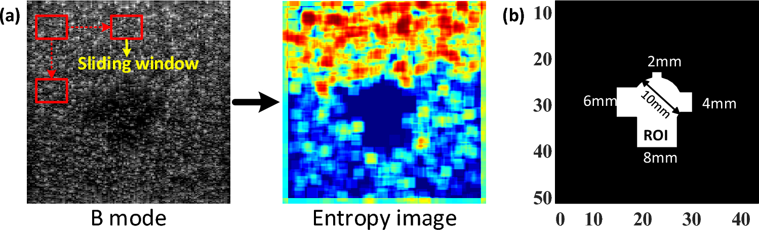
Tsui P-H, Chen C-K, Kuo W-H, Chang K-J, Fang J, Ma H-Y, et al. Small-window parametric imaging based on information entropy for ultrasound tissue characterization. Sci Rep. 2017;7(1):41004. https://doi.org/10.1038/srep41004.
Article
Google Scholar
Hughes MS. Analysis of digitized waveforms using Shannon entropy. J Acoust Soc Am. 1993;93:892–906.
Article
Google Scholar
Hughes MS, McCarthy JE, Marsh JN, Wickline SA. Joint entropy of continuously differentiable ultrasonic waveforms. J Acoust Soc Am. 2013;133(1):283–300. https://doi.org/10.1121/1.4770245.
Article
Google Scholar
Zhou Z, Tai D-I, Wan Y-L, Tseng J-H, Lin Y-R, Wu S, et al. Hepatic steatosis assessment with ultrasound small-window entropy imaging. Ultrasound Med Biol. 2018;44(7):1327–40. https://doi.org/10.1016/j.ultrasmedbio.2018.03.002.
Article
Google Scholar
van Sloun RJG, Demi L, Postema AW, De La Rosette JJMCH, Wijkstra H, Mischi M. Entropy of ultrasound-contrast-agent velocity fields for angiogenesis imaging in prostate cancer. IEEE Trans Med Imaging. 2017;36(3):826–37. https://doi.org/10.1109/Tmi.2016.2629851.
Article
Google Scholar
Ghasemifard H, Behnam H, Tavakkoli J. High-intensity focused ultrasound lesion detection using adaptive compressive sensing based on empirical mode decomposition. J Med Signals Sens. 2019;9(1):24–32. https://doi.org/10.4103/jmss.JMSS_17_18.
Article
Google Scholar
Guiasu S. Grouping data by using the weighted entropy. J Stat Plan Inference. 1986;15:63–9. https://doi.org/10.1016/0378-3758(86)90085-6.
Article
MathSciNet
Google Scholar
Tsui P-H. Ultrasound detection of scatterer concentration by weighted entropy. Entropy. 2015;17:6598–616. https://doi.org/10.3390/e17106598.
Article
Google Scholar
Li X, Jia X, Shen T, Wang M, Yang G, Wang H, et al. Ultrasound entropy imaging for detection and monitoring of thermal lesion during microwave ablation of liver. IEEE J Biomed Health Inform. 2022;26(8):4056–66. https://doi.org/10.1109/jbhi.2022.3167252.
Article
Google Scholar
Chan HJ, Zhou Z, Fang J, Tai DI, Tseng JH, Lai MW, et al. Ultrasound sample entropy imaging: a new approach for evaluating hepatic steatosis and fibrosis. IEEE J Transl Eng Health Med. 2021;9:1800612. https://doi.org/10.1109/jtehm.2021.3124937.
Article
Google Scholar
Richman JS, Moorman JR. Physiological time-series analysis using approximate entropy and sample entropy. Am J Physiol Heart Circ Physiol. 2000;278(6):H2039–49. https://doi.org/10.1152/ajpheart.2000.278.6.H2039.
Article
Google Scholar
Chen W-T, Wang Z, Xie H, Yu W. Characterization of surface EMG signal based on fuzzy entropy. IEEE Trans Neural Syst Rehabil Eng. 2007;15:266–72.
Article
Google Scholar
Zhang S, Shang S, Han Y, Gu C, Wu S, Liu S, et al. Ex vivo and in vivo monitoring and characterization of thermal lesions by high-intensity focused ultrasound and microwave ablation using ultrasonic nakagami imaging. IEEE Trans Med Imaging. 2018;37(7):1701–10. https://doi.org/10.1109/TMI.2018.2829934.
Article
Google Scholar
Hajian-Tilaki K. Receiver operating characteristic (ROC) curve analysis for medical diagnostic test evaluation. Caspian J Intern Med. 2013;4(2):627–35.
Google Scholar
Huang H, Xu H, Wang X, Silamu W. Maximum F1-score discriminative training criterion for automatic mispronunciation detection. IEEE/ACM Trans Audio Speech Lang Process. 2015;23(4):787–97. https://doi.org/10.1109/TASLP.2015.2409733.
Article
Google Scholar


Comments (0)