
Remember me


Orthodontic treatment, a common therapeutic intervention for correcting malocclusion and dental misalignment is often associated with discomfort and pain, especially in the initial stages. Pain during orthodontic treatment is primarily attributed to the inflammatory response triggered by the mechanical forces exerted on the teeth and surrounding tissues.[1,2] Patients commonly report soreness, tenderness, and pain, leading to a potential decrease in treatment adherence and overall patient satisfaction.[3–5] Consequently, efforts to alleviate orthodontic-related pain have garnered significant attention, and one emerging modality showing promise in this context is low-level laser therapy (LLLT).
Orthodontic pain arises due to the inflammatory mediators released during the remodeling processes that occur in response to orthodontic forces applied to the teeth. These forces lead to cellular and molecular changes in the periodontal ligament and surrounding tissues, resulting in the release of prostaglandins, histamines, and other inflammatory substances.[6–8] The subsequent inflammatory response activates pain receptors, contributing to the sensation of pain and discomfort.[9] Understanding the etiology of orthodontic pain is crucial for developing effective strategies to manage and mitigate its impact on patients.
LLLT, also known as photobiomodulation, involves the application of low-intensity lasers or light-emitting diodes (LEDs) to stimulate cellular processes and promote tissue healing.[10–12] LLLT has gained popularity in various medical and dental fields for its anti-inflammatory, analgesic, and biostimulatory effects. The mechanism of action involves the absorption of light energy by cellular chromophores, leading to physiological responses at the cellular and molecular levels. [13–15] In the context of orthodontics, LLLT has emerged as a potential adjunctive therapy to alleviate pain and accelerate the tissue healing process associated with orthodontic adjustments.[16–18]
The application of LLLT in orthodontics aligns with the growing trend of incorporating evidence-based and patient-centered approaches in dental care. Patient comfort and satisfaction are integral components of orthodontic treatment success, and strategies to minimize pain and discomfort contribute to a positive patient experience.[19,20] Considering the potential benefits of LLLT, its integration into orthodontic practice has the potential to enhance patient outcomes and treatment adherence.
Despite the accumulating evidence supporting the efficacy of LLLT in reducing orthodontic pain, variations in study designs, laser parameters, and outcome measures necessitate a comprehensive evaluation of the existing literature. A critical examination of the available evidence will contribute to a deeper understanding of the role of LLLT in orthodontic pain management and guide clinicians in optimizing its use within orthodontic protocols.
This study aims to clinically evaluate the effectiveness of LLLT in reducing pain caused by orthodontic movement in the early stages of treatment.
MATERIAL AND METHODSThe study was designed to clinically assess the effectiveness of LLLT in alleviating pain associated with orthodontic movement during the initial stages of treatment.
Ethical considerations were addressed by obtaining informed consent from all participants, and the study protocol received approval from the relevant ethics committee. The research was conducted in accordance with the Helsinki Declaration and Bharati Vidyapeeth (Deemed to be University) Dental College and Hospital approved the protocol (Protocol number: BV [DU] MC&Sangli/IEC/19–49/21, Date: 03/02/2021). The study adhered to rigorous ethical guidelines to ensure participant safety and confidentiality.
The study included 20 individuals who met specific inclusion criteria: requiring non-extraction orthodontic treatment using pre-adjusted fixed appliances with a 0.022″ slot; initiating the alignment and leveling phase with the 0.012″ thermoactivated nickel-titanium archwire (Libral Traders Pvt. Ltd. India); not taking medication that could impact the study results; maintaining good oral and general health; and providing signed informed consent, indicating their agreement with the research procedures.
Participants were randomly allocated to the laser group (LG) and control group (CG). The LG (n = 10) received low-level laser irradiation (808 nm, 100 mW) immediately after appliance installation, while the CG (n = 10) received no pain control intervention post-installation.
A power analysis was conducted to determine the appropriate sample size for this clinical study. The calculation was based on the expected effect size, significance level, and desired power of the statistical tests. Assuming an effect size of 0.8, a significance level (α) of 0.05, and a desired power (1-β) of 0.80, the estimated sample size was determined to be 20 participants, with 10 participants in each group (LG and CG). This sample size provides adequate statistical power to detect significant differences in pain scores between the two groups, ensuring the reliability and validity of the study findings.
The gallium-aluminum-arsenides infrared laser used in this study operated at a wavelength of 808 nm, with a cross-sectional beam diameter measuring 3 mm. This laser device, identified as Novolase Gold, Novolase Technologies, India, was configured to a power level of 100 mW.
The laser therapy was administered as a single session, promptly following the bonding of brackets and the installation of the initial orthodontic archwire (0.012″ thermoactivated nickel-titanium). Each session of irradiation lasted for an average duration of 19.5 min, focusing on the distal region of the first molar to the corresponding region on the opposite first molar. Specifically, three points situated between the roots and distal spaces of the first molar – cervical, middle, and apical – on both the buccal and lingual/palatal sides, where the orthodontic appliance was affixed, were subjected to 15 s of irradiation each. This equated to 1.5 J/cm2 of energy delivered per tooth, with a total of 12 irradiations administered to each tooth. The cumulative energy dosage per tooth amounted to 18 J.
Pain levels were assessed using a 100 mm horizontal visual analog scale (VAS), segmented into three points, each marked with descriptors (smiley expressions): 0 (zero) denoting no pain, 5 (five) indicating moderate pain, and 10 (ten) signifying severe pain. Patients were instructed to place a vertical mark on the scale corresponding to their pain level at 06, 24, 48, and 72 h up to 7 days at intervals of 24 h post-orthodontic appliance installation.
Participants completed a questionnaire, recording VAS scores at the specified intervals and detailing their pain perception concerning laser therapy, including the initiation, peak intensity, decline, and absence of pain. Patients indicated the day and time when they experienced these pain levels.
All participants were informed that taking painkillers was not prohibited. However, if they chose to use painkillers, they were required to disclose the specific medication in the questionnaire, and those who did so were excluded from the sample.
Statistical analysisThe obtained data were statistically analyzed with the Statistical Package for the Social Sciences version 21.0. The tests applied included Chi-square tests for intergroup comparability and Mann–Whitney tests for pain symptomatology and perception. P < 0.05 was considered as statistically significant.
RESULTSThe demographic details of the study participants are mentioned in [Table 1]. There were 11 male (mean age 19.36 ± 4.13 years) and 9 female subjects (mean age 18.67 ± 1.80 years). [Table 2 and Figure 1] present the comparison of pain on chewing (VAS score) among subjects treated with laser and without laser therapy. After 6 h post-treatment, the median pain score on chewing among subjects receiving laser therapy was significantly lower as compared to the median pain score on chewing among subjects who did not receive laser therapy (P < 0.001). Similarly, the median pain score on chewing among subjects receiving laser therapy was significantly lower as compared to the median pain score on chewing among subjects who did not receive laser therapy for up to 7 days (P < 0.001).
Table 1: Demographic details of the study participants.
Gender n Minimum age Maximum age Mean±SD Male 11 14 27 19.36±4.13 Female 9 16 21 18.67±1.80Table 2: Comparison of pain on chewing (VAS score) among subjects treated with laser and without laser therapy.
Time With laser-on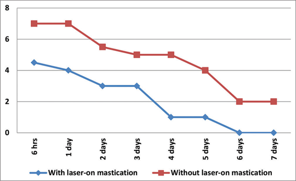
Export to PPT
The comparison of spontaneous pain (VAS score) among the subjects treated with laser and without laser therapy is presented in [Table 3 and Figure 2]. After 6 h post-treatment, the median pain score among the subjects receiving laser therapy was significantly lower as compared to the median pain score on chewing among subjects who did not receive laser therapy (P < 0.001). Similarly, the median pain score among subjects receiving laser therapy was significantly lower as compared to the median pain score on chewing among subjects who did not receive laser therapy after 1 day and 2 days post-treatment. On post-treatment, 3rd day, and onward, the difference in spontaneous pain among the two groups were not statistically significant.
Table 3: Comparison of spontaneous pain (VAS score) among subjects treated with laser and without laser therapy.
Time With laser Without laser P-value Median IQR Median IQR 6 h 4 1 7 0 <0.001* 1 day 3.5 2 7 1 <0.001* 2 days 3 2 4 2 0.001* 3 days 2 1 2 2 0.093 4 days 1 0 1 1 0.069 5 days 1 1 1 2 0.673 6 days 0 1 0 1 0.857 7 days 0 1 0 1 0.522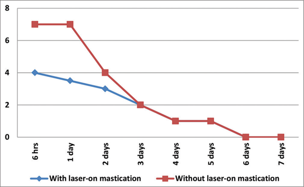
Export to PPT
Results showed that lower median VAS scores for pain on chewing among the subjects with and without laser therapy were zero and two, respectively, and there was a significant difference in the lowest median pain on chewing scores among the two groups. However, a comparison of the lowest spontaneous pain score among the two groups showed a non-significant difference [Table 4]. The maximum median VAS scores for pain on chewing among the subjects with and without laser therapy were five and seven, respectively, and there was a significant difference in the lowest median pain on chewing scores among the two groups. The maximum median VAS scores for spontaneous pain among the subjects with and without laser therapy were four and seven, respectively, and there was a significant difference in the maximum median spontaneous pain scores among the two groups [Table 5].
Table 4: Comparison of least VAS score.
Variable Group Median IQR P-value Pain on chewing With laser 0 0.75 <0.001* W/o laser 2 1 Spontaneous pain With laser 0 1 0.268 W/o laser 0 0Table 5: Comparison of maximum VAS score.
Variable Group Median IQR P-value Pain on chewing With laser 5 1 <0.001* W/o laser 7 1 Spontaneous pain With laser 4 1.25 <0.001* W/o laser 7 0[Table 6] compares the number of subjects with a complete absence of pain. There were 15 subjects in the LG who showed a complete absence of pain on chewing as compared to only three subjects who did not receive any laser therapy. This difference in the number of subjects with a complete absence of pain among the two groups was significant. There was a non-significant difference in the number of subjects who showed a complete absence of spontaneous pain among the two groups.
Table 6: Comparison of the number of subjects with complete absence of pain.
Variable Group Yes (%) No (%) P-value Pain on chewing With laser 15 (75) 5 (25) <0.001* W/o laser 3 (15) 17 (85) Spontaneous pain With laser 17 (77.3) 5 (22.7) 0.510 DISCUSSIONOrthodontic pain is a common concern during treatment, impacting patient comfort and satisfaction. The inflammatory response triggered by mechanical forces plays a pivotal role in this discomfort.[9] While various strategies have been explored to alleviate orthodontic-related pain, LLLT has emerged as a promising modality with anti-inflammatory and analgesic effects.[21–23]
The etiology of orthodontic pain is rooted in the inflammatory mediators released during tissue remodeling in response to applied forces. Prostaglandins, histamines, and other inflammatory substances contribute to the activation of pain receptors, leading to discomfort and tenderness.[24] Traditional approaches to pain management involve the use of analgesics;[25,26] however, the quest for nonpharmacological interventions has led to the exploration of therapies like LLLT.
LLLT, also known as photobiomodulation, operates on the principle of applying low-intensity lasers or LEDs to stimulate cellular processes and enhance tissue healing.[27,28] The mechanism involves the absorption of light energy by cellular chromophores, initiating physiological responses at the cellular and molecular levels.[10] In orthodontics, LLLT’s potential lies in its ability to mitigate the inflammatory response associated with orthodontic adjustments, providing relief to patients.[29,30]
The study’s design adhered to ethical considerations, obtaining informed consent and approval from the relevant ethics committee. The use of a VAS for pain assessment at specified intervals up to 7 days post-treatment ensured a systematic evaluation of pain levels. The participants were selected based on specific criteria, including the absence of medications influencing results and good oral and general health, enhancing the study’s internal validity.
The comparison of pain on chewing (VAS score) among subjects treated with and without laser therapy demonstrated consistent and significant reductions in pain levels in the LG at various time intervals. These findings align with the previous studies highlighting the efficacy of LLLT in pain reduction during orthodontic treatment.[31-34]
Spontaneous pain, another crucial aspect of orthodontic discomfort, also exhibited a significant reduction in the LG, particularly during the initial post-treatment period. This aligns with the proposed anti-inflammatory effects of LLLT, mitigating the molecular and cellular changes that contribute to spontaneous pain.[35,36] However, it is noteworthy that the difference in spontaneous pain between the two groups diminished on the 3rd day post-treatment. This could be attributed to the transient nature of the analgesic effect of LLLT, suggesting that repeated sessions might be beneficial for sustained pain relief.
The analysis of the least and maximum VAS scores for pain on chewing and spontaneous pain provides valuable insights. The significantly lower minimum and maximum scores in the LG underscore LLLT’s potential to not only reduce overall pain but also limit its variability. This consistency in pain reduction is crucial for enhancing the predictability of patient experiences during orthodontic treatment.[37-39]
The number of subjects with complete absence of pain on chewing demonstrated a compelling advantage for the LG. A 75% rate of complete pain absence in the LG compared to 15% in the CG indicates a substantial clinical impact. This suggests that LLLT might not only reduce pain but also contribute to a more comfortable orthodontic experience for a significant proportion of patients.[19,40]
While the findings of this study are promising, it is essential to contextualize them within the broader landscape of existing literature. Previous research on LLLT in orthodontics has shown varying results, with some studies reporting significant pain reduction,[41,42] while others indicate LLLT is not effective in pain reduction following initial orthodontic archwire placement.[43–47] The variations could be attributed to differences in study designs, laser parameters, and outcome measures.
The duration and timing of LLLT application are critical factors influencing its efficacy. In the present study, LLLT was applied once, post-bracket bonding and archwire installation. This protocol aligns with some studies demonstrating positive effects with a single application.
The findings of this study hold significant clinical implications for orthodontic practice. The demonstrated effectiveness of LLLT in reducing orthodontic pain suggests its potential as a valuable adjunctive treatment during the early stages of orthodontic interventions. Orthodontists can consider integrating LLLT into their treatment protocols to enhance patient comfort and satisfaction. By alleviating pain associated with orthodontic adjustments, LLLT may contribute to improved treatment adherence, positively influencing overall patient experiences. This non-invasive and low-risk therapeutic modality could be particularly beneficial for patients who are more sensitive to pain or those with lower pain tolerance. In addition, the study’s exploration of different pain parameters, including pain on chewing and spontaneous pain, provides a nuanced understanding of LLLT’s effects, allowing clinicians to tailor its use based on specific patient needs. While further research is warranted, the positive outcomes of this study suggest that LLLT has the potential to become a valuable tool in the hands of orthodontic practitioners seeking to optimize patient care and treatment outcomes.
This study exhibits several strengths, contributing to the credibility and reliability of its findings. A key strength lies in the rigorous methodology employed, featuring a randomized controlled trial design. This robust approach enhances the study’s internal validity, providing a solid foundation for concluding the impact of LLLT on orthodontic pain. Pain assessment using a VAS at multiple time points, up to 7 days post-treatment, offers a detailed and nuanced understanding of the temporal dynamics of pain relief. By examining various aspects of pain, such as pain on chewing and spontaneous pain, and analyzing least and maximum VAS scores, the study provides a comprehensive evaluation of LLLT’s effects on different facets of orthodontic discomfort.
The study’s limitations include a relatively small sample size, which could potentially limit the generalizability of findings. In addition, the use of a single laser type and parameters might not capture the broader spectrum of LLLT modalities employed in clinical practice.
The promising outcomes of this study pave the way for intriguing future perspectives in the realm of orthodontic pain management. As the field of LLLT continues to evolve, future research could delve into optimizing laser parameters, such as wavelength, power, and duration, to establish standardized protocols that maximize efficacy. Exploring the long-term effects of LLLT on orthodontic pain and patient-reported outcomes could provide valuable insights into its sustained benefits over extended treatment periods. In addition, investigating the mechanisms behind LLLT’s impact on inflammatory processes in orthodontic tissues may unravel novel pathways for pain modulation. Comparative studies evaluating LLLT against other pain management modalities, such as analgesic medications or alternative physical therapies, could further inform evidence-based decision-making in orthodontic care. As technology advances, the development of portable or at-home LLLT devices may offer convenience to patients, potentially enhancing treatment compliance. Collaborative efforts between orthodontic practitioners and researchers can collectively contribute to refining and expanding the role of LLLT in orthodontic practice, ultimately shaping more patient-centered and effective approaches to orthodontic pain management.
CONCLUSIONThis study provides valuable insights into the effectiveness of LLLT in reducing orthodontic pain during the early stages of treatment. The findings, coupled with a comprehensive analysis of existing literature, underscore the potential of LLLT as a valuable adjunctive therapy in orthodontic practice. While further research is warranted to refine protocols and address existing variations, the current evidence suggests that LLLT holds promise in enhancing patient comfort and satisfaction during orthodontic treatment.
Comments (0)