
Remember me


Many individuals with COVID-19 continue to experience symptoms well beyond the acute phase of the disease, which can last for months or even years [,]. This persistent condition, known as post–COVID-19 condition (PCC), affects an estimated 6% to 15% of patients with COVID-19, and conservative estimates suggest that approximately 65 to 75 million people worldwide are experiencing PCC [,]. Consequences go beyond individual health, impacting social systems and the economy. For instance, the financial strain on the German health care and pension system was projected to reach €1.7 billion (US $1.96 billion) in 2021, with additional production losses and gross value-added losses amounting to €3.4 billion (US $3.91 billion) and €5.7 billion (US $6.56 billion), respectively [].
PCC manifests as a multisystemic disorder with diverse clinical symptoms and can be categorized into 3 clusters: physical and cognitive impairments and mental disorders [,]. The most common symptoms among all the clusters are fatigue, hyperventilation, limitations in daily activities, cognitive impairments, and mental disorders []. Overall, 8% to 16% of patients with PCC meet the diagnostic criteria for myalgic encephalomyelitis or chronic fatigue syndrome (ME/CFS) because they experience both chronic fatigue and postexertional malaise (PEM) [-]. Chronic fatigue, defined by fatigue persisting for at least 6 months, and PEM, characterized by physical, mental, or emotional stress intolerance with exacerbation of symptoms even during everyday activities, are the leading symptoms of ME/CFS []. In addition to physical, mental, or emotional stress, PEM in patients with PCC is often triggered by hypersensitivity to food, chemicals, noise, light, extreme temperatures, inadequate sleep, or illness [].
Regardless of the severity of acute COVID-19, the symptoms listed can persist for months and lead to an impaired quality of life and ability to work [,]. Female sex, older age, overweight status, smoking habits, preexisting comorbidities, and previous hospitalization or intensive care unit admission are significantly associated with a higher risk of developing PCC []. The etiology of PCC is believed to involve a combination of organ damage caused by the pathogen; a misdirected immune response; and additional mechanisms such as viral persistence, autoimmunity, endothelial dysfunction, and dysautonomia []. However, the specific physiological and biochemical pathomechanisms remain elusive [,].
Consequently, the focus of PCC treatment is on symptom management via multidisciplinary therapeutic approaches [,]. A survey in German rehabilitation centers for PCC showed the effectiveness of such measures in alleviating symptoms and improving health-related quality of life and work capacity []. Notably, 96 (72%) of 134 rehabilitation facilities reported longer durations of follow-up care for individuals with PCC than for those with other diseases. The most important therapeutic areas include exercise and respiratory therapy, psychotherapy, and therapeutic relaxation. A recent review discussed the positive physiological effects of endurance and strength exercise programs, such as immunomodulatory optimization and anti-inflammatory effects, which could reduce the long-term and health-related effects of PCC []. Several systematic reviews have examined the effects of exercise and respiratory therapy, particularly on physical capacity, hyperventilation, and quality of life, in patients with PCC [-].
Hyperventilation has been identified as a potential contributor to dyspnea in patients with COVID-19 and ME/CFS [] as it may result in reduced carbon dioxide levels, leading to respiratory alkalosis and related symptoms such as breathlessness, dizziness, and fatigue. This mechanism is particularly relevant for patients with PCC experiencing persistent dyspnea as dysfunctional breathing patterns, including hyperventilation, may exacerbate respiratory symptoms and negatively impact overall quality of life []. Consequently, addressing hyperventilation through targeted respiratory therapy could be an important approach to improving hyperventilation and enhancing rehabilitation outcomes. In general, exercise and respiratory therapy have been shown to be safe and effective; however, physical activity or structured exercise may also trigger PEM in some patients [].
Understanding PEM is important, especially for safe exercise therapy, and requires an analysis of the responses triggered by exercise. Objective pathological changes at the tissue level, particularly in blood and muscle tissue, can be observed following intense physical exertion in patients with PCC []. The cardiopulmonary exercise test (CPET), an objective assessment of cardiorespiratory fitness (CRF), oxygen uptake (VO2), and ventilatory efficiency during exercise, is used to diagnose dysfunction at the pulmonary, cardiac, and muscle level []. Patients with PCC who are severely affected have decreased levels of CRF, lower VO2 and ventilatory efficiency, and an impaired metabolic response during exercise and recovery [-]. Thus, the CPET serves as a valuable tool for the physiological characterization of patients with PCC, facilitating the prescription of personalized, symptom-titrated exercise programs.
Promising strategies for handling PEM in daily activities include pacing and the symptom-titrated approach to exercise therapy [-]. Both strategies focus on patient-centered care. Pacing therapy primarily aims to prevent the exacerbation of symptoms, especially fatigue and the onset of PEM, while promoting optimal activity levels for patients in their daily lives, whereas exercise therapy aims to improve their physical capacity. While pacing is widely used in the therapeutic treatment of patients with PCC and involves treatment as usual with a healthy lifestyle (eg, diet and sleep behavior) and drug treatment for accompanying symptoms, exercise therapy is controversially discussed, especially in patients with PCC who are severely affected, and is only used with reservations in standard care [].
Given the widespread prevalence of PCC and its substantial impact on the health care system, telerehabilitation has emerged as a potentially cost-effective solution [] for accessing a high number of patients irrespective of location and time [,]. Current systematic reviews on exercise and respiratory therapy–based telerehabilitation in patients with PCC [,] have reported improvements in quality of life and hyperventilation, as well as an increase in physical capacity.
ObjectivesTherefore, we developed a multimodal, personalized, and symptom-titrated telerehabilitation program that includes weekly teleconsultations, a pacing approach, and exercise and respiratory therapy for patients with PCC who are severely affected and unable to work. The primary aim of this study is to investigate the superiority of our telerehabilitation program compared to treatment as usual in terms of CRF (highest VO2 rate achieved during the CPET [VO2peak; mL/min/kg]) and hyperventilation (minute ventilation [VE]/carbon dioxide production [VCO2] slope [full slope]). In addition, the secondary aim is to explore the clinical and exercise physiology processes associated with PCC. A better understanding of postviral fatigue conditions can improve not only medical treatment but also the management of return to work for patients with PCC and ME/CFS.
This study was prospectively registered in the German Clinical Trials Register on July 28, 2023 (registration: DRKS00032394).
Ethical ConsiderationsThis study was reviewed and approved by the ethics committee of the Rhineland-Palatinate Medical Association at the University Medical Center Mainz (registration: 2023-17082) in June 2023. The research was conducted in full compliance with institutional and ethical standards of the Declaration of Helsinki for research with human participants. All participants provided written informed consent before participation in the study. Participants were informed about the study purpose, procedures, potential risks and benefits, the voluntary nature of their participation, and their right to withdraw from the study at any time without consequences. Regarding any secondary analyses using existing data, the original consent and ethics approval explicitly covered the possibility of secondary analysis without requiring additional consent from participants. All collected data were fully anonymized before analysis. No personally identifiable information was stored or used in any publications or supplementary materials. Appropriate technical and organizational measures were implemented to protect participants’ confidentiality and data security. Participants did not receive any monetary compensation for taking part in this study. Participation was entirely voluntary without financial incentive. No identifiable images of study participants are included in the manuscript or supplementary materials.
Study DesignOverviewThis study is designed as a single-center prospective randomized controlled trial (RCT) with a waitlist control design () and is conducted by the Department of Sports Medicine, Prevention, and Rehabilitation at Johannes Gutenberg University Mainz in cooperation with the German social accident insurance provider for nongovernmental health and social care institutions (German: Berufsgenossenschaft für Gesundheitsdienst und Wohlfahrtspflege [BGW]). The total study duration is 16 weeks and consists of an 8-week intervention phase and an 8-week follow-up phase with 3 examination time points: baseline assessment (T0) before the intervention, primary assessment (T1) after the intervention, and follow-up assessment (T2) after the follow-up phase. Participants will be informed about the objectives and risks of the study 1 week before T0 in written form (information document and informed consent form via email) and verbally at T0. They are required to provide their written consent before the baseline examination and, therefore, are not blinded to the study intervention. Patients will then undergo a comprehensive baseline assessment at T0 (questionnaires, clinical assessment, CPET, and muscle strength testing). A summary of the study phases, examination time points, and assessments is provided in . After the baseline assessment, participants are stratified by sex, age, and peak power output (PO; W/kg of body weight) during the CPET and then randomly assigned to either the intervention group (IG) or the control group (CG) by a researcher (BH) using a self-written R script (R Foundation for Statistical Computing; ).
The CPET is used to allocate the participants from the IG to 1 of 3 training groups (TGs; TG 1-TG 3) based on the allocation criteria (). After the intervention phase, participants in both groups (IG and CG) will undergo the same comprehensive examination at T1 as at T0 to evaluate the effects of the intervention on the IG and assign CG participants to the intervention and allocate them to 1 of the 3 TGs. In the follow-up phase, the CG participants undergo the same intervention as the IG during the intervention phase, whereas the IG (reallocated if necessary) continues the exercise and respiratory therapy in a self-directed manner. To evaluate the long-term effects of the intervention on the IG and the short-term effects on the CG, both groups will undergo the same comprehensive examination at T2.
 Figure 1. Study flowchart. The study consists of 3 assessment time points (T0-T2), including an 8-week intervention and an 8-week follow-up phase. Participants were allocated or reallocated to 3 training groups (TGs) based on baseline cardiorepiratory fitness parameters. The intervention included telerehabilitation (teleconsultation and web-based training), while the control group received usual care. CG: control group; Endurance: aerobic interval training; IG: intervention group; Pacing: pacing strategy for self-management of energy; POTS: postural orthostatic tachycardia syndrome training; Respiratory: respiratory training; Strength: strength training. Table 1. Overview of the study phases and groups, examination time points, and assessments.
aT0: baseline assessment.
b8 weeks.
cT1: primary assessment.
dT2: follow-up assessment.
eIG: intervention group.
fCG: control group.
gDSQ-SF-PEM: DePaul Symptom Questionnaire–Short Form for Postexertional Malaise.
hConducted by a study physician.
iWeight, height, and body composition.
jECG: electrocardiography.
kThe complete list of blood biomarkers can be found in .
lOptical peripheral microcirculatory analysis.
mcfDNA: cell-free DNA.
nCapillary lactate (mmol/L) concentration and venous cfDNA concentration (ng/mL).
oWeekly teleconsultations and web-based exercise and respiratory therapy.
Table 2. Criteria for training group (TG) allocation based on the algorithm for prognostic and diagnostic stratification criteria for patients with chronic lung and heart disease described in the study by Guazzi et al []a.GroupVO2peakb (mL/min/kg)PPOc (W/kg of body weight)VE/VCO2d slope (full slope)TG 1<16<1>45TG 216-201-1.530-45TG 3>20>1.5<30aIf 2 out of 3 variables correspond to one of the TGs, the study participant is assigned to that group. If all 3 variables are aligned with different groups, the group allocation is discussed individually with the study physician.
bVO2peak: highest oxygen uptake rate achieved during the cardiopulmonary exercise test.
cPPO: peak power output.
dVE/VCO2: minute ventilation/carbon dioxide production.
ParticipantsA total of 80 participants will be recruited by the Department of Sports Medicine, Prevention, and Rehabilitation at Johannes Gutenberg University Mainz in cooperation with the BGW and selected according to the inclusion and exclusion criteria defined in [,]. The study population will consist of employees in the German health care or social services sector who are unable to work full or part time due to PCC. All participants will have a documented SARS-CoV-2 infection acquired through their work and experience persistent PCC symptoms lasting >12 weeks [], leading to substantial impairment in daily functioning and occupational disability.
Textbox 1. Inclusion and exclusion criteria.Inclusion criteria
Age of ≥18 yearsSignature of the consent formProven SARS-CoV-2 polymerase chain reaction test provided by medical personnelPost–COVID-19 condition (PCC) diagnosis by the family or company physician (main diagnosis+ International Classification of Diseases diagnosis code U09.9)Occupational disability (full or part time) due to PCCInsured by the German social accident insurance provider for nongovernmental health and social care institutionsExclusion criteria
No internet access or inability or unwillingness to participate in the studyLack of capacity to consent or doubts about capacity to consentParticipation in another rehabilitation program based on exercise and respiratory therapy (this means that individual passive physical therapy [eg, massage or thermotherapy], psychotherapeutic treatments, and visits to the physician are permitted)Red flags [] and absolute contraindications for exercise training and physical activity []Participation in another studyInability to undergo performance diagnostics on the cycle ergometerOther unspecified factors discouraging study participationOutcomesThe primary objective of this study is to investigate the effects of the intervention on CRF and hyperventilation. Evaluation of changes from baseline to primary assessment with respect to the IG and CG will involve (1) changes in CRF (VO2peak [mL/min/kg]) and (2) changes in hyperventilation (VE/VCO2 slope [full slope]).
The secondary objective is to investigate the effects of exercise on other physiological, clinical, and psychosocial variables. Evaluation of changes from baseline to primary assessment with respect to the IG and CG will involve (1) changes in PO at maximal exertion (peak PO [W/kg of body weight]) and at the first and second ventilatory threshold (PO [W/kg of body weight]; if the ventilatory thresholds cannot be determined, the lactate thresholds will be used alternatively); (2) changes in VO2 efficiency, measured using the VO2 efficiency slope (OUES [VO2/VElog]), oxygen pulse (O2 pulse [mL/beat]), and oxygen cost of work (ΔVO2/ΔWR [mL/min/W]); (3) changes in ventilatory efficiency, evaluated using the cardiorespiratory optimal point (COP [VE/VO2min]), ventilatory equivalent for carbon dioxide (ECO2) and for oxygen (EO2), end-tidal carbon dioxide tension (PETCO2 [mmHg]) and end-tidal oxygen (PETO2 [mmHg]) tension, and Nijmegen questionnaire score; (4) changes in handgrip strength (HGS; kg); (5) changes in blood biomarker levels, evaluated using venous cell-free DNA (cfDNA; ng/mL) and capillary lactate (mmol/L) concentrations; (6) adherence to the intervention protocol (percentage of the prescribed intervention days); (7) changes in clinical condition and functional disability (frequency and severity of PEM, measured using the rating scale for perceived symptom severity [RPSS; described in the Monitoring of Clinical Condition and Control of Training Load section], the score on the DePaul Symptom Questionnaire–Short Form for Postexertional Malaise, the Canadian Consensus Criteria for diagnosis of ME/CFS, and the Bell chronic fatigue and immune dysfunction syndrome disability scale); and (8) changes in selected psychosocial variables (somatization, depression, and anxiety [Brief Symptom Index–18]; severity of insomnia [Insomnia Severity Index]; social support [short form of the Perceived Social Support Questionnaire]; optimism and pessimism [Optimism-Pessimism Short Scale–2]; and self-efficacy [General Self-Efficacy Scale]).
It should be noted that, except for the VE/VCO2 slope (full slope), which is calculated from the start of load to maximum exertion, and cardiorespiratory optimal point, which is the minimum ventilatory equivalent for oxygen during the CPET, all spiroergometric parameters are measured at rest, at the ventilatory (lactate) thresholds, and at maximal effort.
Outcome MeasuresQuestionnairesValidated questionnaires are completed at each examination time point before the clinical assessment, and the CPET records various disease- and health-related aspects. The questionnaires are administered via the WordPress Gravity Forms Plugin (Rocketgenius Inc) and are accessible to participants from the day of consent until T0 or 7 days before for T1 and T2. To minimize response fatigue and bias, participants are explicitly instructed to distribute questionnaire completion over several days rather than completing them all at once, ensuring better energy management and more accurate responses. However, the completion period is limited to a maximum of 7 days to maintain the validity of the responses at each test time point. Any questionnaires not completed within this time frame are finalized on-site before the clinical assessment and CPET.
The Nijmegen questionnaire will be used to evaluate symptoms of breathing pattern disorders and hyperventilation syndrome []. The frequency, duration, and severity of PEM are determined using the DePaul Symptom Questionnaire–Short Form for Postexertional Malaise []. In addition, 2 already established questionnaires for ME/CFS will be used. These are the Canadian Consensus Criteria [], which represent a definition scheme for the clinical diagnosis of ME/CFS, and the Bell chronic fatigue and immune dysfunction syndrome disability scale [], which represents a determination of functional disability. Due to the strong similarity in symptoms between PCC and ME/CFS [,], both questionnaires are also suitable for patients with PCC. The Brief Symptom Index–18 will be used to quantify somatization, depression, and anxiety []. The Insomnia Severity Index will be used to quantify the severity of insomnia symptoms []. The General Self-Efficacy Scale will be used to assess general optimistic self-belief []. The short form of the Perceived Social Support Questionnaire will be used to assess social support []. The Optimism-Pessimism Short Scale–2 will be used to measure the psychological trait of optimism-pessimism []. To assess participant satisfaction with our multimodal telerehabilitation program, we created and applied a satisfaction questionnaire based on previous process evaluations of our last 4 clinical trials [].
Clinical AssessmentAll participants undergo a clinical assessment at all 3 examination time points before the CPET. The aim is to perform risk stratification, particularly to identify participants at high risk of PEM, and perform specific analyses to better understand PCC. Measurements of height, body composition (InBody 3.0; Biospace), blood pressure (digital sphygmomanometer; ERKA), 12-lead electrocardiography (CardioPart 12 PC; AMEDTEC Medizintechnik GmbH), and pulmonary function (Body Box 5500; Medisoft GmbH) will be conducted by medical assistants at rest. Anamnesis and clinical examinations will be conducted by a blinded study physician (EL [internist]) according to the inspection, percussion, palpation, auscultation, and functional check algorithm. The results of the clinical assessments will be discussed with the physician and sports scientist to decide whether the participant is physically able to perform the CPET or whether they will be excluded from the study. In addition, venous blood analysis (), blood gas analysis (Stat Profile Prime Plus; Nova Biomedical GmbH), and optical peripheral microcirculatory analysis with hyperspectral imaging (TIVITA Tissue System; Diaspective Vision GmbH) will be conducted. For venous blood analysis, 4.9 mL of blood are collected from the antecubital vein into a K3-EDTA Monovette (Sarstedt) 5 minutes before the CPET. For blood gas analysis, 50 µL of capillary blood are collected from the earlobe 5 minutes before and immediately after the CPET.
CPET ComponentSpiroergometryTo evaluate changes in CRF, hyperventilation, and other physiological exercise variables, spiroergometry will be conducted on an ER 900 PC cycling ergometer (Ergoline GmbH) using the Blue Cherry software (Geratherm Respiratory GmbH). The test protocols are designed to consist of 4 to 6 stages, each lasting 2 minutes, during which the exercise intensity (in W) is continuously increased at the end of each stage (). In total, 2 test protocols with different exercise durations will be used. These protocols are tailored to the patient’s physical capacity and ensure a test duration of 8 to 12 minutes, thus maximizing the reliability of the VO2peak determination [,]. All CPETs is carried out by a blinded sports scientist (DTO).
A 12-lead electrocardiogram is continuously monitored during the tests. VE, VO2, and VCO2 will also be continuously measured and analyzed using the breath-by-breath method. The rating of perceived exertion (RPE) will be assessed using the Borg scale (6-20) in the last 30 seconds of each exercise stage []. Testing is stopped when absolute or relative termination criteria () are met. Primary termination criteria or subjective symptoms (eg, fatigue, leg fatigue, angina, and dyspnea) are documented immediately after completion of the CPET.
To provide accurate information on the data processing strategy to determine the VO2peak, we will follow the current guidelines for reporting of data processing strategies to determine maximum VO2 () []. The raw, unmodified data are filtered using a third-order Butterworth forward-backward low-pass filter with a 0.04-Hz cutoff implemented in the spiro package for R (version 0.0.4) [] to determine the VO2peak. The VE/VCO2 slope (full slope) is calculated using linear regression analysis of VE and VCO2 with unmodified raw data throughout the load phase of the exercise test protocol () []. For the analysis of submaximal CRF and energy metabolism, the ventilatory thresholds [] are defined based on established criteria ().
 Figure 2. Illustration of the cardiopulmonary exercise test (CPET) protocols. The CPET protocol includes 2-minute stages starting at 25 W with load increments of 10 or 20 W per stage. Venous blood samples (filled drops), capillary blood samples (unfilled drops), blood pressure measurements, and rating of perceived exertion assessments are indicated. Textbox 2. Absolute and relative termination criteria [54,57].
Absolute termination criteria
Decrease in systolic blood pressure of >10 mm Hg compared to the initial blood pressure despite an increase in stress with other signs of ischemiaDefinite angina pectorisIncreasing cerebral symptoms (eg, ataxia, confusion, or presyncope)Signs of reduced peripheral perfusion (cyanosis or pallor)Technical reasons that make it impossible to adequately analyze the electrocardiogram or systolic blood pressureThe study participant’s desire to terminate the exercisePersistent ventricular tachycardiaST segment elevation of at least 0.1 mV in leads without pathological Q waves (not augmented vector right or precordial lead V1)Relative termination criteria
Decrease in systolic blood pressure of >10 mm Hg compared to the initial blood pressure despite an increase in stress without other signs of ischemiaIncreasing angina pectorisArrhythmias other than sustained ventricular tachycardia, including multifocal ventricular extrasystoles, triplets, supraventricular tachycardia, blockages, or atrial fibrillationFatigue, breathlessness, wheezing, leg cramps, or claudicationDevelopment of a bundle branch block or intraventricular conduction delay that cannot be distinguished from ventricular tachycardiaReduced cadence of <40 revolutions per minuteArterial hypertension (250 mm Hg systolic or 115 mm Hg diastolic)ST segment or QRS complex changes, such as horizontal or descending ST segment depression (>0.2 mV) or pronounced change in position typeLactate DiagnosticsFor lactate diagnostics, capillary blood samples (20 μL) are taken from the earlobe 5 minutes before, during, and at the end of each exercise stage (last 30 seconds) and 3 minutes after completion of the CPET. Blood samples are measured immediately after the CPET through the polarographic determination method using an EKF Biosen S-Line measuring device (EKF Diagnostics GmbH) and analyzed using the winlactat 4.0 software (mesics GmbH). Lactate diagnostics include the measurement of the lactate concentration (mmol/L) and the determination of individual aerobic and anerobic thresholds for each participant using the baseline +1.5 mmol/L method according to Dickhuth et al [].
cfDNA DiagnosticsAt T0, venous blood samples are taken from the antecubital vein 5 minutes before, immediately after, 30 minutes after, and 60 minutes after the CPET using 4.9 mL of K3-EDTA Monovette (Sarstedt). In contrast to those at T0, the blood samples are only taken before and after the CPET at T1 and T2. The samples are centrifuged at 2500 × g for 15 minutes at room temperature. In total, 2 mL of plasma are taken and centrifuged a second time at 2500 × g for 15 minutes at room temperature. Plasma aliquots (150 and 550 µL) are collected in 1.5-mL tubes and stored at −20 °C according to established guidelines [].
Concentrations of cfDNA will be measured using quantitative real-time polymerase chain reaction with unpurified plasma, as described previously []. The pipetting steps will be conducted using an automated pipetting system (ASSIST PLUS; INTEGRA Biosciences GmbH).
Muscle Strength TestingHGS will be quantified during participants’ clinical assessments (before and after the CPET) by medical assistants using a Jamar hydraulic hand dynamometer (SH5001; Saehan Industries GmbH) following an established protocol by Nacul et al []. Participants are instructed to sit in an upright position and squeeze the handgrip as hard as possible, lasting 3 seconds, in 3 consecutive trials with 30 seconds of rest between trials. The entire procedure takes approximately 3 minutes to complete, including instructions. To examine changes in HGS over the course of the study, HGSmax is defined as the highest value of the 3 consecutive trials. In addition, we calculate the HGSdiff to determine the decrease in HGS between the 3 consecutive trials by subtracting the mean value between the second and third trials from the value of the first HGS trial, as described elsewhere [].
InterventionOverviewFollowing the baseline examination (T0) and a 1-week washout period (recovery after the CPET), the IG will engage in therapist-led telerehabilitation for 8 weeks (intervention phase) followed by 8 weeks of self-directed therapy (follow-up phase). Concurrently, the CG will undergo treatment as usual for the initial 8 weeks, transitioning to the same therapist-led telerehabilitation as the IG in the follow-up phase. At each examination time point (T0-T2), after the clinical assessments and the CPET, all participants will undertake a sports medicine consultation with a sports scientist (DTO) to discuss the examination results, and they will be motivated toward a healthy lifestyle (eg, nutrition and stress control) and receive daily pacing recommendations. The results of the randomization (IG or CG) will be communicated to all participants within a few days after the baseline examination by a researcher (BH) via email.
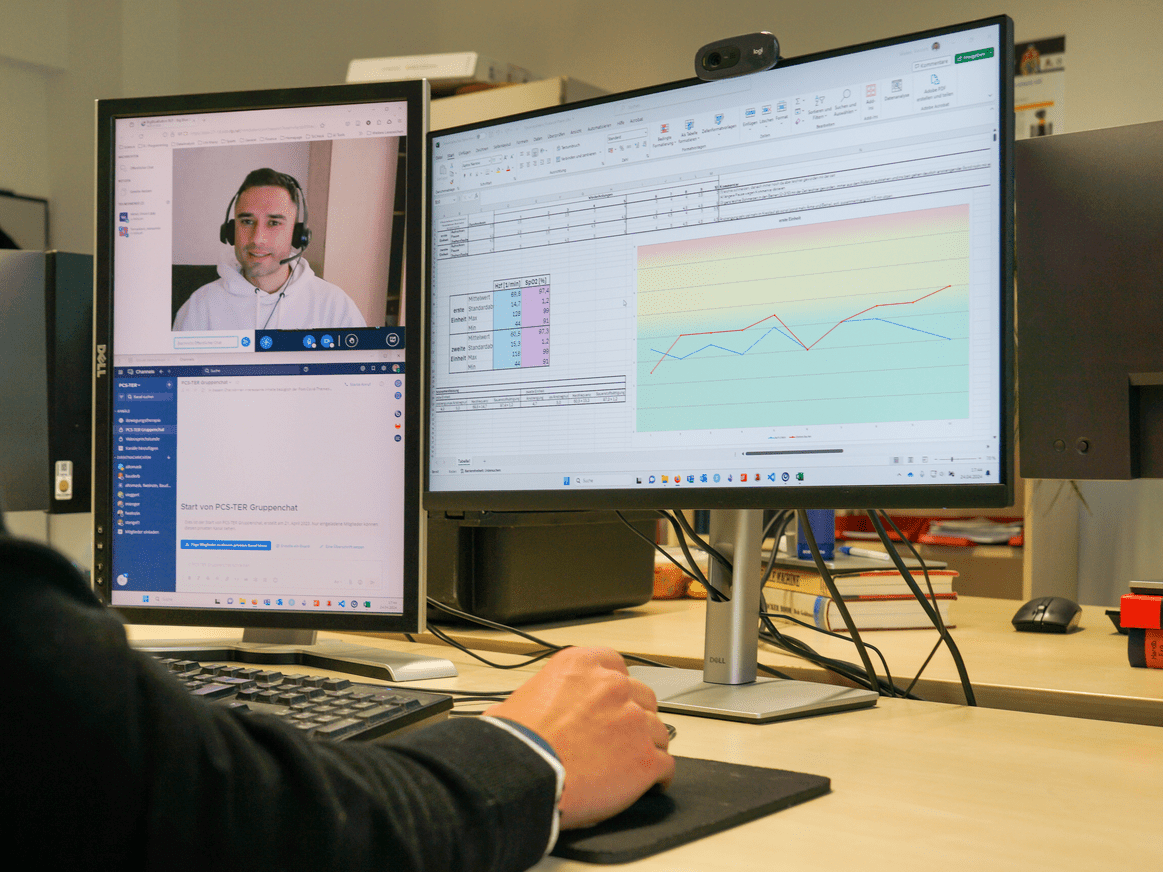
Digital InfrastructureThe digital infrastructure that will be used during the intervention phase is divided into 2 different parts. The first part is a central website designed for mobile and desktop use (WordPress; WordPress Foundation). The second part is the software Mattermost (mobile app and desktop application; Mattermost Inc), which is used for digital communication between therapists and patients ().
The website consists of three main areas: (1) a diary for monitoring and controlling clinical condition and training load in addition to the questionnaires already described in the Questionnaires section; (2) an education area with short educational videos about PCC and exercise and respiratory therapy, including a detailed explanation and the pacing approach; and (3) a therapy program area including written materials (eg, weekly therapy plans, instructions for each type of training, and control of training intensity depending on the clinical condition).
All important information on the use of the website and Mattermost is provided in the study manual and communicated to the participants via email. In the intervention phase, the IG participants can use both the website and Mattermost. In contrast, CG participants can use only a restricted version of the website (without the education or therapy program areas) for daily reporting via the clinical condition and training load diary (CCTD).
 Figure 3. Digital infrastructure—(A) website for mobile and desktop use (WordPress; WordPress Foundation) and (B) mobile app and desktop application (Mattermost; Mattermost Inc). The author consents to the use of their image in this publication. IG Setup
On the basis of the CPET results and the algorithm listed in , IG participants are assigned to 1 of 3 TGs (TGs 1-3). Before the start of the intervention, all participants will take part in 2 video-based introductory sessions in the first week to get to know their personal sports therapist, learn how to use the digital infrastructure, and understand the therapy program (eg, therapy plan and monitoring of clinical condition).
Therapy ProgramDue to the short duration of the intervention (8 weeks), and to achieve an optimal balance between the disease condition and the workload, a therapy plan () was created consisting of a combination of exercise and respiratory training types and training and recovery days and following the principle of periodization and cyclization. The intervention and follow-up phases during the 16-week period are divided into 2 mesocycles (8 weeks) and 16 microcycles (1 week). A change in TGs (eg, from TG 1 to TG 2) is planned for the end of the mesocycle if an objective improvement (or deterioration), measured through the CPET and evaluated using the described TG allocation algorithm (), occurs. Depending on the clinical conditions and the RPE during the training (eg, no worsening of clinical symptoms, and the subjectively perceived training intensity is too low), an individual group change within the mesocycle can also take place in consultation with the head physician (PS). The therapy plan can be individually adapted after each microcycle in teleconsultations with the sports therapist.
The cyclical alternation () between training and recovery days aims to consider the workload from the previous training session and provide the body with time to adapt. For example, if the intensity of the previous training session was too high, the study participants have time to reflect on this training stimulus and adapt, thus preventing the occurrence of PEM or not increasing its severity in a new training session. The training days include both exercise and respiratory training sessions held in the morning and afternoon. Because of the low exercise intensity, only TG 1 participants engage in daily training involving respiratory and postural orthostatic tachycardia syndrome (POTS) training, one in the morning and the other in the afternoon. The training sessions are conducted asynchronously for all participants (ie, without real-time therapist supervision).
 Figure 4. Exemplary weekly therapy plan for training group 3. The intervention and follow-up phases during the 16-week period are divided into 2 mesocycles (8 weeks) and 16 microcycles (1 week). The therapy plan consists of a combination of exercise and respiratory training types, training days, and recovery days. Participants are required to keep a daily clinical condition and training load diary (CCTD).
 The 3 TGs () follow an individual therapy plan. For TG 1 participants, the therapy plan includes respiratory and POTS training every day for at least 5 times a week. Respiratory training is based on the apnea training method [] for increasing hypoxemia and ischemia tolerance. One respiratory training session consists of 3 sets of 4 repetitions, with a 2- to 3-minute pause between sets. One repetition consists of a deep inspiration maneuver followed by 4 maximum expirations and 4 submaximal inspirations to the level of resting inspiration. A POTS training session involves position change exercises for the arms, legs, and total body (eg, an exercise for standing up). The structure of each exercise consists of 2 to 3 sets with 5 to 10 repetitions, with a 1-minute pause between sets and 2 to 3 minutes between each exercise. For TG 2 and TG 3 participants, the therapy plan includes exercise training every other day but at least 3 times a week. TG 2 engages in endurance training as a substitute for POTS training. Moreover, TG3 performs both endurance and strength training instead of POTS training. Endurance training, designed as aerobic interval training and patient-centered training (eg, walking and cycling), entails 1 to 2 sets for TG 2 and 2 to 3 sets for TG 3, with a set pause of 2 to 3 minutes. Each set involves 8 to 10 repetitions with a work-to-rest ratio of 30:30 seconds or 30:60 seconds, with intensity control based on the modified Borg scale (0-10) for the RPE. Work intervals aim for an intensity between 4 and 6 (less strenuous to moderately strenuous), whereas (active) rest intervals target an intensity between 1 and 3 (very light to light). The individual progression or regression of the training load (eg, number of sets, intensity, and active or passive rest) depends on the patient’s exercise tolerance and can be carried out in consultation with a personal sports therapist.
All participants, both from the IG and CG, will be asked not only to practice additional pacing in their daily lives to be active but also to use their energy resources as efficiently as possible. To this end, based on the Royal College of Occupational Therapists pace, plan, and prioritize principle pacing approach [] and the CPET, we developed a new objective pacing approach based on PO at the individual anaerobic threshold—ventilatory threshold 2/lactate threshold 2 (VT2/LT2)—as the threshold for continuous physical capacity and the metabolic equivalent of task ( []) [,].
 Figure 5. Objective pacing approach based on metabolic equivalent of task (MET) data from the work by Jetté et al developed by the Department of Sports Medicine, Prevention, and Rehabilitation at Johannes Gutenberg University Mainz. Objective pacing approach using cardiopulmonary exercise testing (CPET)–derived thresholds, illustrated with a 30-year-old female pilot participant. A continuous physical capacity threshold of 55 W (approximately 3 METs) corresponds to the individual’s ventilatory threshold 2/lactate threshold 2 (VT2/LT2). Household activities are categorized by intensity and color coded for pacing guidance (red: 30:30 seconds; yellow: individual pacing; green: continuous work). PPO: peak power output; VO2peak: highest oxygen uptake rate achieved during the cardiopulmonary exercise test.
 Our pacing strategy is based on the following six foundations:
Prevention of worsening of symptoms and occurrence of PEM: this requires the identification of extrinsic and intrinsic factors that trigger PEM, such as physical, cognitive, and emotional stressors.Planning and prioritization of activities: this entails combinations of activity and rest phases and alternations among different activities (physical, cognitive, and emotional).Pacing: all participants will receive a list of different physical activities sorted according to their metabolic equivalent of task.Continuous physical activity is permitted only within the limits of dominant aerobic energy metabolism (<VT2/LT2) and must be subjectively well tolerated by the patient. If the desired physical activity is >VT2/LT2 in intensity, it should be performed only using the 30:30-second interval method (work-to-rest ratio of 1:1).A cautious and gradual increase in activity can only occur if the clinical condition is good (refer to the Monitoring of Clinical Condition and Control of Training Load section).The duration of work intervals and the work-to-rest ratio can be individually adjusted in consultation with a personal sports therapist depending on the patient’s clinical condition, exercise tolerance, and regeneration capacity.During the intervention phase, all participants will have weekly teleconsultations (30-minute video calls) via Mattermost with their personal sports therapist. The teleconsultation is designed as a motivational and problem-solving interview with the following structure: (1) review of the previous week, highlighting all the positive aspects; (2) discussion of the current clinical and training-related form; and (3) outlook or intervention modifications for the upcoming week. In addition to the clinical condition (clinical symptoms) and training-related variables, the participants are asked to record any problems that arise during training or throughout the day. During the intervention phase, participants have the opportunity to use a chat function to consult with their personalized sports therapist for immediate support. In addition to weekly teleconsultations between therapists and participants, the therapy team should meet once a week to discuss the progress of therapy and current challenges, among other things. The therapy team consists of 5 therapists and a head physician (PS), and they are not blinded.
Monitoring of Clinical Condition and Control of Training LoadThe daily recording of the clinical condition and the interactive adjustment of the exercise therapy aim to reduce or stabilize symptoms and adjust the training load to achieve the best training effects.
The training load is regulated based on clinical condition using a clear algorithm (), which includes the assessment of the clinical condition based on the RPSS and the recommendation of the training intensity based on the RPE. The RPSS was developed through our clinical experience with PCC at the sports medicine outpatient clinic, Mainz [], and through insights from international literature on symptom occurrence in PCC [,]. The successful application of the RPSS in practice constitutes its inaugural use in a scientific context. Both the RPSS and RPE will be documented in the CCTD. The RPSS comprises the following items: fatigue (influenza-like symptoms or general fatigue), dyspnea (breathing difficulties, headache, visual disturbance, or chest pain), POTS (high pulse, inability to stand, and need to lie down), anxiety (anxiety, panic, or avoidance), and hypersensitivity (to noise, light, temperature, touch, and muscle or nerve pain). Each symptom group is accompanied by a free-text field for individual comments that is rated on a scale from 0 to 10 (0=no symptoms; 1-3=mild symptoms; 4-6=moderate symptoms; 7-10=severe to maximum symptoms).
Participants are asked to complete the RPSS every day in the morning to monitor their clinical condition. Every day starting with even 1 symptom cluster between 7 and 10 (PEM condition) is counted as a PEM episode. On training days, the RPSS should be completed by the start of training at the latest. To avoid exercise-induced overload and exacerbation of clinical symptoms, patients should only exercise when each symptom cluster has a value of ≤6 (). Ideally, each symptom cluster should have a value between 0 and 3 (good clinical condition); then, patients can train with an RPE of 4 to 6 to maintain good clinical condition and provide an effective training stimulus. Even if only 1 symptom cluster has a value between 4 and 6 (pre-PEM condition), the RPE value must be set to 1 to 3 during training. However, if any of the RPSS symptom clusters have a value between 7 and 10 (PEM condition), the upcoming exercise training session should be canceled. In addition, if symptoms increase by ≥7 values during training, the current training session should be stopped. Finally, an increase in symptoms of ≥2 values in more than one symptom cluster should also be considered for training load management. The possible cause for this increase should be investigated via teleconsultation. After the training session or at the end of the day at the latest, participants are required to document their training (type; duration; intensity; and, if applicable, commentary) in the CCTD.
 Figure 6. Clinical condition is assessed based on symptom severity (rating scale for perceived symptom severity), and training recommendations (rating of perceived exertion [RPE]) are derived accordingly. If any symptom cluster indicates a more severe condition (eg, pre–postexertional mala
Comments (0)