This study demonstrated that adding 3D FLASH sequences enabled reliable detection of OPLL in cervical spine MRI with higher sensitivity and accuracy than that observed with conventional T1- and T2-weighted TSE sequences alone. Furthermore, reader confidence and interobserver agreement were better when 3D FLASH was added to the TSE sequences. The OPLL cases detected after reviewing 3D FLASH sequences tended to be thinner than those detected on TSE only.
Recent studies evaluated the osseous structures causing neural foraminal stenosis or central canal stenosis on cervical spine MRI using CT-like MR images [4, 16,17,18]. Most of these studies utilized ultrashort-echo time (UTE) or zero-echo time (ZTE) sequences, demonstrating the usefulness of these sequences for assessing bony components of neural foraminal stenosis or distinguishing disk herniation from posterior osteophytes [4, 16,17,18]. Schwaiger et al. compared two CT-like MR sequences, 3D GRE and UTE, for evaluating fractures and degenerative bone changes in the lumbar spine, using CT as a reference [19]. They reported that 3D GRE had higher agreement with CT and better image quality. UTE, while providing good tissue contrast, required twice the slice thickness for a similar acquisition time and was more sensitive to motion artifacts and B0 inhomogeneity, whereas GRE-based images were more affected by metallic artifacts. Based on their findings, 3D GRE was suggested to be more advantageous for clinical use under comparable imaging conditions.
Several reports have demonstrated the use of CT-like MR with ZTE and synthetic CT images in diagnosing OPLL [18, 20,21,22]. In a study by Tran et al., no significant difference was observed in the detection of OPLL with or without ZTE (OPLL was identified in 10 cases without ZTE and 11 cases with ZTE), which could be due to the small number of OPLL cases included [18]. Jeong et al. reported that reviewing synthetic CT increased sensitivity (reader 1: 47 to 90%, reader 2: 63 to 93%) but decreased specificity (reader 1: 98 to 89%, reader 2: 94 to 84%). In contrast, our study showed increases in both sensitivity and specificity, with a notable improvement in specificity. Similar to our findings, their study also reported improved inter-reader agreement, rising from moderate to good after adding CT-like images [22]. To the best of our knowledge, no study has investigated the utility of 3D T1 GRE-based CT-like images for detecting OPLL. In this study, using CT as a reference standard for all cases demonstrated that 3D FLASH could aid in reliable detection of OPLL.
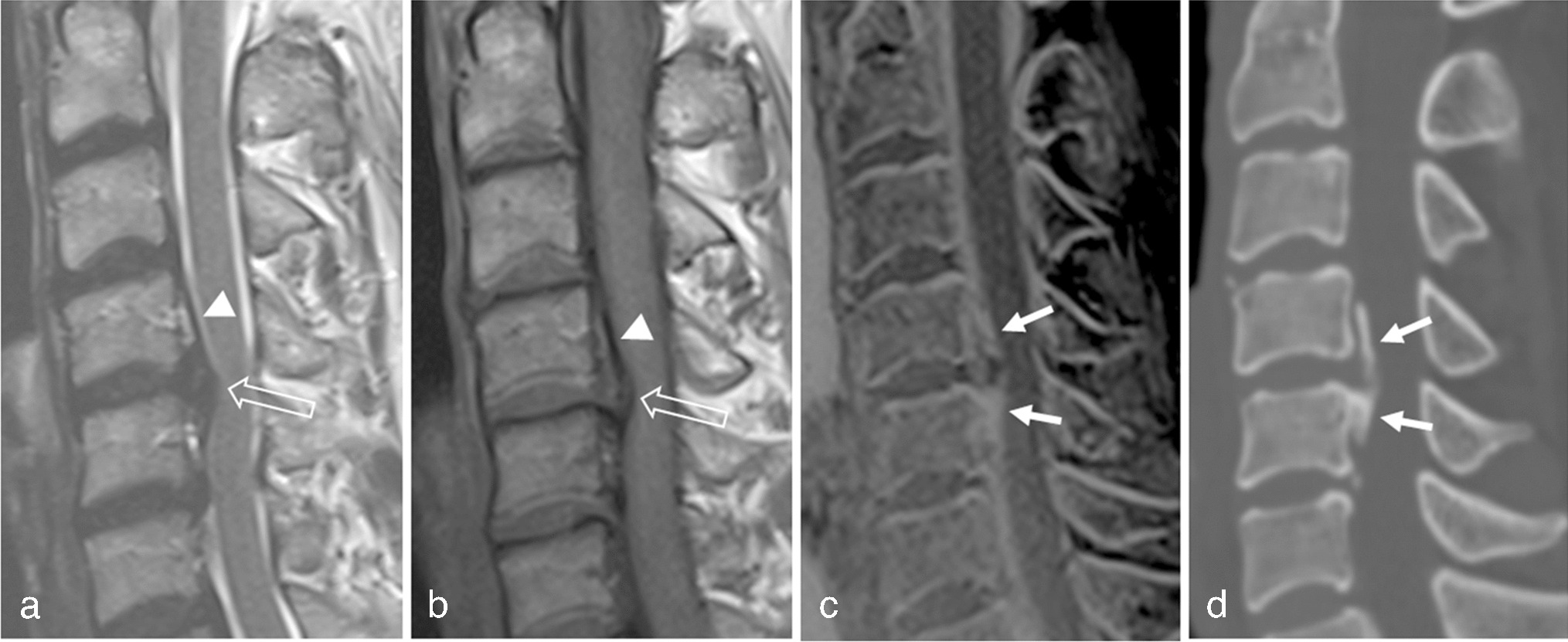
Thick OPLL is well demonstrated on conventional MRI, with T1 intermediate to high SI in the OPLL reflecting fatty bone marrow. However, thin OPLL appearing as a thin hypointense band is difficult to distinguish from thickened PLL, disk herniation, or prominent anterior epidural vascularity [21]. Figure 1 shows a case of OPLL that was missed on the TSE. Both readers observed disk herniation, and a thin hypointense band was considered to indicate elevated PLL. After reviewing the 3D FLASH images, both readers detected OPLL which was confirmed by CT. In contrast, Fig. 2 illustrates an over-diagnosed case, where a low-signal intensity band posterior to the vertebral body on TSE imaging was mistaken for calcified PLL. This was disproven by 3D FLASH and CT images. The authors speculate that the hypointense band could be due to the PLL elevation from disk herniation and chemical shift artifact. Both cases highlight the difficulty of accurately assessing OPLL on TSE images and demonstrate the added value of 3D FLASH for clarification. Furthermore, considering the result that the OPLL in the TSE group was thicker than that in the 3D FLASH group, 3D FLASH appears to be especially helpful for difficult cases with thin OPLL. Reader confidence and interobserver agreement also improved after viewing the 3D FLASH. Therefore, 3D FLASH could serve as a problem-solving tool when an ambiguous osseous lesion is observed on TSE, thereby reducing the need for additional CT scans.
The implementation of CT-like MRI sequences in clinical practice could potentially streamline decision-making processes for cervical spine surgeries. Currently, many patients undergo both MRI and CT scans preoperatively. By incorporating these advanced MRI techniques, we may be able to simplify the diagnostic workflow, potentially reducing the need for separate CT scans in some cases. While further research is needed, this approach could lead to decreased healthcare costs, efficient resource utilization, and reduced radiation exposure for patients.
This study had several limitations. First, 3D GRE-based CT-like images are sensitive to susceptibility and motion artifacts. Although we excluded patients with a history of cervical spine surgery, 3D FLASH could provide inadequate image quality in such patients [19, 20]. However, this sequence is commonly available and has low hardware and software requirements [20]. Second, unfamiliar tissue signals can increase the difficulty of image interpretation when compared to CT. Therefore, experience with this particular sequence would likely be needed for accurate interpretation. Third, although posterior osteophytes are an important cause of central canal stenosis, we did not evaluate them because we intended to focus on OPLL. We plan to focus on posterior osteophytes in future studies. Fourth, this study used both per-patient and per-level analyses. OPLL-type classification required assessing overall morphology, necessitating per-patient analysis. For OPLL thickness, differences in angulation between CT and MRI made per-level measurements challenging, so we measured at the level with the greatest thickness and the most similar angulation between modalities. Although less detailed than per-level analysis, this approach addressed broader questions. Future studies with standardized imaging protocols could provide more detailed insights. Lastly, this retrospective design limited our ability to calculate the sample size prospectively. A post hoc power analysis using G*Power, however, confirmed that our sample size met the requirement for a power of 0.7 [23]. Furthermore, the statistically significant differences observed, in conjunction with the post hoc power analysis, suggest the sample size was adequate for the study objectives. Future studies are encouraged to aim for higher statistical power with sample sizes calculated during the planning phase for greater robustness.
In conclusion, 3D FLASH can be helpful for detecting OPLL when combined with conventional T1- and T2-weighted images. These results support increasing evidence that using CT-like MR to assess osseous structures can avoid further work-up with CT imaging.


Comments (0)