
Remember me
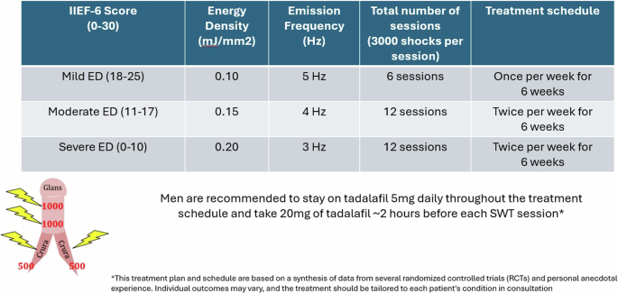


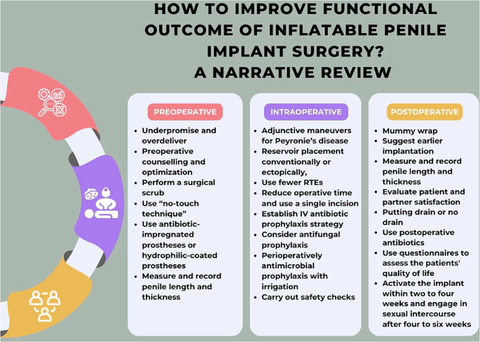


Figure 1 describes the study inclusion process in the form of a flow chart following PRISMA (2020) [13]. The initial search on the 26th of January 2023 identified 1303 records. Title and abstract screening were performed, after which 24 studies fulfilled the PICOS criteria, which then underwent further evaluation. From these studies, an additional 18 papers were excluded due to insufficient reporting on the details of FSE, and its concordance with final paraffin histology, which rendered them unsuitable for our systematic review. An updated search was performed on the 12th of September 2024 which retrieved 124 papers, 1 of which was included for analysis. Overall, 7 studies were included, and the extracted data are summarised in Table 1. There were no randomised or non-randomised controlled studies, and all 7 studies included were observational studies. The total number of patients with intraoperative FSE was 574.
Fig. 1
Flow diagram of the search following PRISMA (2020) [13].
Table 1 Studies histopathology and outcomes results.Accuracy of frozen section examination margin reportsFSE for PeCa is not routinely used for initial diagnostic purposes. Instead, margins are taken to confirm the complete excision of the tumour. The sensitivity, specificity, PPV, NPV, and accuracy results are summarised in Table 2. In both Li et al. and Morelli et al.’s studies, there was 100% concordance, the PPV and sensitivity calculation cannot be calculated due to 0 patients with positive margin values [18, 19]. All studies showed a high percentage accuracy of intraoperative FSE with a range from 92.9 to 99.4% and a mean accuracy of 95.4%. Additionally, the mean sensitivity, specificity, PPV, and NPV were 71.4%, 99.9%, 98.8%, and 96.5%, respectively [18,19,20,21,22,23,24].
Table 2 Accuracy of reports comparing FSE of margins results with final histopathology.Indications of frozen section examination in penile cancer surgeryRadical penectomy and total penectomy can have intraoperative FSE indications for ensuring that the excision margin is clear of PeCa. Intraoperative FSE during total penectomy is performed at the discretion of the surgeon or in line with the departmental policy. Danakas et al. showed the benefit of FSE during total penectomy with 4 cases showing negative conversion of margins with initial positive or atypical FSE. However, they concluded that there was not a significant impact on final surgical margin status nor long-term oncologic outcomes with the use of FSE [20].
PPS can be performed in cases where it is possible to completely excise the tumour and maintain a functional penile length. Studies utilising partial penectomy, radical circumcision, wide local excision (WLE), glansectomy and glans resurfacing were reviewed. Parnham et al. presented 171/177 patients from February 2005 to January 2016 who underwent glansectomy and split-thickness skin graft (STSG) with intraoperative FSE. They described biopsies of the complete circumference of the urethra and of each corporal tip which was sent for FSE. These improved outcomes in at least 10 patients who had further resection following a positive FSE result [23]. A further 15 cases of glansectomy were shown to prove 100% accuracy [19]. Additionally, FSE can be performed in total glans resurfacing where spongiosal biopsies of the deep margin are taken. O’Kelly et al. presented total glans resurfacing, showing 95% accuracy of FSE where one case had an FN result [22].
Final margin status and risk of recurrenceLocal recurrence (LR) was a secondary outcome measure assessed. One study suggested that LR most likely occurs within 6 months post-operation [18], whereas another suggested that it occurs within 2 years [21]. Two studies provided the median time of recurrence at 8.7 and 10 months [21, 23]. The range of LR in all studies was 0–16%, although patients were not followed up for the same amount of time in the selected reports, the mean risk of patients having a local recurrence was 7.9% across all studies [18,19,20,21,22,23,24]. Danakas et al. suggested that patients with positive surgical margins tend to have a higher risk for disease recurrence, compared with those with negative surgical margins [20]. This suggested that the risk of a positive margin is greater when PeCa is removed without the use of intraoperative FSE. Pang and Yunis et al. examined the local recurrence rate in 137 patients undergoing PPS surgery for penile SCC. In this study, the overall LR rate was 10.2%. Among patients with positive FSE margins, the recurrence rate was higher at 25%, compared to 8.5% in those with negative FSE margins [24]. This suggested that positive surgical margins, even when identified and addressed with further resection during surgery, may still carry a higher risk of LR.
Postoperative complications and patient-reported outcomesAcross 4 studies, where complications were reported, there was an average complication rate of 17.7% [18, 19, 22, 23]. Li et al. presented 3/32 (9.4%) complications where 2 patients had wound dehiscence, and 1 patient had the formation of a local abscess [18]. Whilst Morelli et al. presented a higher rate of complications at 26.7% with 2 partial graft loss’, 1 meatal stenosis, and 1 phimosis [19]. The highest complication rate in one study was 34.5%, where 29 patients had partial graft loss, 5 had complete or near-complete graft loss, 12 underwent regrafting, and 4 reported meatal stenosis [23]. Depending on the type of complication, the patients underwent further surgery, required pharmacological intervention to manage the complication or had conservative management. However, one study reported 0% complications following 19 total glans resurfacing operations [22]. Additionally, 3 studies assessed changes in sexual function. One study showed that 24.1% of patients maintained poor sexual function, and the other 75.9% had no change after surgery [18]. Whereas another study presented an 81% improvement in sexual function [19]. The final study maintained sexual functionality in all patients [22].
Two studies from this review analysed metastatic disease following surgery. Pang and Yunis et al. show 24.1% of patients with regional or distant metastasis post-surgery with a median follow-up to metastasis of 5 months [24]. Li et al.’s cohort of patients had 5 (15.6%) with lymph node metastases, none of whom had adjuvant therapy before or after surgery. All of their patients reported satisfactory urination [18].
Validity assessmentThe RoB was evaluated in each of the papers, results are outlined in Supplementary Table 3. Danakas et al. received a high-quality rating, whilst the other 6 studies received a moderate-quality rating [18,19,20,21,22,23,24]. Confounders were identified in each of the studies, which included tumour stage, tumour grade, nodal stage, quality of excision (surgeon preference or case complexity), and prior therapy, however, Danakas et al. showed no statistically significant difference in the histopathological characteristics of the patients [20].
Comments (0)