
Remember me


The prognosis of lung metastasis is better than that of other visceral metastases except for skin or distant node metastases. Therefore, lung metastasis is classified separately from other distal metastases in the melanoma staging of the American Joint Committee on Cancer [12, 17]. Several studies showed a poor prognosis of mucosal melanoma compared with that of cutaneous melanoma. Prognosis by clinical subtypes may depend on available agencies [18]. Since BRAF V600E/K mutation in mucosal melanoma is less frequent than that in cutaneous melanoma, most of the patients with mucosal melanoma have no treatment options with BRAF/MEK inhibitors. Furthermore, the efficacy of immune checkpoint inhibitors is lower in mucosal melanoma than in cutaneous melanoma [19].
In this study, GGNs were observed in 14.9% of patients with pulmonary metastases from malignant melanoma. We observed five case reports of lung metastases from malignant melanoma that appeared as GGN in previous studies [5,6,7,8,9]. To our knowledge, this is the first study to report on the frequency of lung metastases from malignant melanomas appearing as GGNs. In contrast, lung metastases from pancreatic cancer reportedly have a high frequency of air-space patterns [1,2,3,4]. Aissaoui et al. reported that among 76 patients with pulmonary metastasis from pancreatic cancer, three (4%) comprised pure GGN [3]. In this study, four patients (4.6%) had only pure GGN metastasis (Type 4), which is similar to that reported by Aissaoui et al. [3]. At least 10 patients exhibited GGNs in Aissaoui's report (7 with halo signs and 3 with pure GGNs; 10/76 = 13.2%). The frequency of GGN metastasis was 14.9% in our study. Although the classifications used in his study and ours differ, making detailed comparisons challenging, we believe that lung metastases from malignant melanoma may present GGNs approximately as frequently as lung metastases from pancreatic cancer. As previously mentioned, 54.5% of 11 patients with enlarged nodules experienced a change from GGN to solid nodules. Depending on when the follow-up CT was performed, the GGN may have already changed into a solid nodule, which may have affected the frequency of GGNs observed in these studies.
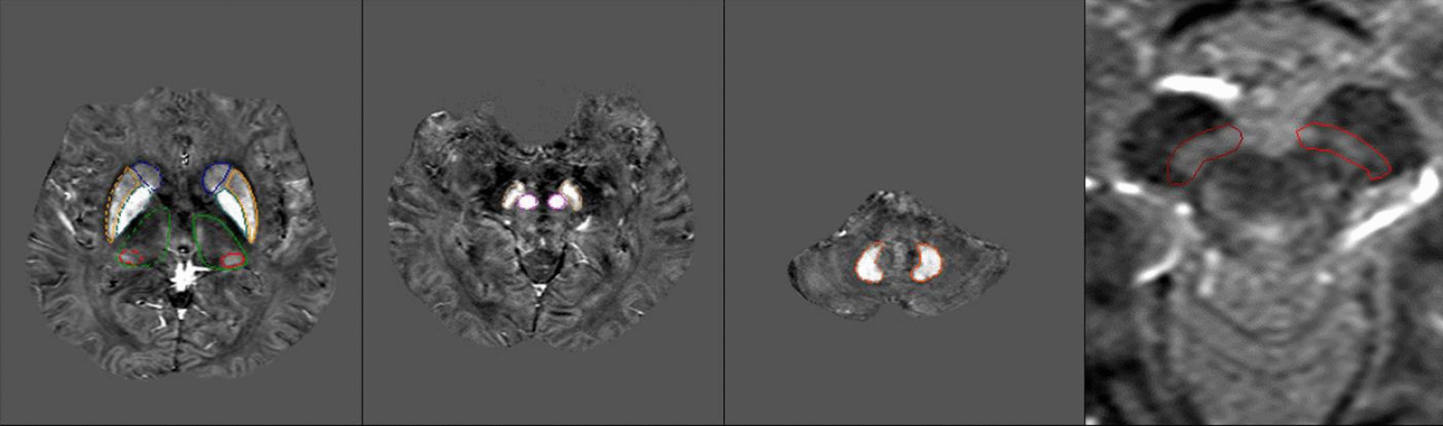
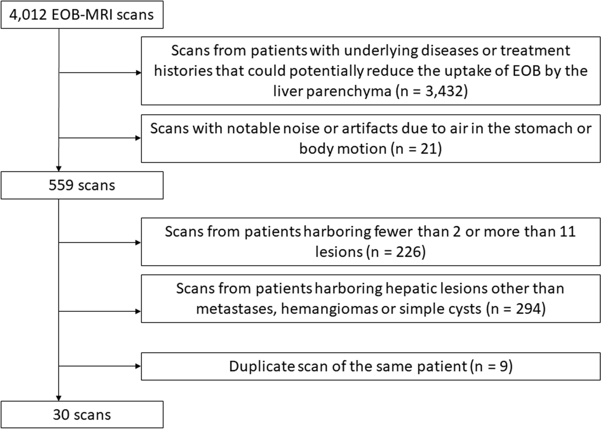
Various pathologies, including inflammation, focal fibrosis, atypical adenomatous hyperplasia, lung adenocarcinoma, and, rarely, metastatic tumor, present with ground-glass opacity. The most important differential diagnosis for GGN is lung adenocarcinoma. It is difficult to differentiate GGN from lung cancer and metastases from malignant melanoma on CT without follow-up CT (Fig. 6). We believe that the difference in the tumor doubling time is a point of differentiation between metastatic malignant melanoma and lung adenocarcinomas. In our study, the tumor doubling time was 52.0 ± 33.5 days (SD)/50.9 days (median) (range: 10.9–111 days). As for lung metastases of malignant melanoma, Okita et al. reported that multiple GGO lesions increased after 1 month [5]. Mizuuchi et al. reported a solitary GGN with a tumor doubling time of 230 days [6]. Kang et al. reported a solitary GGN that enlarged from 7 to 11 mm in diameter over 3 months [8]. Masuda et al. and Daliaz et al. reported a case of metastatic malignant melanoma identified as a rapidly increasing GGN (the exact tumor size was not described) [7, 9]. In contrast, regarding the rate of tumor growth in lung adenocarcinoma, Aoki et al. reported that Noguchi classifications A, B, and 48% of C had a tumor doubling time of > 1 year [20]. Hasegawa et al. reported mean volume doubling times of 813 and 457 days for pure GGN and part-solid nodules, respectively [21]. Kakinuma et al. reported that 90% of GGNs < 5 mm were stable. They also reported that the duration of the appearance of solid components in adenocarcinomas was 3.6 years [22]. The tumor growth rate of malignant melanoma metastasis is faster than that of lung adenocarcinoma, which may be a differentiating factor.
Fig. 6
A 67-year-old woman with mucosal melanoma. a. CT 24 months after the operation shows 3 mm-sized pure GGN in the right lower lobe (white arrow). b. After 6 months, the GGN had grown to 8 mm and turned into a part-solid nodule (white arrowhead). This nodule resembled a lung adenocarcinoma; however, it grew at a faster rate than a lung adenocarcinoma. In addition to this nodule, there were multiple fast-growing GGNs and solid nodules in bilateral lungs (not shown). This nodule was diagnosed as lung metastasis
However, differences in the tumor doubling time should also be reflected in the follow-up period. According to Lung Imaging Reporting and Data System (Lung-RADS) Version 2022, CT examination within 12 months is recommended for non-solid nodules less than 30 mm and part-solid nodules with a total diameter < 6 mm, whereas CT examination within 6 months is recommended for non-solid nodules with a diameter greater than 30 mm, part-solid nodules with a total diameter greater than 6 mm, and solid components with a diameter less than 6 mm [23]. A GGN detected in the lungs of a patient with a history of malignant melanoma indicates a possible metastasis, and using the same follow-up time as in the Lung-RADS Version 2022 may be considerably late. In our study, the tumor doubling time was 52.0 ± 33.5 days; thus, we recommend short-term follow-up at 1–2 months.
Pathologically, hemorrhage and lepidic tumor growth are the most common causes of pulmonary metastases with GGO [1,2,3,4,5,6,7,8,9, 24, 25]. There are reports of GGN metastases from hemorrhage, including malignant melanoma, angiosarcoma, choriocarcinoma, osteosarcoma, and Kaposi’s sarcoma [1, 24, 25], and those from lepidic tumor growth, including malignant melanoma, pancreatic cancer, colon cancer, breast cancer, and gastric cancer [1,2,3,4,5,6,7,8,9]. We have only two pathologically proven cases, and there was no hemorrhagic metastasis.
Our findings suggest that lung metastasis with GGN is more likely to occur in mucosal melanomas than in cutaneous or acral melanomas (Table 1). Melanoma types in previous reports were variable and included one mucosal, two cutaneous, one acral, and one uveal melanoma [5,6,7,8,9]. More cases are warranted to determine whether GGNs are more likely to be present in mucosal melanoma.
This study had certain limitations. First, CT examinations were performed using various protocols owing to the retrospective nature of the study. Second, many patients in our study did not have pathologically confirmed pulmonary metastases from malignant melanoma. Third, the number of patients with GGN metastasis was small.
In conclusion, 14.9% of patients with lung metastases of malignant melanoma showed GGNs. Tumor doubling time is useful for differentiating between lung metastasis of malignant melanoma and lung adenocarcinoma. We suggest that short-term follow-up CT should be used to differentiate lung metastases from malignant melanoma and other malignancies that appear as GGNs.
Comments (0)