
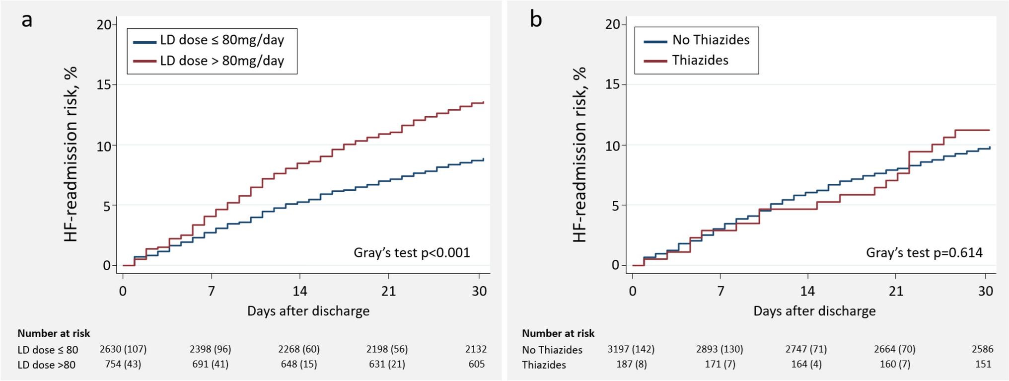
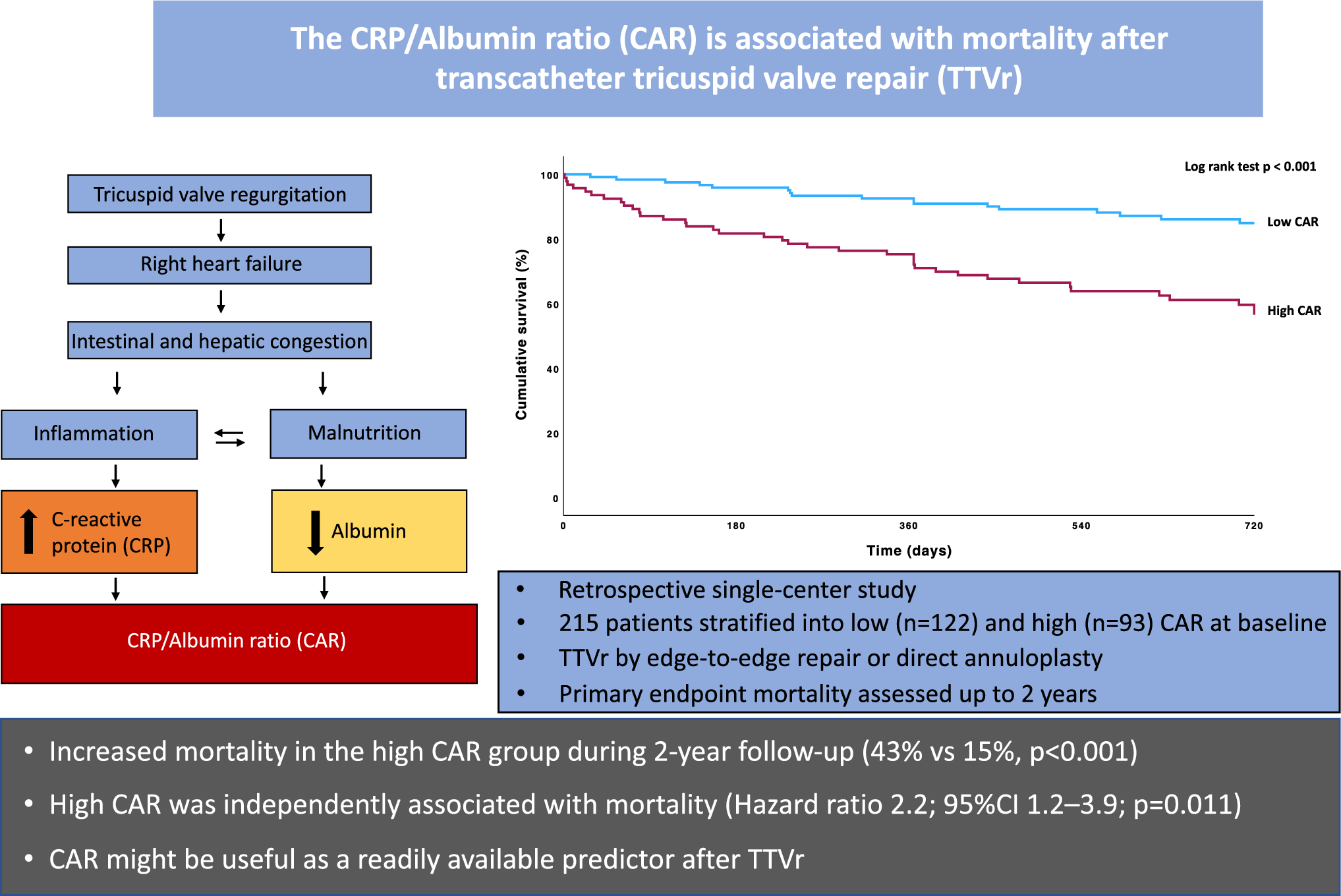
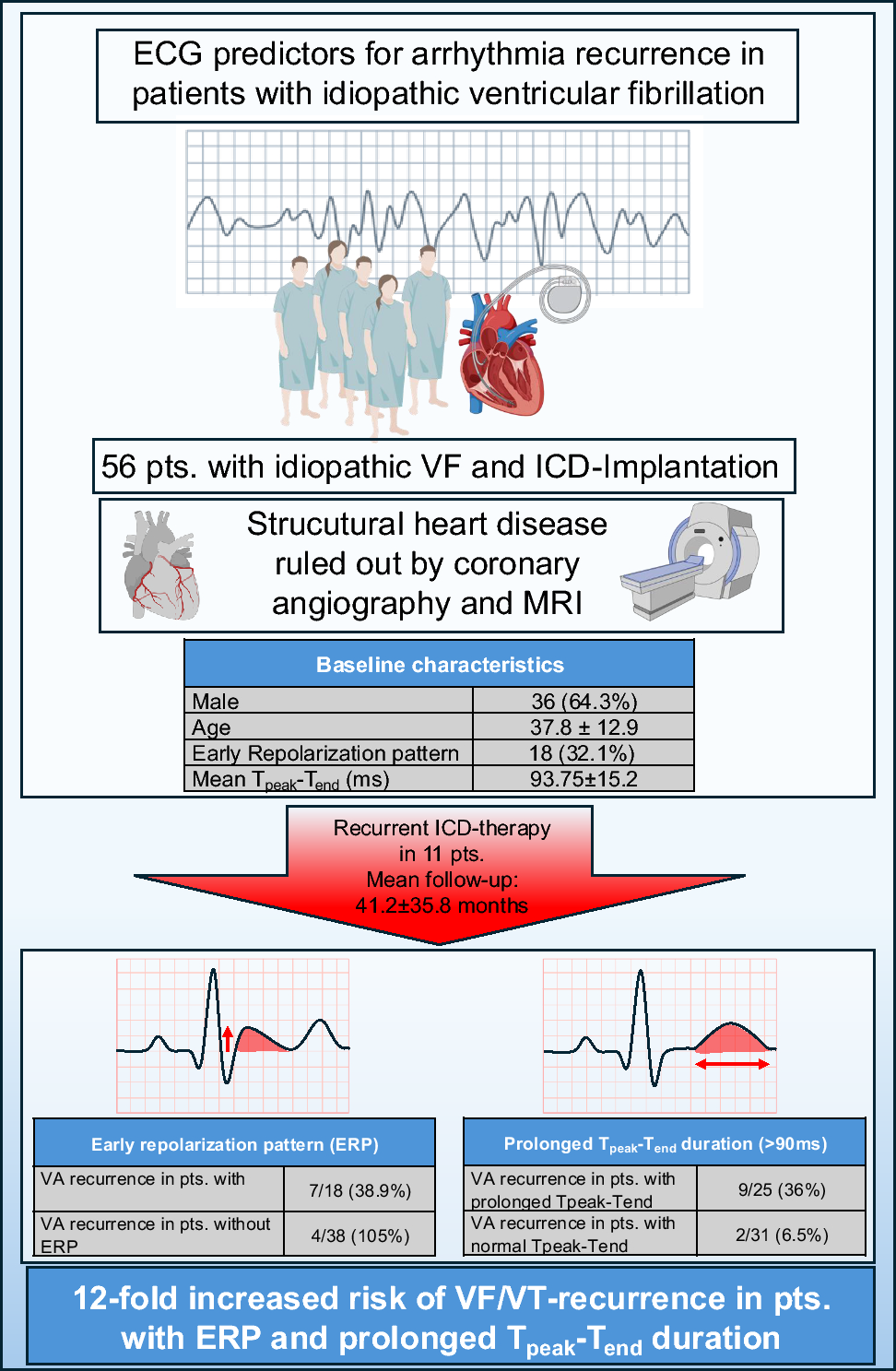
Remember me


Of a total of 304 patients with DMR and complex valve anatomy enrolled in the prospective registry, 36 patients (11.8%) were lost to follow-up. There were no significant differences in baseline characteristics between patients lost to follow-up and those not lost to follow-up (Supplemental Table 2). Of the 169 patients eligible for 3-year follow-up, TTE was complete for 127 (75.1%, Fig. 1). The median follow-up duration was 1214 days. Patients were elderly (mean age 80 ± 7), predominantly male (61.5%) and 69.7% were in NYHA class III/IV. There was a high burden of comorbidities including coronary artery disease (48.4%), atrial fibrillation (60.2%), chronic kidney disease (50.3%) and diabetes mellitus (28%), as shown in Table 1.
Fig. 1
Flowchart of patients with anatomically complex DMR. Illustration of enrollment of patients with anatomically complex DMR in the MitraUlm Registry and follow-up at 1, 2 and 3 years; DMR degenerative mitral regurgitation, TTE transthoracic echocardiography
Table 1 Baseline characteristicsEchocardiographic and invasive hemodynamic assessmentEchocardiographic assessment at baseline revealed a mean left ventricular ejection fraction of 57 ± 8%, the mean mitral valve area was 5.4 cm2 and the mean mitral gradient was 2.5 mmHg (Table 1). The most common anatomical complexities were the presence of ≥ 2 independent jets (33.5%), mitral valve orifice area < 4 cm2 (12.3%) followed by multisegmental prolapse (12%) and commissural lesions with wide/multiple jets (10.9%), as shown in Central Illustration A and Supplemental Table 1. 21.2% of the patients had very complex valve anatomies including cleft, severe calcification in the grasping zone, short leaflet length < 8 mm or large tissue defect in the leaflet. Complexity criteria did not change significantly over time except for the prevalence of cleft, which increased significantly from 2.8% in patients treated with the first, second and third Generation MitraClip to 10.7% in patients treated with the fourth-Generation MitraClip, Supplemental Table 1. 241 patients (79.3%) had one anatomical complexity, whereas 20.7% of patients had two anatomical complexities. Invasive hemodynamic measurements showed elevated systolic pulmonary artery pressures (50 ± 11 mmHg) and mean left atrial pressures (23 ± 6 mmHg).
Periprocedural outcomes in anatomically complex DMRProcedural and 30-day outcomes are shown in Table 2. Successful device implantation was achieved in 92.4% of patients. The mean number of implanted clips per patient was 1.6, with a mean device time of 84 minutes and a mean fluouroscopy time of 25 minutes. MitraClip Generation 1, 2 and 3 were implanted in 66.8 % of patients, whereas 33.2% of patients received MitraClip Generation 4. A total of 75.2 % of patients treated with MitraClip Generation 4 received wide clips, either NTW or XTW alone or in combination with the other 2 clips (NT or XT). The 30-day MACCE rate was 8.9 % and the 30-day all-cause mortality rate was 4.3%.
Table 2 Procedural and 30 days outcomesEchocardiographic outcomesAt discharge, 93.8% of patients presented with MR ≤ 2 and 58.6% had MR ≤ 1. At 3 years, these proportions were 85.9% and 45.7% (Fig. 2A). Patients treated with the fourth-generation MitraClip system had a significantly higher proportion of MR ≤ 1 compared to patients treated with the first-, second- and third-generation MitraClip devices both at discharge (74.3% vs 50.7%, P < 0.001) and at 3-year follow-up (56.5% vs 39.5%, P = 0.064). Patients with multisegmental prolapse and commissural lesions with wide/multiple jets had the lowest MR reduction at discharge (MR ≤ 1 in 43.2% and 45%, respectively) and at 3-year follow-up (31.6% and 33.3%, respectively), whereas MR reduction to grade ≤ 1 at discharge for all other MV pathologies ranged from 56.8% in patients with large flail to 66.7% in patients with MV orifice area < 4 cm2 (Central Illustration B, C). Patients with multisegmental prolapse and commissural lesions treated with the fourth-generation MitraClip had better MR reduction compared to patients treated with the first-, second- and third-generation MitraClip devices (MR ≤ 1 in 63.6% vs 37.9%, P = 0.135 for commissural lesions and 58.8% vs 40.7%, P = 0.280 for multisegmental prolapse).
Fig. 2
MR Grade and Transmitral Valve Gradient in Anatomically Complex DMR. Figure shows MR grade and transmitral gradient at discharge and at follow-up. A. Percentual distribution of MR grade at discharge, 1-year and 3-year follow-up; B Transmitral gradient at discharge and at 3-year follow-up; C and D Mitral regurgitation and transmitral valve gradient at discharge and at 3-year follow-up; Proportions represent patients within the MR severity and gradient groups of: MG < 5 mmHg with MR grade ≤ 1, MR grade 2, MR grade ≥ 2 and MG ≥ 5 mmHg with MR grade ≤ 1, MR grade 2, MR grade ≥ 2. DMR degenerative mitral regurgitation, MG mitral gradient, MR mitral regurgitation
The mean mitral gradient increased from 2.5 mmHg at baseline to 4.3 mmHg at discharge, with no significant increase at 3-year follow-up (Fig. 2B). Patients with commissural lesions with wide/multiple jets had the highest mitral gradients at discharge (40% of patients had gradients ≥ 5 mmHg) and at 3-year follow-up, whereas only 31.2% of patients with MV orifice area < 4 cm2 at baseline had mitral gradient ≥ 5 mmHg at discharge, as shown in Fig. 3A, B. MR ≤ 2 with mean gradient < 5 mmHg was achieved in 66.1% of all patients at discharge and in 57.5% of patients at 3-year follow-up. MR ≤ 1 and mean gradient < 5 mmHg was achieved 40.8% of all patients at discharge and 30.7% at 3-year follow-up, as shown in Fig. 2C,D.
Fig. 3
MR Grade and Transmitral Valve Gradient by specific anatomical complexities. Mitral regurgitation and transmitral valve gradient by anatomical complexity at discharge. Proportions represent patients within the MR severity and gradient groups of: MG < 5 mmHg with MR grade ≤ 1, MR grade 2, MR grade ≥ 2 and MG ≥ 5 mmHg with MR grade ≤ 1, MR grade 2, MR grade ≥ 2. * Other includes the following anatomical complexities: significant perforation or missing leaflet tissue in the grasping area, moderate to severe calcification in the grasping area and leaflet mobility length < 8 mm. MG mitral gradient, MR mitral regurgitation
Functional and clinical 3-year outcomesA significant improvement in NYHA functional class from baseline to 3-year follow-up was observed for all patients with anatomically complex DMR (Fig. 4). The proportion of patients with NYHA functional class I/II increased from 30.3% at baseline to 69.6% at 1-year and remained stable (62.3%) at 3 years.
Fig. 4
Functional Outcomes in anatomically complex DMR. NYHA functional class at baseline, 1-year and 3-year follow-up. NYHA new-york heart association
Major adverse events, all-cause mortality, rehospitalization and reintervention rates at 3 years in patients with anatomically complex DMR are provided in Table 3. Major adverse events were reported in 37.5% of patients and hospitalization for heart failure in 28% of patients. Cardiovascular death was the most frequent major adverse event, followed by severe bleeding. The 3-year Kaplan–Meier estimate of freedom from all-cause mortality in the overall cohort was 67.4%. Survival rates by specific anatomical characteristics are depicted in the Central Illustration D. In the entire cohort, patients with multisegmental prolapse and commissural lesions had the lowest survival rates at 3–year: 59.1% and 55% respectively. Mortality rates in patients with other anatomical complexities ranged between 28.9% in patients with MV orifice area < 4 cm2 and 33.3% in those with ≥ 2 independent jets. Patients with very challenging valve anatomy such as cleft or severe calcification in the grasping area had a 3-year mortality rate of 33.3%. Patients treated with the fourth-generation MitraClip system had a significantly higher 3-year survival rate compared to patients treated with the first-, second- and third-generation MitraClip devices (80.2% vs 61.6% Log Rank P = 0.002, Central Illustration E). Significant improvements in 3-year survival with the fourth MitraClip Generation were also observed in patients with commissural lesions with wide/multiple jets (72.7% vs 51.7%, P = 0.230) and multisegmental prolapse (70.6% vs 51.9%, P = 0.218). Patients with MR ≤ 1 at discharge had significantly higher survival rates at 3 years compared to patients with MR > 1 at discharge (82% vs 47.6%, Log Rank P < 0.001, Central Illustration F). 9.5% of patients with anatomically complex DMR required reintervention after a median time interval of 5.5 months.
Table 3 Major adverse events at 3-year follow-upPredictors of all-cause mortality and reinterventionClinical, echocardiographic and hemodynamic characteristics related to all-cause mortality by univariate and multivariate analysis are detailed in Supplemental Table 3 and Table 4. The presence of at least two anatomical complexities, post-procedural left atrial pressure ≥ 20 mmHg and pulmonary hypertension at baseline are significantly predictive of 3-year all-cause mortality, whereas MR reduction to grade ≤ 1 was independently associated with increased 3-year survival (HR 5.55, 95% CI 2.38–12.5, P < 0.001). Supplemental Table 4 and Table 4 summarize the results of the univariate and multivariate analyses for the prediction of reintervention. The presence of more than two anatomical complexities, large flail and MR grade > 2 at discharge were the strongest independent predictors of reintervention after the index procedure.
Table 4 Multivariable predictors for all-cause mortality and reintervention
Comments (0)