
Remember me


A cohort comprising 197 patients was enrolled, with 88 of these patients having accessible mNGS results, clinical information, and residual BALF samples, which were included in the ultimate analysis. This cohort comprised 54 immunocompromised individuals and 34 immunocompetent patients. The median age was 64 years, with 47 (53.41%) males enrolled. Twenty-four patients (27.27%) received long-term corticosteroid therapy for solid-organ transplantation or autoimmune diseases, and nine patients (10.2%) were treated with chemotherapy for solid tumors or hematologic malignancies. Demographic features and baselines characteristic are detailed in Table 1. There were no significant differences between the two groups in terms of gender, age, history of antibiotic therapy, or outcome (p > 0.05). However, immunocompetent patients group had a higher incidence of comorbidities than immunocompromised patients group (64.71% vs. 42.59%, p = 0.04), and a lower proportion of severe disease than immunocompromised patients (17.65% vs. 35.19%, p = 0.07).
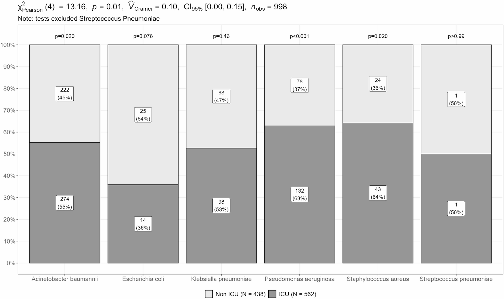
Table 1 Characteristics of patients and baseline of two groupsComparing the diagnostic value of tNGS, mNGS, and CMT in all patientsTo assess the diagnostic value of tNGS, it was compared with culture, CMT (all samples included culture for bacteria and fungi, and PCR for virus), composite reference standard, and mNGS. Among the 88 patients, tNGS showed a similar diagnostic agreement rate to mNGS (88.64% and 87.50%), but higher than CMT and culture (60.23% and 44.32%) (Fig. 2A). In 85.23% of the patients, at least two methods demonstrated diagnostic concordance, while 7.95% of the patients showed concordance only with one NGS method (Fig. 2B). Using culture as a reference, tNGS had a sensitivity of 94.87% and specificity of 10.20%, with an overall concordance rate of 47.73%. When compared to CMT, tNGS demonstrated a sensitivity of 94.55% and a specificity of 15.15%, with an agreement rate of 64.77%. Compared to the composite reference standard, tNGS demonstrated a sensitivity of 90.48%, specificity of 50.00%, and an 88.64% agreement rate (Table 2). Similarly, mNGS demonstrated an 87.50% consistency with the composite reference standard, with a sensitivity of 88.09% and specificity of 75.00% (Supplementary Table 2). tNGS detected pathogens in 28/33 samples that were negative in CMT, showing an 78.79% consistency rate with the composite reference standard (Table 2). Then, tNGS compared with mNGS demonstrated a sensitivity of 96.43% and specificity of 75.00%, with an agreement rate of 95.45%. The kappa value between tNGS and mNGS was 0.58. The comparative assessment of mixed infection detection capability across the three methodologies showed that tNGS and mNGS had sensitivities of 80.56% and 61.11%, respectively, significantly surpassing the sensitivity rate of 36.11% demonstrated by CMT (Fig. 2C). This superiority in sensitivity for detecting mixed infections was also observed in cases of single-pathogen infections (tNGS vs. mNGS vs. CMT: 83.33% vs. 79.17% vs. 58.33%). Both tNGS and mNGS showed high sensitivity for detecting bacterial (92.06% and 84.13%) and fungal (86.64% and 92.11%) infection samples, while tNGS exhibited higher sensitivity for detecting viral infections (95.83% vs. 50.00%). Both tNGS and mNGS methods demonstrated higher sensitivity than CMT for bacterial and fungal infection samples (Fig. 2C).
Fig. 2
Diagnostic performance of tNGS in 88 samples. (A) Diagnostic agreement rate of tNGS, mNGS, CMT, and culture. (B) Pie chart showing the proportion of diagnoses determined by different methods. CMT, conventional microbiological testing. (C) Comparison of detection sensitivity of mixed pathogens and various pathogens infections between tNGS, mNGS, and CMT. (D) Comparison of pathogen spectrum detected between CMT and tNGS. (E) Comparison of pathogen spectrum detected between mNGS and tNGS. (F) Comparison of pathogens detected between tNGS, mNGS, and CMT. G+: Gram-positive bacteria; G-: Gram-negative bacteria. *: p < 0.05, **: p < 0.01,***: p < 0.001
Table 2 Performance characteristics of targeted NGS in 88 clinic samplesThe pathogens detection was compared between tNGS with mNGS and CMT. In total, 130 causative pathogens were confirmed, with tNGS detecting 123 of them, mNGS identifying 111, and CMT finding 58. tNGS and mNGS showed comparable and significantly higher detection rates for pathogens than CMT, regardless of whether the pathogens were fungi, DNA viruses, or bacteria (Fig. 2D-F). The most prevalent bacteria were Acinetobacter baumannii, Pseudomonas aeruginosa, and Klebsiella pneumoniae, while the most common fungi were Pneumocystis jirovecii and Aspergillus fumigatus. SARS-CoV-2 and Influenza A virus were common viruses, however, mNGS did not detect them as it only identified DNA. tNGS not only detected SARS-CoV-2 and Influenza A virus but also successfully identified the subtypes for all 12 pathogens, with subtypes that are completely consistent with PCR (Supplementary Table 3). While both tNGS and CMT concurrently detected 57 pathogens, tNGS identified an additional 66. Notably, tNGS detected a higher prevalence of DNA viruses than CMT, due to the absence of appropriate testing for these pathogens in CMT. Concurrently, six pathogens, comprising three fungi and three bacteria, remained undetected by both methods but were detected by mNGS. Compared to mNGS, tNGS revealed an additional 19 pathogens. When comparing the fungi and bacteria not detected by tNGS with those detected by both NGS methods, it was found that the Reads Per Million (RPM), an important indicator of the relative abundance of microbial nucleic acids in NGS detection, was lower in mNGS for fungi (p = 0.07), while there was no significant difference for bacteria (p = 0.54). Meanwhile, the RPM of bacteria additional detected by tNGS was lower than those detected by both methods (Supplementary Fig. 1).
Comparing diagnostic value of tNGS, CMT and mNGS in immunocompromised patientsWe compared the diagnostic agreement rates of difference methods in immunocompromised and immunocompetent patients. The rates are illustrated in Fig. 3A. There were significant differences in the diagnostic agreement rates between NGS methods and CMT (p < 0.001). However, no difference was found between tNGS and mNGS. Higher mixed pathogens rates and higher proportion of fungi infections was found in immunocompromised patients (44.44% vs. 32.35%, p > 0.05; 46.30% vs. 32.35%, p > 0.05, Supplementary Fig. 2). Then, we evaluated the diagnostic performance of tNGS in two groups. Compared to the composite reference standard, tNGS demonstrated sensitivity of 83.87% and 94.34% in both immunocompetent and immunocompromised groups (Table 3), respectively, while mNGS showed 80.65% and 92.45% sensitivity. Furthermore, the pathogen spectrum varied between immunocompetent and immunocompromised individuals (Fig. 3B). The most common pathogens in immunocompetent patients were Acinetobacter baumannii, Influenza A virus, and Klebsiella pneumoniae, whereas immunocompromised patients more frequently exhibited Pneumocystis jirovecii, Aspergillus fumigatus, Candida albicans, and non-tuberculous mycobacteria. In the immunocompromised group, a higher number of fungal species were detected (29 vs. 11), including an increased prevalence of Aspergillus spp (12 vs. 5). and Pneumocystis jirovecii (7 vs. 1).
Fig. 3
Comparison diagnosis of tNGS with mNGS and CMT in immunocompromised patients. (A) The comparisons of diagnostic accuracy between three methods in immunocompetent patients and immunocompromised patients. (B) Pathogen spectrum of immunocompetent and immunocompromised patients. (C) Comparison between tNGS with CMT and mNGS in the detection of pathogens in immunocompromised patients. (D) Comparison between tNGS with CMT and mNGS in the detection of pathogens in immunocompetent patients. The pathogen detection rate is depicted in the figure. *: p < 0.05, **: p < 0.01,***: p < 0.001
Table 3 Diagnostic performance of NGS in difference groups compared with composite reference standardWe conducted a further comparative analysis of the pathogen detection among immunocompromised and immunocompetent individuals using tNGS, mNGS, and CMT (Fig. 3C&D). Consistent with the aforementioned findings, tNGS demonstrated higher pathogen detection rates compared to CMT across both immunocompromised and immunocompetent patients (97.62% vs. 44.05%, p < 0.0001; 89.13% vs. 45.65%, p < 0.0001). Particularly in immunocompromised patients, tNGS showed a higher detection rate for all types of pathogens (Fig. 3C). In immunocompromised patients, tNGS demonstrated a higher pathogen detection rate than mNGS (97.62% vs. 86.90%, p = 0.009), primarily due to its detected for RNA viruses. tNGS almost identified all pathogens detected by mNGS, with the exception of one Nocardia farcinica and one Leuconostoc pseudenterum not within the coverage range. In immunocompetent individuals, the pathogen detection rates were comparable (89.13% vs. 82.61%, p = 0.36), with tNGS detecting more RNA viruses while mNGS had a higher detection rate for fungi.
Clinical impacts of NGS on etiological diagnosis and antibiotic adjustmentBased on the retrospective impact of mNGS testing on etiologic diagnosis and antibiotic decision-making, the effect of NGS was categorized into three groups. As shown in Tables 4, 73.86% (65/88) of mNGS and 72.72% (64/88) of tNGS had a positive impact on etiologic diagnosis and antibiotic decision-making, with 17.05% leading to appropriate antibiotic adjustments. Based on the results of mNGS, 12 patients underwent escalation of therapy, one patient underwent de-escalation of therapy, and two patients had their antibiotic treatment discontinued after infection was ruled out. Compared to mNGS, tNGS could also lead to similar adjustments for these 15 patients. Furthermore, the effectiveness and prognostic outcomes of antibiotic treatments were compared. In patients with positive impact from mNGS, 88.71% (55/62) improved after treatment, and 85.71% in the antibiotic adjustment group showed improvement. The same result was observed in the tNGS assay.
Table 4 Clinical impacts on antibiotic adjustment and prognosisComparison tNGS with mNGS in the detection of microbial nucleic acidsGiven the unbiased nature and ultra-sensitivity of NGS testing, the detection of non-pathogenic microorganisms in BALF samples was inevitable. Among the 88 patients, mNGS detected 283 microorganisms, while tNGS identified 402 microorganisms. In comparison of the RPM between pathogens and other microorganisms of tNGS, no significant differences were observed. The consistency of microbial nucleic acid reporting between mNGS and tNGS was analyzed. Both NGS methods shared the detection of 249 microorganisms, accounting for 87.99% of those identified by mNGS. The positive predictive value for the shared microorganisms was 41.77%, while the positive predictive values for microorganisms exclusively detected by mNGS or tNGS were 20.59% and 12.42%, respectively. Thirty-four microorganisms only detected by mNGS, and 20.59% (7/34) were not covered in the tNGS assay. Among them, 7/34(20.59%) were identified as causal pathogens, including three of fungi and four of bacteria. Comparing the RPM of these microorganisms with those shared by both NGS, it was found that the RPM for Gram-positive bacteria and fungi in the group detected only by mNGS was lower (Supplementary Fig. 3). One hundred fifty-three microorganisms were detected exclusively by tNGS, including 39.87% bacteria, 9.80% fungi, 33.99% DNA viruses, and 16.34% RNA viruses (Fig. 4C). Among them, 19/153(12.42%) were identified as causal pathogens, including 15 viruses and 4 bacteria. It is worth noting that the majority of these microorganisms are composed of common respiratory tract colonizing microorganisms. (Fig. 4D). Further comparison of the RPM of these additional microorganisms detected by tNGS with those shared by both NGS revealed that the RPM of the shared microorganism group was higher, a trend observed across bacteria, viruses, and fungi.
Fig. 4
Comparison between tNGS and mNGS in microorganisms detections. (A) Venn diagram showing the results of microorganisms detected in tNGS and mNGS. (B) Microorganisms positive predictive values of different groups. (C) The proportion of microorganisms types in different groups. G+: Gram-positive bacteria; G-: Gram-negative bacteria. (D) The proportion of common microorganisms among the additional detections by tNGS includes Candida species as the common fungi, Human herpesvirus1/4/5 among DNA-viruses, Streptococcus pneumoniae among G+, and Escherichia coli, Acinetobacter baumannii, Klebsiella pneumoniae, Stenotrophomonas maltophilia, Burkholderia cepacian, and Pseudomonas aeruginosa among G-. (E) Comparisons the RPM of difference microorganisms in tNGS grouped in consistently detection (detected by both NGS) and tNGS additional detection. RPM: read per million. *: p < 0.05, **: p < 0.01,***: p < 0.001
Comments (0)