Intraoperative Cytological Assessment of Bone Marrow from Resected Mandible in Analyzing the Surgical Bone Margins in Oral Squamous Cell Carcinoma: A Promising Tool
Introduction
Complete local excision of the primary tumor should be the mainstay in the surgical treatment of oral squamous cell carcinoma. If the extent of carcinoma growth indicates bone resection, determining the bone margins is difficult intraoperatively. Hence, an appropriate method is needed to assess the bone margins intraoperatively to prevent tumor recurrence.
Materials & methods
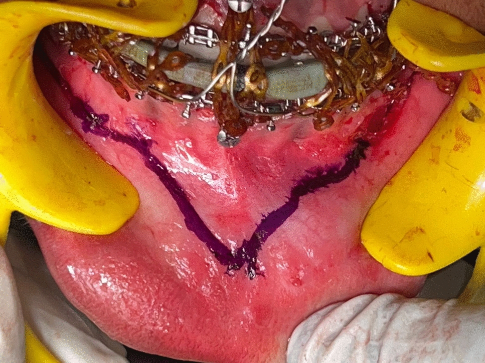
A prospective study was carried out in an institutional setup with 35 patients who were diagnosed for oral squamous cell carcinoma involving mandible. After the resection, marrow samples were collected using a bone curette from three locations: margin of the resected mandible (site-A), margin of the residual mandible (site-B), and marrow immediately beneath the tumor (site-C). Following the collection, the samples were processed, and stained using rapid-pap technique. Examination was done under a light microscope to detect the presence of malignant cells. The resected mandible was also processed for assessment in the usual manner following the decalcification. It was used as the gold standard for comparison with cytological findings. Analysis: The results were evaluated by determining sensitivity, specificity, positive predictive value (PPV), negative predictive value (NPV), and accuracy.
Results
Among 35 patients (30 males, 5 females), the mean age was 52 years. The average time taken for the technique was 26 min. The sensitivity of cytological assessment from site-A is 100%, specificity: 95.83%, PPV: 91.67%, NPV: 100%, and the accuracy: 97.14%. The sensitivity of the assessment from site-B is 90.91%, specificity: 91.67%, PPV: 83.33%, NPV: 95.65%, and the accuracy: 91.43%.
Conclusion
The evaluated intraoperative cytological assessment has excellent diagnostic variables in detecting tumor cells. The accuracy of the test is phenomenal, and it can be performed regularly in any surgical setup managing oral squamous cell carcinoma where bone resection is needed. This technique can be considered as a feasible diagnostic tool for controlling and optimizing the adequacy of bone resection.


Comments (0)