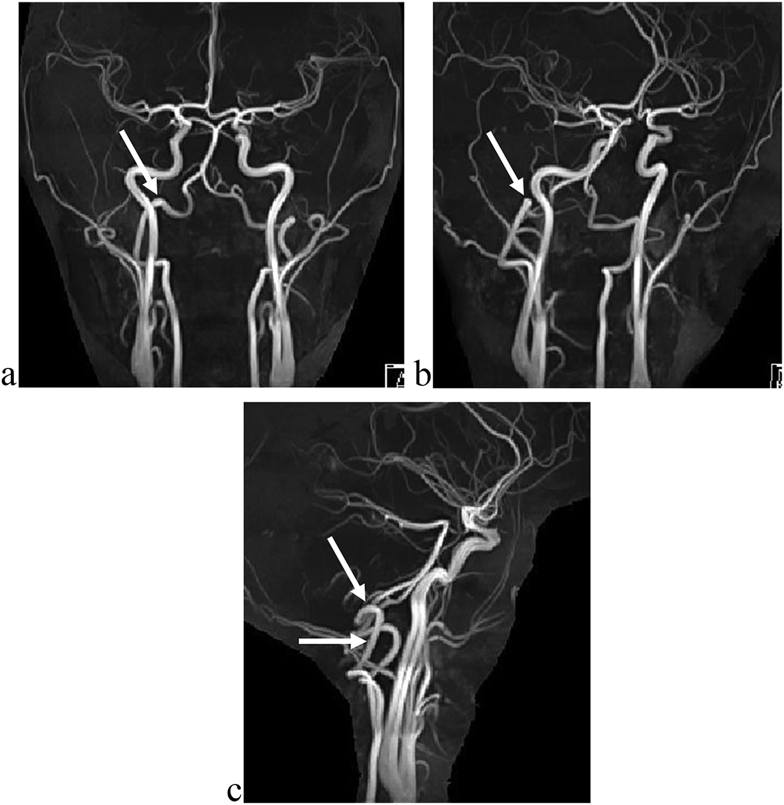
From an anatomical viewpoint, the vertebral level of the CB could supply the general concept of anatomic variability. However, although the height of the CB is referred to vertebrae, this definition is impractical during surgery as neither the patient is placed in an anatomic position nor the vertebrae are accessible [26]. Anterior anatomic landmarks are more practical to locate the CB during surgery [26]. However, although some authors always found the CB posterior inferior to the tip of the greater horn of hyoid bone [20], their finding is contradicted by the present results demonstrating that the greater horn should not be regarded as a unique landmark of the CB.
Different studies assessed the vertical position of the CB bilaterally and found no bilateral asymmetry [17], or the side-to-side differences in the level of CB were not statistically significant [3]. Although not explicitly documented, such vertical asymmetry of the CBs results from the study of McAfee (1953) [24]. In 1979, Smith and Larsen wrote that perusal of the literature had not revealed any report concerning a possible symmetry of the CB, and therefore, they performed their study on 100 angiograms [34]. The authors found that in 22% of the cases, the right CB was superior to the left CB, while 50% had a higher left CB [34]. In a bilateral study of the vertical location of the CB, the right CB was significantly at a higher level than the left one [12]. Lo et al. (2006) found the bilateral asymmetry of the CB in 48% of cases [22]. Woldeyes (2014) found bilaterally asymmetrical CBs in 61.5% of cases [36]. Kurkcuoglu et al. (2015) detected the bilateral asymmetry of CB in 33% of cases [19]. Mompeo and Bajo (2015) found the bilateral asymmetry of the CB in just 10.52% of the 19 studied cases [28]. The present study found bilaterally asymmetrical CBs in 48.3% of cases for the vertebral level and 40.14% of cases for the anterior cervical landmarks. However, it was found here that CB’s bilateral symmetry is equally significant for vertebral levels and anterior cervical landmarks. The anatomical correlation between the vertebral and anterior cervical levels is somewhat unexpected, although different significant associations were established. Other authors did not evaluate the bilateral symmetry of the CB [5].
Different vertical locations of the CB were found in the present study and others [1, 3,4,5, 9, 11, 12, 14, 17, 19, 21, 24, 25, 27,28,29,30,31,32,33,34,35,36,37]. As presented in Table 7, different studies observed only the vertebral level of the CB; other authors studied the CB in relation to different anterior cervical landmarks, while a few authors observed both vertebral and anterior cervical landmarks. Numerous studies were performed by dissections. Few studies, including the present one, studied > 100 cases. Some of these studies estimated the level of the CB as normal, high or low, just referring the CB to the superior margin of the thyroid cartilage [2, 29].
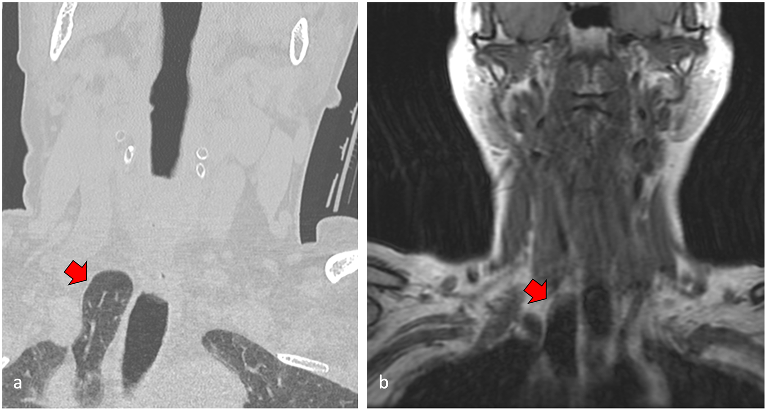
Table 7 Previous studies of the vertical topography of carotid bifurcation determined either vertebral levels, anterior cervical landmarks, or both. Methods and lots are listed. Few studies investigated both anterior and posterior landmarks of the carotid bifurcation. A: angiography; CT: computed tomography; D: dissectionMcAfee et al. (1953) performed vertical measurements to detect the CB bilaterally in 70 dissected cadavers [24]. The authors found that CB was in the upper 5th of the neck in 82% of dissections, in an area about 2.5 cm long, measured inferiorly from the inferior margin of the mandible, and in 18% it was located in the upper 2/5 of the neck [24]. The mean distance between the mandible and CB was 2.14 cm [24]. The lowest CB was 7.2 cm superior to the clavicle [24]. McAfee found in 7.1% the CB above the gonial angle. This type 5 variant was found here in 6.12%.
We found by the present study that: (1) the inter-thyro-hyoid and hyoid levels are rather encountered in men, on any side; (2) on the right side, the normal and the gonial types are rather found in women; on the left side in women the CB is rather found in the interval between the upper margin of the thyroid cartilage and the mandible; (3) there is a higher variability of the vertebral level of CB in men; in women, most cases had a C3-C4 level on the right and a C2-C4 level on the left.
The present study found significant associations between certain vertebral levels and anterior cervical landmarks, such as C2-types 1 and 2, C3/C4-type 3, C4-type 5 on the right side and C2/C3-type 1, C3 and C4-type 2, C3/C4-types 3, 4, 5 on the left side. As referred to in typical anatomy, these associations may surprise if one relates the hyoid bone and the thyroid cartilage to a specific vertebra. The vertical position of the laryngeal apparatus and the geometry of the cervical vertebrae should be regarded as variable. Mirjalili’s study [27] also shows that the correspondence between the anatomical position of the anterior cervical landmarks and the vertebral landmarks is not absolute: both the hyoid and the thyroid cartilage can be located anywhere between the C3 vertebra and the C5/C6 disc. However, Mirjalili et al. found no statistically significant differences related to age or sex, and they did not determine the CB topography versus gonial angle [27]. Demirtas et al. recently determined a statistically significant correlation between CB levels and CB angles on both sides: CB angles narrow as bilateral CB levels decrease [13].
Our results converge with the conclusions of a previous study of the anterior and posterior landmarks of the CB. Cihan and Deveci (2022) concluded that estimating the CB’s location according to the gonion and hyoid bone will give a more accurate result [11]. Cobiella et al. (2021) used the body of the hyoid bone level as a single landmark for the CB [12]. The values obtained for the level of the CB ranged from 4 cm below the hyoid body and 2.5 cm above the respective landmark. The authors found no significant differences in relation to the distribution of CB by sex [12].
Hayashi et al. (2005) found the CBs most frequently at the level of the middle 3rd of the C3 vertebra, but the mean position of the CB was located at the lower 3rd of the C3 vertebra [17]. A more recent study of 100 angiograms determined vertebral levels of CB [19]. The highest CB level was at the C2 vertebra, and the lowest was at the C6/C7 intervertebral disc in both sexes [19]. In the general group, CB was found most frequently, in 29% of cases, at the level of the C4/C5 intervertebral disc on the right side of the neck and at the level of the C4 vertebra (26%) on the left side of the neck [19]. A low level of the CB was found just above the C6/C7 intervertebral space by Gulsen et al. (2009) [16]. The intrathoracic CB is extremely rare [18], so it is not surprising that the present study did not find this level.
Another study classified the level of CB into three types: normal, at the level of the upper margin of the thyroid cartilage (60%); high, superior to the thyroid cartilage (40%); and low, not found then [5]. In the present study CBs were found lower than the superior margin of the thyroid cartilage in type 6, in 2.72%.
Anangwe et al. (2008) considered two possibilities of CB: high, above the C3/C4 junction and low, below it [3]. The authors assumed that the C3/C4 junction corresponds to the upper margin of the thyroid cartilage [3]; this vertebral level of the CB is also considered by others [10, 33]. However, they did not perform dissections to validate this topographic correlation; the dissections were limited to the carotid triangle [3], so the vertebral level classification appears speculative. Ferracci et al. (2022) documented that the CB is generally located near the superior border of the thyroid cartilage but in front of the C4–C5 disk [15].
McNamara et al. tested the accuracy of a straight-line distance (SLD) between the skull base and the CB to identify that bifurcation [25]. The authors found that the greater horn of the hyoid had the most significant correlation with the SLD quartile group [25]. According to McNamara et al., a standardised definition of high CB is still lacking as there are different potential approaches to defining it [25]. However, one can classify the level of CB using a purely statistical definition, such as the shortest quartile of a normal distribution, as used by McNamara, or a clinical definition, the level that makes the intervention more difficult for the vascular surgeon and with a higher risk of complications for the patient [8].
Anu et al. (2007) found the CB at C3 in 50% on the right and 55% on the left, at C4 (40% on the right, 35% on the left), at C2 in 10% of cases and at C5 in 1% [4]. Woldeyes (2014) studied by dissection only 13 cadavers and located the CB at C2/C3 (3.85%), C3 (42.31%), C3/C4 (15.38%) and at C4 (38.46%) [36]. Chalise et al. (2021) studied the vertebral level of CB in 18 cadavers; it ranged from C2/C3 to C4 [9]. Cihan and Deveci (2022) found the highest vertebral level of CB in the lower 1/3 of the C2 vertebra and the lowest level in the upper 1/3 of the C6 vertebra [11].
Devadas et al. (2018) followed anterior cervical landmarks in the study of vertical topographic possibilities of CB: superior border of thyroid cartilage (75%), inter-thyro-hyoid level (10%), hyoid level (13.75%) and inter-hyo-mandibular level (1.25%) [14]. These authors did not find the CB below the superior margin of the thyroid cartilage. Their landmarks correspond respectively to levels 1 (13.95%), 2 (24.49%), 3 (39.12%) and 4 + 5 (19.73%) in the present study. The differences in type prevalences between these two studies are consistent.
The limitations of this study are related to the simultaneous variations of the ECA and ICA. Thus, for a specific level of the CB, the branching pattern of the ECA could differ from one case to another. Moreover, a certain topographical level of the CB may not give additional information on the deviated courses of the carotid arteries due to their coiling, kinking, or different axial rotations.


Comments (0)