
Remember me
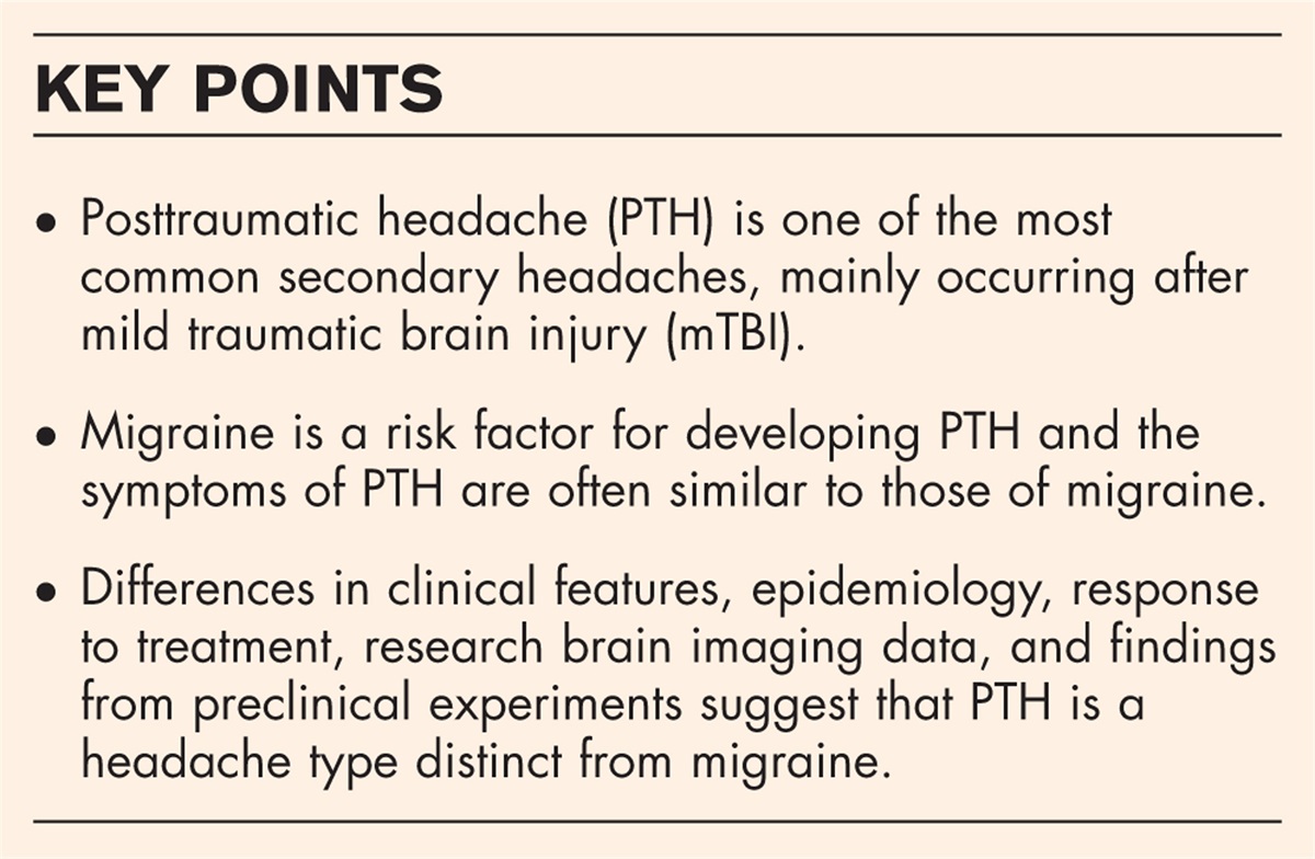
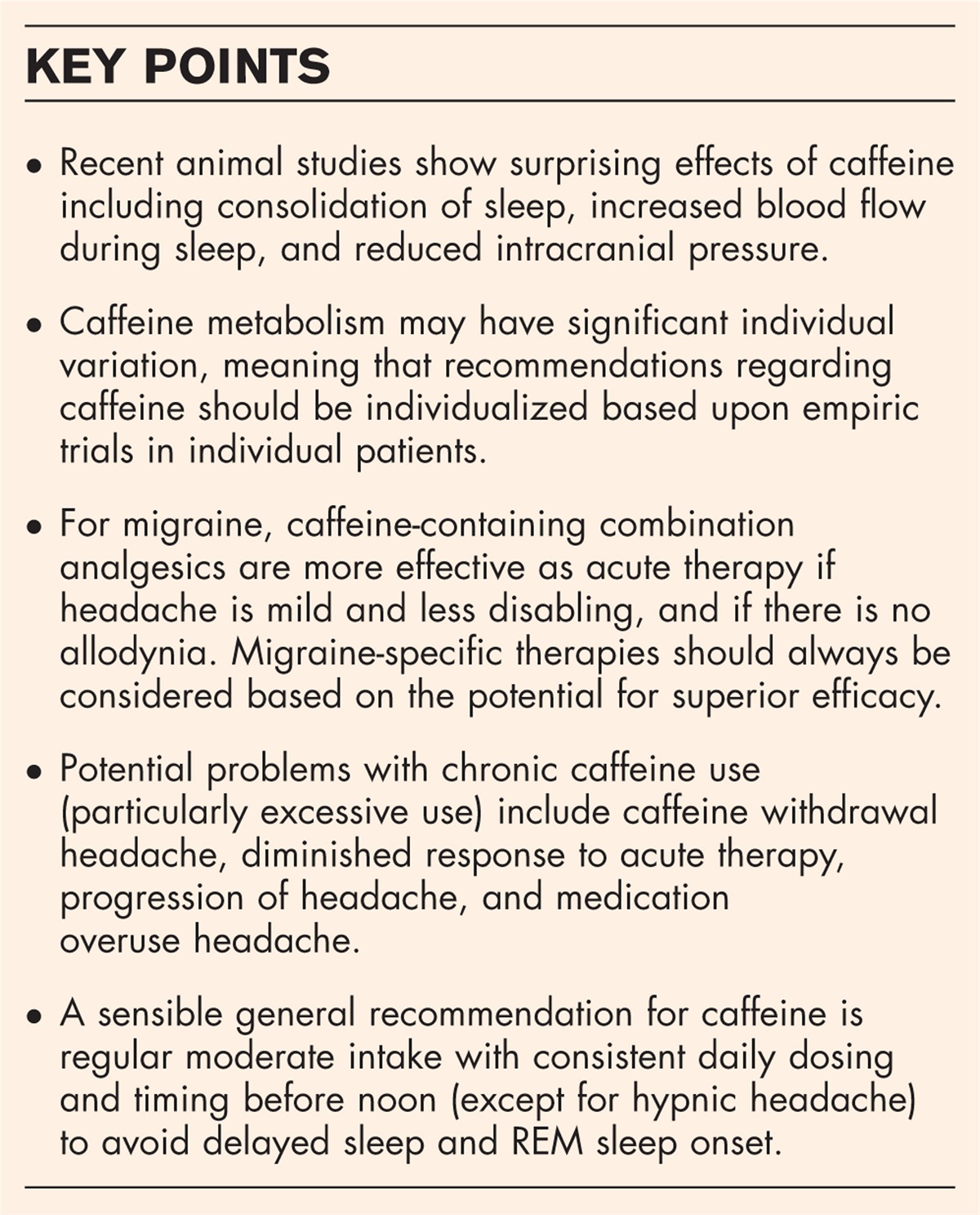


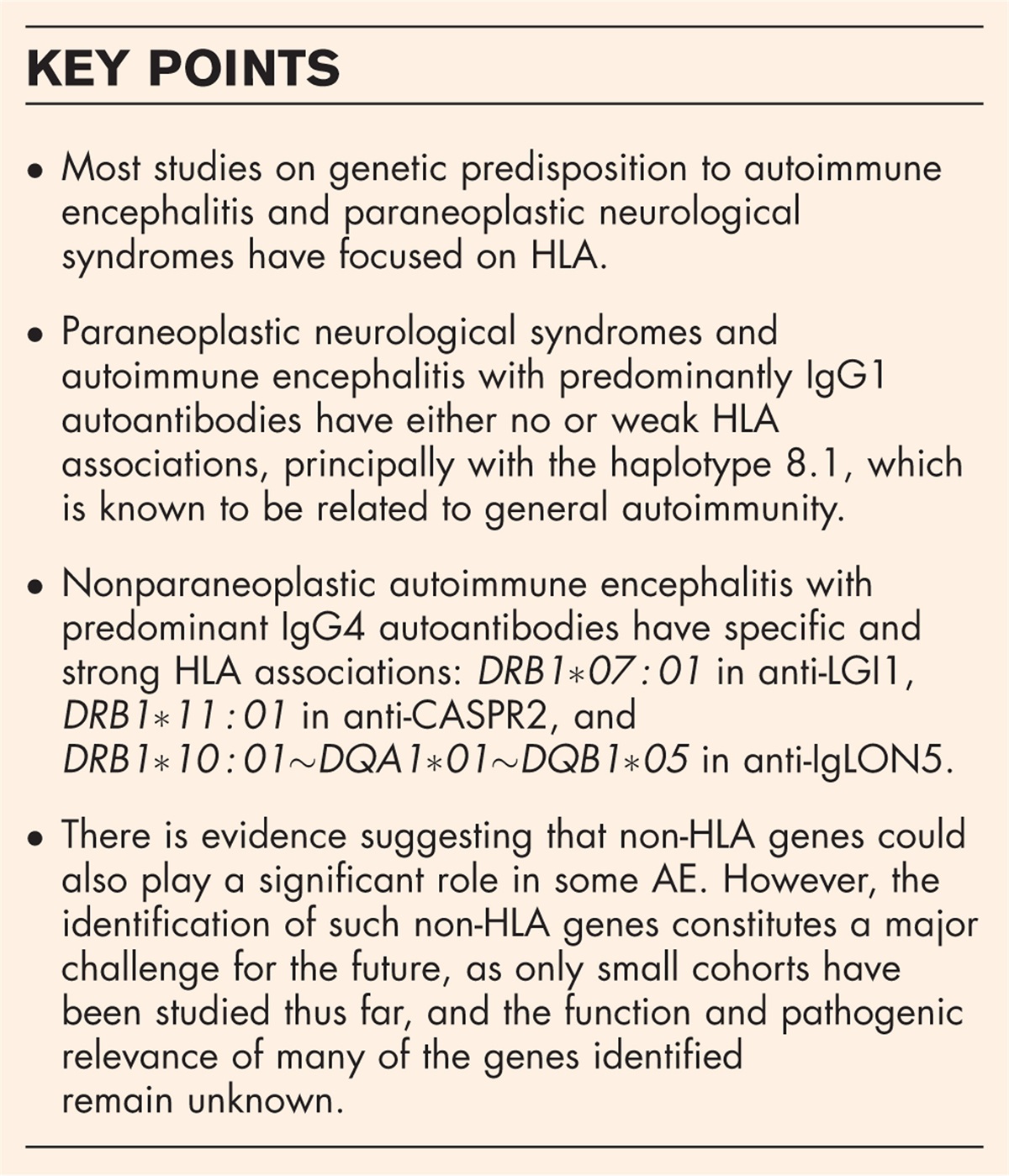
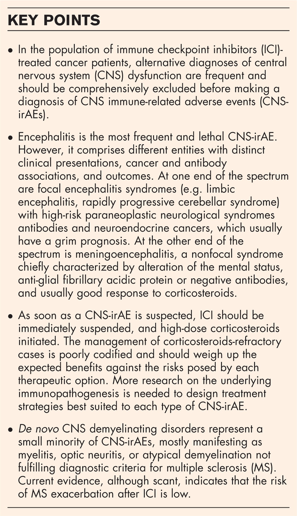


Anti-IgLON5 disease is characterized by a variety of symptoms involving multiple areas of the central nervous system. Patients typically develop a severe and distinctive sleep disorder with parasomnias involving both rapid-eye movement (REM) and non-REM sleep and stridor with obstructive sleep apnoea (OSA) along with more disabling symptoms like bulbar dysfunction, gait instability, movement disorders and cognitive impairment [1–3]. The hallmark of the disease is the presence of antibodies against IgLON5, a cell adhesion molecule of unknown function. The clinical course of anti-IgLON5 disease is mainly chronic or insidious (although up to 25% of patient may show a subacute presentation in weeks or a few months) and immunotherapy is usually less effective than in other encephalitides with antibodies targeting proteins of the neuronal surface, like NMDAR or LGI1 encephalitis [4]. However, an earlier recognition of the disease and prompt initiation of the immunotherapy can be beneficial, because some patients improved significantly with a response ranging from 13 to 41% in different series [5▪▪]. The initial neuropathological studies on two autopsies found abnormal hyperphosphorylated tau aggregates mainly in neurons of the hypothalamus and the tegmentum of brainstem with a rostro-caudal gradient of severity [1]. However, cases without tauopathy have been recently described suggesting that neurodegeneration can be a late event [6▪,7] and pointing to an immune-mediated disease process as the primary cause of the disease.
 Box 1:
 no caption available
EXPANDING THE CLINICAL PROFILE OF ANTI-IgLON5 DISEASEMost patients (∼ 80%) will present, during the course of the disease, a combination of symptoms including bulbar dysfunction (mainly dysphagia, dysarthria or episodes of respiratory failure), plus sleep disorder (REM and NREM parasomnias and stridor with obstructive sleep apnoea), gait instability and other movement disorders (mainly generalized chorea and craniofacial dyskinesias). The definite diagnosis of the disease is based on the presence in serum and/or CSF of antibodies targeting IgLON5. However, the diagnosis of anti-IgLON5 disease is challenging because the presenting symptoms may be heterogeneous and the syndrome, which usually have a chronic progression, may be restricted at onset to isolated clinical manifestations mimicking neurodegenerative diseases. For instance, patients who present with gait instability and gaze palsy as the most prominent symptoms, may be misdiagnosed of progressive supranuclear palsy (PSP), or Huntington's disease can be suspected if chorea and cognitive impairment are the presenting symptoms. The complicated clinical spectrum of symptoms associated with anti-IgLON5 disease has been recently expanded to neuromuscular manifestations compatible with motor neuron disease (NMD)-like phenotype when symptoms of bulbar dysfunction are combined with muscle fasciculations and weakness [8▪▪,9,10], but also, to stiff-person syndrome spectrum disorder (SPSD) when hyperexcitability, stiffness and muscle spasms are present [3]. Therefore, concomitant infrequent symptoms in classical MND or SPSD like acute recurrent respiratory distress requiring tracheostomy, dysautonomia or sleep problems should lead to suspect of anti-IgLON5 disease. Paraclinical studies are usually negative or noninformative, and only in a few patients, the MRI shows evidence of inflammatory changes (<5%), mild pleocytosis in the CSF (20–30%), especially in those patients with a short delay from onset to lumbar puncture, or a mild increase in CSF proteins (40–50%) [5▪▪,11]. Thus, the chronic course plus absence of inflammatory findings in the CSF and brain MRI are other reasons that can lead to confound anti-IgLON5 disease with neurodegenerative disorders. Recently, neuronal surface antibodies were screened in a large cohort of 920 patients with a clinical diagnosis of neurodegenerative dementia, and 3 of them tested positive for IgLON5 antibodies. The three patients had been diagnosed of Alzheimer dementia but showed atypical features such as nonamnestic presentations (e.g. progressive aphasia or visuospatial and perceptual disorders) subacute deterioration or fluctuating disease course, mild pleocytosis or normal amyloid beta42 or tau and phospho-tau levels in CSF. This study indicates that a small proportion of patients suspected to have neurodegenerative dementias have neuronal antibodies indicative of autoimmune encephalitis, including anti-IgLON5 disease [12▪▪]. In this sense, awareness of this possibility is important to differentiate anti-IgLON5 disease from neurodegenerative disorders that would not benefit from immunotherapy (Fig. 1).
 FIGURE 1:
 Graphical representation of clinical symptoms observed in anti-IgLON5 disease in combination with subtypes and possible mimics. In grey circles, IgLON5 mimics disorders and diseases that can resemble anti-IgLON5 disease. MSA, multiple system atrophy; OSAS, obstructive sleep apnoea syndrome; PSP, progressive supranuclear palsy; RBD, REM sleep behaviour disorder. Dark grey rectangles: Subtypes. Original figure.
BIOMARKERS AND PROGNOSTIC SCALES IN ANTI-IGLON5 DISEASEThere are currently no longitudinal studies of biomarkers to predict response to immunotherapy. A multicentric retrospective study with 53 patients proposes that a high serum level of neurofilament light chain at diagnosis would predict a worse prognosis and that earlier diagnostic and initiation of the immunotherapy are beneficial. Preliminary evidence indicates that patients do not have an increment of phospho-tau and a decrease of beta-amyloid-42 protein in the CSF like is characteristic in Alzheimer dementia [5▪▪]. A recent relevant study indicates that tau depositions of patients with anti-IgLON5 disease can be visualized with [18F]PI-2620 PET. Although validation in a larger cohort is needed, this is an important observation because this type of neuroimaging could be useful to assess the extent of tau deposits in vivo[13▪▪]. Scarcity of studies about biomarkers for monitoring the progression and wide differences on applied immunotherapies make challenging to propose guidelines but, according to our experience, a trial of immunotherapy (e.g. intravenous methylprednisolone or immunoglobulins followed by rituximab) is worth to be considered given the possibility of improvement (particularly if the patient is treated early in the disease course), the evidence supporting an underlying autoimmune pathophysiology, and the disability and mortality associated with the disease. The severity of anti-IgLON5 disease and response to immunotherapy in the previous studies has been evaluated with the modified Rankin scale, but recently a specific scale, the IgLON5 composite score (ICS), has been developed to improve the assessment of the severity of the different symptoms occurring in patients with IgLON5 antibodies. The ICS includes 17 symptoms distributed in five domains that cover the most common manifestations observed in anti-IgLON5 disease (Table 1). Each clinical domain provides a partial score obtained from the quantitative assessment of the corresponding symptoms, resulting in a total ICS score ranging from 0 to 69. The ICS is a valid tool to assess the extension and severity of the different clinical manifestations of anti-IgLON5 disease, able to capture clinical stability or changes (either symptom worsening or improvement) and can be a useful in future studies evaluating the effect of immunotherapy or monitoring evolution of symptoms during the clinical course of the disease (C. Gaig et al.[14] accepted in Neurology).
Table 1 - Anti-IgLON5 disease composite score (ICS) Domain Points Bulbar Partial score: 0--21 Stridor 0-1-2-6a Central hypoventilation 0-1-2-6 a Dysphagia 0-1-2-6 a Dysarthria 0-1-2-3 Sleep Partial score: 0--12 Abnormal movements/behaviours-vocalizations 0-1-2-3 Insomnia 0-1-2-3 Excessive daytime sleepiness 0-1-2-3 Obstructive sleep apnoea 0-1-2-3 Movement disorders Partial score: 0--15 Gait difficulties and falls 0-1-2-6 a Chorea 0-1-2-3 Orofacial dyskinesias 0-1-2-3 Other movement disorders. Specify: 0-1-2-3 Cognition Partial score: 0--12 Cognitive impairment 0-1-2-6 a Neuropsychiatric (psychosis, delirium) 0-1-2-6 a Other Partial score: 0--9 Oculomotor abnormalities 0-1-2-3 Dysautonomia 0-1-2-3 Fasciculations 0-1-2-3 Total composite score 0-690: Absent/normal; 1: Mild; 2: Moderate; 3 (or 6∗): Severe.
aThe score was rated 6, instead of 3, when stridor, central hypoventilation, dysphagia, gait difficulties, cognitive impairment or neuropsychiatric manifestations were considered severe, to weight better the more severe disability caused by these symptoms.From https://www.neurology.org/doi/10.1212/WNL.0000000000208101.Initial evaluation of available autopsies revealed the presence of an atypical tauopathy with evidence of 3R and 4R tau deposits restricted to neurons, mainly in hypothalamus and tegmentum of the brainstem [1,15]. In subsequent studies, patients without the described brainstem tauopathy have been reported, suggesting that this can be a late or secondary event in the disease [7]. Interestingly, a recent study with nine cases has broadened the neuropathological features observed in the anti-IgLON5 disease [6▪]. The initially described neuronal brainstem tauopathy was present in the majority of them (five patients) but one patient fulfilled PSP criteria showing a 4R neuronal and glial tauopathy, while the remaining three cases that precisely had a short duration of the disease, only showed primary age-related neurofibrillary disease in the limbic area probably due to age-related comorbidity. These findings further support that the distinctive brainstem tauopathy associated to the disease may be a late event. Additionally, and concomitant to tau pathology, abnormal deposition of transactive response DNA-binding protein 43-kDa (TDP-43) was found in two cases in neurons and in microglial cells [6▪,16]. Although the distribution of these TDP-43 inclusions and the cellularity involved was reminiscent of those seen in ALS, these findings should be categorized as unusual TDP-43 disease according to current consensus guidelines [17]. Perivascular and parenchymal inflammatory infiltrates of B and T cells, in addition to MHC class I upregulation in neurons and microglial activation have been described recently in autopsies supporting a crucial role for autoimmunity in the disease (Fig. 2). Furthermore, prominent deposition of IgG4 in the neuropil has been observed in areas of the brain that coincide with regions of high IgLON5 expression and those described as affected by the distinctive tau disease in anti-IgLON5 disease like tegmentum of brainstem [6▪].
 FIGURE 2: Cellular inflammation in anti-IgLON5 disease. Cellular inflammation was mild to moderate and mainly composed of perivascular and parenchymal CD3 (a) and CD8 positive T cells (b) and few perivascular CD79a positive B cells/plasma cells (c). Parenchymal CD8 T cells were granzyme B positive and granules showed a polarization towards neurons (d; arrows). In addition, neurons showed an upregulation of MHC class I in the reticular formation and olivary nuclei (e, rectangle enlarged in f). Marked microglia activation was found in the HLA-DR staining in tegmentum of medulla oblongata and nucleus olivaris (g), including formation of microglial nodules (h; rectangle in g enlarged in h). Images are depicted from patient 1. Scale bars: 50 μm. Adapted from [6▪].INSIGHTS IN THE PATHOPHYSIOLOGY OF THE DISEASE
The autoimmune hypothesis of the pathogenesis of the disease is supported by numerous evidences. First, all the patients with anti-IgLON5 disease have antibodies against IgLON5 which is a cell surface protein. Although IgG4 is the predominant subclass of IgLON5-abs, almost all the patients harbour also variable amounts of IgG1 [2], and indeed, in-vitro experiments using cultures of rat hippocampal neurons demonstrated that IgG1 IgLON5-abs were responsible for the irreversible internalization of the clusters [18]. Furthermore, long-term incubation with purified total IgG with different percentage of IgG1/IgG4 led indistinctly to alterations in the cytoskeleton with dystrophic neurites, axonal swellings, ring and bulb-like structures and early termination after crosslinking and internalization in these cultures [19]. These features are compatible with a pathogenic effect of IgLON5-abs leading to the development of early signs of neurodegeneration. Proposed pathogenic mechanism for IgG4 antibodies subclass in other autoimmune diseases, like the case of anti-MusK antibodies in myasthenia gravis or anti-LGI1 antibodies in autoimmune encephalitis, is the disruption of protein-protein interactions [20]. IgLON5 belongs to the IgLON family of cell adhesion molecules [21] and like other members of the family is spontaneously shed from the cell surface of neurons, interacting with other members of the IgLON family in cis (in the same cell) and in trans (between cells). Patient's antibodies interfere with this binding, but this potential pathogenic mechanism was not specific to IgG4 [22▪]. Second, 60% of patients present the same HLA class II haplotype (DRB1∗10 : 01-DQB1∗05 : 01), which is infrequent in the normal population (1–3%) [5▪▪,11]. DRB1∗ alleles have also been associated to another autoimmune diseases like rheumatoid arthritis [23]. Importantly, carrying the HLA-DRB1∗10 : 01 allele in patients with anti-IgLON5 disease correlated with bulbar dysfunction in one study [11] and with the typical sleep disorder in two independent studies [5▪▪,11]. Third, the characteristic neuronal brainstem/hypothalamic tauopathy is not present in all the patients and signs of inflammation has been described in patients with a short disease duration as already stated above [6▪,7]. Fourth, early treatment with immunotherapy seems to ameliorate some symptoms (a feature that would be unusual in a primary neurodegenerative disease ([5▪▪]). Finally, despite strong evidence pointing to an autoimmune origin of the disease, the few published studies attempting the passive transfer of the antibodies of the patients in rodent animal models did not confirm unequivocally the pathogenicity of the antibodies because they did not reproduce the typical clinical symptoms of the patients (e.g. sleep parasomnia and sleep breathing difficulties) or demonstrate the described in-vitro effects of the antibodies. A pilot study with mice expressing humanized tau (htau) protein and wildtype mice as controls were intraventricularly infused with purified IgLON5-IgG by osmotic pumps for 14 days. Behavioural studies showed no differences even when the sleep pattern was studied in detail but curiously accumulation of abnormal tau deposits was preferentially found in the brainstem and hippocampus of female hTau and wildtype mice but not in males infused with purified IgLON5-IgG. However, these results require further confirmation [24▪]. In a second study of passive transfer of patient's antibodies, mice were either injected daily during 10 days into the lateral ventricle or into the parenchyma in the hippocampal area with IgLON5 IgG or control IgG. Animals receiving IgLON5-IgG exhibited memory deficits, anxiety-like behaviour and progressively accumulation of human IgG immunostaining in the area of injection but the irreversible downregulation of membrane IgLON5 clusters observed in vitro was not investigated, which is a limitation [25▪]. Similarly, another study that injected the purified IgG into the substantia nigra pars compacta of mice for 7 days, showed a decline in motor balance that persisted 3 months after the last infusion, dopamine decrease content in the area of injection, microglial activation, and a moderate increase of p-tau measured by western blot after three months [26▪].
CONCLUSIONAnti-IgLON5 disease is characterized by a heterogeneous spectrum of clinical symptoms; therefore, patients can be misdiagnosed initially with other autoimmune or neurodegenerative diseases. In addition, lack of awareness, the chronic disease progression and the absence of any other evidence of inflammation in some patients add complexity to the diagnosis. In the future, longitudinal studies of biomarkers in follow-up samples will be necessary to predict outcome and response to therapies. Assessment of neurofilament light chain levels in serum together with Tau PET imaging appear to be a promising strategy to evaluate the patient. The discovery of inflammatory infiltrates in new neuropathological studies reinforces the link between inflammation and neurodegeneration. New studies in animal models are needed to fully comprehend the pathological events in anti-IgLON5 disease.
AcknowledgementsThe authors thank Dr Francesc Graus for the critical review of the manuscript.
The authors want to thank European Joint Programme on Rare Diseases (EJPRD) for funding a Networking event contract number: 463001015.
Financial support and sponsorshipThis study has been funded by Instituto de Salud Carlos III through the project FIS21/00165 (Co-funded by European Regional Development Fund ‘Investing in your future’). This project has received funding form the European Union's Horizon 2020 research and innovation programme under the EJP RD COFUND-EJP N°825575. IDIBAPS belongs to CERCA (Centres de recerca de Catalunya).
Conflicts of interestThere are no conflicts of interest.
REFERENCES AND RECOMMENDED READINGPapers of particular interest, published within the annual period of review, have been highlighted as:
▪ of special interest
▪▪ of outstanding interest
REFERENCES 1. Sabater L, Gaig C, Gelpi E, et al. A novel nonrapid-eye movement and rapid-eye-movement parasomnia with sleep breathing disorder associated with antibodies to IgLON5: a case series, characterisation of the antigen, and postmortem study. Lancet Neurol 2014; 13:575–586. 2. Gaig C, Graus F, Compta Y, et al. Clinical manifestations of the anti-IgLON5 disease. Neurology 2017; 88:1736–1743. 3. Wenninger S. Expanding the clinical spectrum of IgLON5-syndrome. J Neuromuscul Dis 2017; 4:337–339. 4. Dalmau J, Graus F. Antibody-mediated encephalitis. N Engl J Med 2018; 378:840–851. 5▪▪. Grüter T, Möllers FE, Tietz A, et al. Clinical, serological and genetic predictors of response to immunotherapy in anti-IgLON5 disease. Brain 2022; 146:600–611. 6▪. Berger-Sieczkowski E, Endmayr V, Haider C, et al. Analysis of inflammatory markers and tau deposits in an autopsy series of nine patients with anti-IgLON5 disease. Acta Neuropathol 2023; 146:631–645. 7. Erro ME, Sabater L, Martínez L, et al. Anti-IGLON5 disease: a new case without neuropathologic evidence of brainstem tauopathy. Neurol Neuroimmunol Neuroinflamm 2020; 7:e651. 8▪▪. Sista SR, Crum B, Aboseif A, et al. Motor-neuron-disease-like phenotype associated with IgLON5 disease. J Neurol 2022; 269:6139–6144. 9. Tao QQ, Wei Q, Song SJ, Yin XZ. Motor neuron disease-like phenotype associated with anti-IgLON5 disease. CNS Neurosci Ther 2018; 24:1305–1308. 10. Werner J, Jelcic I, Schwarz EI, et al. Anti-IgLON5 disease: a new bulbar-onset motor neuron mimic syndrome. Neurol Neuroimmunol Neuroinflamm 2021; 8:e962. 11. Gaig C, Ercilla G, Daura X, et al. HLA and microtubule-associated protein tau H1 haplotype associations in anti-IgLON5 disease. Neurol Neuroimmunol Neuroinflamm 2019; 6:e605. 12▪▪. Bastiaansen AEM, van Steenhoven RW, Te Vaarwerk ES, et al. Antibodies associated with autoimmune encephalitis in patients with presumed neurodegenerative dementia. Neurol Neuroimmunol Neuroinflamm 2023; 10:e200137. 13▪▪. Theis H, Bischof GN, Brüggemann N, et al. In vivo measurement of Tau depositions in anti-IgLON5 disease using [18F]PI-2620 PET. Neurology 2023; 101:e2325. 14. Gaig C, Grüter T, Heidbreder A, et al. Development of a Composite Score for the Clinical Assessment of Anti-IgLON5 Disease. Neurology 2024; 102:e208101. 15. Gelpi E, Höftberger R, Graus F, et al. Neuropathological criteria of anti-IgLON5-related tauopathy. Acta Neuropathol 2016; 132:531–543. 16. Cagnin A, Mariotto S, Fiorini M, et al. Short communication microglial and neuronal TDP-43 pathology in anti-IgLON5-related tauopathy. J Alzheimer Dis 2017; 59:13–20. 17. Nelson PT, Lee EB, Cykowski MD, et al. LATE-NC staging in routine neuropathologic diagnosis: an update. Acta Neuropathol 2023; 145:159–173. 18. Sabater L, Planagumà J, Dalmau J, Graus F. Cellular investigations with human antibodies associated with the anti-IgLON5 syndrome. J Neuroinflammation 2016; 13:226. 19. Landa J, Gaig C, Plagumà J, et al. Effects of IgLON5 antibodies on neuronal cytoskeleton: a link between autoimmunity and neurodegeneration. Ann Neurol 2020; 88:1023–1027. 20. Dalakas MC. Autoimmune neurological disorders with IgG4 antibodies: a distinct disease spectrum with unique IgG4 functions responding to anti-B cell therapies. Neurotherapeutics 2022; 19:741–752. 21. Ranaivoson FM, Turk LS, Ozgul S, et al. A proteomic screen of neuronal cell-surface molecules reveals IgLONs as structurally conserved interaction modules at the synapse. Structure 2019; 27:893–906. e9. 22▪. Landa J, Serafim AB, Gaig C, et al. Patients’ IgLON5 autoantibodies interfere with IgLON5-protein interactions. Front Immunol 2023; 14:1151574. 23. Alvarez I, Collado J, Daura X, et al. The rheumatoid arthritis-associated allele HLA-DR10 (DRB1∗1001) shares part of its repertoire with HLA-DR1 (DRB1∗0101) and HLA-DR4 (DRB∗0401). Arthritis Rheum 2008; 58:1630–1639. 24▪. Alvente S, Matteoli G, Molina-Porcel L, et al. Pilot study of the effects of chronic intracerebroventricular infusion of human anti-IgLON5 disease antibodies in mice. Cells 2022; 11:1024. 25▪. Ni Y, Feng Y, Shen D, et al. Anti-IgLON5 antibodies cause progressive behavioral and neuropathological changes in mice. J Neuroinflammation 2022; 19:140. 26▪. Gao Y, Li H, Luo H, et al. Purified serum IgG from a patient with anti-IgLON5 antibody cause long-term movement disorders with impaired dopaminergic pathways in mice. Biomedicines 2023; 11:2483.
Comments (0)